-
Alcohol And Health
Wikipedia
Native American populations exhibit genetic differences in the alcohol-metabolizing enzymes alcohol dehydrogenase and ALDH , [70] [71] although evidence that these genetic factors are more prevalent in Native Americans than other ethnic groups has been a subject of debate. [72] [73] [74] According to one 2013 review of academic literature on the issue, there is a "substantial genetic component in Native Americans" and that "most Native Americans lack protective variants seen in other populations." [72] Many scientists have provided evidence of the genetic component of alcoholism by the biopsychosocial model of alcoholism, but the molecular genetics research currently has not found one specific gene that is responsible for the rates of alcoholism among Native Americans, implying the phenomenon may be due to an interplay of multiple genes and environmental factors. [75] [76] Research on alcoholism in family systems suggests that learned behavior augments genetic factors in increasing the probability that children of alcoholics will themselves have problems with alcohol. [77] Genetics and amount of consumption [ edit ] Having a particular genetic variant (A-allele of ADH1B rs1229984) is associated with non-drinking and lower alcohol consumption.ALDH2, AVPR1B, SLC29A1, GGT1, ADH1B, NCALD, PECR, PLGRKT, FSTL5, ANKS1B, PKNOX2, LHPP, SLC39A8, FTO, CSRNP3, IGSF22, AGBL4, C15orf32, RHBDL2, SERINC2, CCSER1, STX18-AS1, LINC02268, LINC01818, LINC02661, AOX3P, MREG, C16orf72, ARL15, NRXN1, ADH4, CTNNA2, ESRRG, GCKR, GRK5, KCNJ6, MICB, ADH1C, BRAP, MBNL2, SEMA3A, STAG3, KLF12, IGSF9B, PPP1R16B, FAM162A, UTP20, AOX3P-AOX2P, GSTM2, GSTM1

-
Testicular Germ Cell Tumor
OMIM
Available serum markers such as alphafetoprotein (104150) and human chorionic gonadotropin have allowed clinicians to make important and accurate treatment-related decisions. Testicular cancer is a model for multidisciplinary care, as surgical resection of postchemotherapy radiographically persistent disease can improve the cure rate. ... Exclusion Studies Murty et al. (1996) excluded 4 genes on chromosome 12q22 as candidates for familial testicular cancer: mast cell growth factor (184745), B-cell translocation gene-1 (109580), thymopoietin (188380), and neural precursor cell expressed, developmentally down-regulated-1 (600372). Animal Model In laboratory mice, testicular germ cell tumors (TGCTs) arise from primordial germ cells (PGC) in only the inbred 129 strain, and susceptibility is under multigenic control (Stevens and Hummel, 1957). ... Youngren et al. (2005) stated that Dnd1 was the first protein known to have an RNA recognition motif directly implicated as a heritable cause of spontaneous tumorigenesis, and they suggested that TGCT development in the 129-Ter mouse strain models pediatric TGCTs in humans. Collin et al. (1996), in a genome scan of tumor-bearing progeny from backcrosses between the 129/Sv-Ter/+ and MOLF/Ei strains provided modest evidence that MOLF-derived alleles on mouse chromosome 19 enhance development of bilateral TGCTs.
-
Postural Orthostatic Tachycardia Syndrome
Wikipedia
. ^ Miller AJ, Doherty TA (October 2019). "Hop to It: The First Animal Model of Autoimmune Postural Orthostatic Tachycardia Syndrome" . ... PMID 26846691 . ^ Miller AJ, Doherty TA (October 2019). "Hop to It: The First Animal Model of Autoimmune Postural Orthostatic Tachycardia Syndrome" .

-
Beckwith–wiedemann Syndrome
Wikipedia
These symptoms may include macroglossia, organomegaly, periorbital fullness, and hernias. Knockout models for CDKN1C in mice do exist; in fact, many of the affected offspring exhibit fetal and neonatal lethality and have most of the features related to Beckwith-Weidemann Syndrome. [12] Management [ edit ] Abdominal wall defects are common in newborns with BWS and may require surgical treatment.CDKN1C, IGF2, H19, KCNQ1OT1, KCNQ1, NSD1, H19-ICR, SPTBN1, PIK3CA, CIB2, TGM1, CTCF, WDR20, UROD, RSS, AFP, IGF1, CORO1A, SMS, PLAGL1, H3P47, GPC3, HRAS, ELN, GTF2I, GNAS, IGF2R, DNMT1, GTF2IRD1, GRB10, DLK1, MYOD1, CALCA, HIC1, PALB2, CD48, NLRP2, ZDBF2, IFT46, IGF1R, ZNF44, ATAD5, TRPM5, C11orf21, PHPT1, PGAP2, TBL2, SMUG1, CEND1, FRA18C, ZNF436, CBS, PERCC1, ASAH1, CBSL, DEL11P13, IH, INS-IGF2, POU5F1P4, POU5F1P3, ZACN, ZFP57, ZNF763, VPS51, NLRP7, ZNF569, AMER1, CMTR1, ZNF501, ZNF629, TBPL1, CD81, DDX11, S100A8, PTH, POU5F1, PLXNA2, PAX6, SLC22A18, NRAS, MTHFR, TRPM1, MAT1A, SMAD3, LMO1, LGALS1, KCNJ11, JBS, SNRPN, SOX2, SOX3, ZNF214, DIO3, HTC2, IL1RL1, SGCE, RASSF7, ZNF215, ZNF143, ABCC8, WT1, WEE1, APC, TP53, TFPI, TERT, TYRP1

-
Williams Syndrome
Wikipedia
References [ edit ] ^ a b Nikitina, EA; Medvedeva, AV; Zakharov, GA; Savvateeva-Popova, EV (January 2014). "Williams syndrome as a model for elucidation of the pathway genes - the brain - cognitive functions: genetics and epigenetics" . ... "Defining the social phenotype in Williams syndrome: A model for linking gene, the brain, and behavior" . ... (eds.). Williams Syndrome . Seattle (WA): University of Washington, Seattle.GTF2IRD1, BAZ1B, GTF2I, LIMK1, DLG4, ELN, MLXIPL, FKBP6, CLIP2, FZD9, EIF4H, LOX, SRC, FBN1, STX1A, ADAMTS17, GTF2IRD2, ADAMTS10, NCF1, RFC2, CALCA, FZD3, NSUN5, BCL7B, LAT2, WRN, S100A12, ARSD, PDLIM1, GTF2IP1, CDKN1C, BUD23, H2AX, ATR, ZRS, DECR1, CLDN3, CLDN4, CTCF, ZNF763, DNAJC30, PDLIM5, ADAMTSL2, ZNF629, TRIM73, TRIM74, PLF, STAG3, CBLL2, PRPF31, EBPL, ZNF569, TBL2, SIRPA, PCLO, TRIM50, ZNF501, ZNF44, TRPV6, PRDM9, NSD1, MUL1, ZNF436, RCC1L, ACTB, VDR, HSPB3, ELK3, LTBP2, HTC2, HSPB2, HSPB1, GABRA1, FRA7G, FMR1, DMBT1, FZD1, DDX11, COX8A, CALCR, CACNA2D1, SERPING1, BCL7A, AVP, SMAD1, MBNL1, NCF4, PRKN, FZD5, ZNF143, ZNF24, TRPC3, TG, HNF1B, SYN1, SPRR2A, CLIP1, POMC, PMS2, PMP22, PLS3, SERPINA1, PCNA, WBSCR23
-
Lynch Syndrome I
OMIM
Wijnen et al. (1998) used these results to devise a logistic model for estimating the likelihood of a mutation in MSH2 and MLH1. ... Loukola et al. (1999) reported an evaluation of a logistic model based on family history data for the detection of HNPCC patients with germline mutations. ... In the erratum, the authors stated that the logistic model was able to detect 6 out of 10 (with first-degree pedigrees) and 8 out of 10 (with extensive pedigrees) mutation carriers. ... Most MLK3 somatic mutations identified were of the missense type (62.5%), and more than 80% of them affected evolutionarily conserved residues. A predictive 3D model demonstrated that MLK3 missense mutations clustered in the kinase domain, but probably affected scaffold properties rather than kinase activity.MSH2, MLH1, PMS2, MSH6, MLH3, EPCAM, TGFBR2, FAN1, APC, KRAS, PMS1, MSH3, EXO1, PALB2, CHEK2, ATM, PIK3CA, CDH1, PTPRJ, CTNNA1, CD44, EPHX1, CDKN1B, NFKBIZ, SMARCA4, BARD1, SEMA4A, XRCC4, RPS20, MUTYH, FBXO11, CTNNB1, BRCA2, MRC1, BRCA1, BRAF, TP53, PTEN, FAP, CDKN2A, CCND1, RINT1, PTGS2, H3P10, CD274, REEP5, NHS, BAX, BAAT, SMAD4, NAT2, STK11, COX2, IGF1, MTHFR, HRAS, POLE, HDAC2, ARID1A, MTCO2P12, XRCC6P5, PMS2CL, ARSA, CEACAM5, RAD51C, GSTT1, CYP1A1, MYC, BRIP1, EGFR, POLD1, GSTM1, GSTP1, LRRFIP2, XPA, ANXA10, VHL, RASSF1, TIMP2, AIP, TGFB1, SOCS1, CTCF, RNF43, SEC63, EMB, MIR18B, MIR31, MIR23B, MIR223, MIR152, MIR148A, LINC01194, GSTK1, ARSI, CISD3, HEPACAM, SLCO6A1, MUC16, ZHX2, ATAD1, STN1, MYH14, CPAT1, NLRP2, CDHR2, CDHR5, F11R, SGSM3, NPTN, GREM1, SPEN, ZEB1, NAT1, ST8, SPTBN1, KAT2A, FMR1, ETS1, ERCC2, ERBB2, EPHB1, ENG, ELK3, DNMT3B, DCC, CYP17A1, CYP1B1, CTLA4, KLF6, COMT, COL11A2, CEACAM7, CEACAM3, CDX2, CASP2, BLM, APEX1, APBA1, ALK, AKT1, PARP1, ACVR2A, GJA8, HFE, IGF2, PCNA, SPRR2A, SLC6A2, RNASEL, RAF1, PTPRG, PSG2, MAP2K7, PRB1, PIK3CG, PIK3CD, PIK3CB, SERPINA1, NDUFAB1, IGF2R, NBN, MMP7, MMP1, MGMT, MEN1, MDM2, MCC, MAX, MAT2A, SMAD7, SMAD2, ITGA9, PDCD1

-
Long Qt Syndrome
Wikipedia
Some lines of evidence suggest that repeated afterdepolarisations from many sources contribute to the continuing arrhythmia. [32] However, some suggest that the arrhythmia sustains through a mechanism known as re-entry. According to this model, the action potential prolongation occurs to a variable extent in different layers of the heart muscle with longer action potentials in some layers than others. [32] In response to a triggering impulse, the waves of depolarisation will spread through regions with shorter action potentials but block in regions with longer action potentials. ... "Timothy Syndrome". GeneReviews . Seattle (WA): University of Washington, Seattle.KCNE1, KCNQ1, SCN5A, KCNH2, RYR2, KCNE2, SCN4B, KCNE3, POMC, PTEN, CYP1A1, CACNA1C, CAV3, ANK2, SNTA1, MYBPC3, AKAP9, KCNQ1OT1, DSP, TECRL, SLC2A5, KCNQ1-AS1, SSUH2, KCNJ2, CALM2, HRAS, NOS1AP, KCNA5, KCNA4, KCNK3, CALM1, CALM3, KCND3, SCD, LINC01672, KCND2, KCNQ2, RWS, PICALM, SCN1B, KCNQ4, SNAP91, HCN4, KCNB1, MINK1, PELI1, ACSBG1, TAT, SEC1P, CUZD1, MIR19B1, MIR133A2, KCNE5, MIR133A1, RNF207, CAVIN1, TRDN, LYPD4, ALG10B, TDP2, ABCC8, ALG10, FSD1L, TBX20, ABCC9, FSD1, PRDM6, ACADM, SQLE, HSPA1A, GJB3, GJA5, GAST, FXN, FLNC, DMBT1, CRX, COL4A5, COL4A2, CD59, CAV1, CAD, CACNB3, CACNA1D, AR, HFE, HSPA1B, SLN, HSPA2, SLC18A2, SCN10A, RPGR, PPARD, ADRB1, CNTN3, MYH7, MUC2, ND1, LDLR, KCNQ3, KCNJ11, KCNJ9, KCNJ5, HSPA4, PMP22

-
Angelman Syndrome
Wikipedia
"Understanding the Pathogenesis of Angelman Syndrome through Animal Models" . Neural Plasticity . 2012 : 1–10. doi : 10.1155/2012/710943 .UBE3A, CDKL5, MECP2, GABRB3, SNRPN, SNHG14, SYNGAP1, SLC6A1, PRKN, MUL1, ASXL3, CBLL2, KCNQ3, UBE2K, SNURF, OCA2, MKRN3, SLC9A6, PRNP, GABRG3, TCF4, HERC2, SNORD116@, HDAC1, RNF2, PVALB, ATP10A, UROD, STOML3, GABRA5, DYRK1A, MC1R, NIPA1, HTC2, BDNF, CYFIP1, H3P12, COPS2, ATP8A1, ZNF197, HDAC9, EEF1E1, MIR708, LMLN, NPAP1, DDI1, ATRAID, DYM, EPHA6, LAMTOR1, CYP26B1, SLC5A7, HAP1, DERL1, ASPM, NIPA2, TPPP2, TMPRSS13, TUBGCP5, NPAS3, ACTB, HERC1, MEF2C, CAMK2A, CDKN2C, CFL1, CREBBP, CYP11A1, DBI, DNAH8, EP300, ERBB4, ESR1, FOXG1, FMR1, HDAC2, HTR1A, MSMB, CASK, MST1, MTHFR, NDN, NNAT, NR4A2, PAFAH1B1, PSMD4, SCG5, SLC6A3, APP, TOP1, TP53, UBE2I, MAFK, TJP1

-
Hidradenitis Suppurativa
Wikipedia
This name for the disease reflects the former pathogenetic model of acne inversa, which is considered inflammation of sweat glands as the primary cause of hidradenitis suppurativa.NCSTN, PSENEN, NLRP3, MEFV, NOD2, PSTPIP1, GJB2, PSEN1, IL17A, TNF, HYOU1, KDF1, IL1B, IL1A, IFNG, CRP, IL23A, IL10, IL22, CXCL8, PAPPA, IL6, IL20, GLI3, IL36RN, IL17B, DCD, IL37, IL13, AGO1, YME1L1, ADIPOQ, IL32, ACAD8, ADM, SND1, AGO2, C5AR2, SULT1B1, KRT20, IL26, RETN, ELOVL7, IL1F10, MTDH, RBM45, TET3, IL1RL2, SAA1, TLR4, IFNA1, BCL2, CAMP, MS4A1, CD27, CHI3L1, CTNNB1, CTNND1, EPHB2, ERBB4, HLA-A, HLA-DRB1, IDH1, IDH2, IDH3B, IFNA13, TIE1, IL12RB1, ITGAL, ITGB2, KLRB1, LCN2, CYP4F3, NHS, P2RX7, PDE4A, ANXA5, SAA2, SULT1E1, TARBP2, TGFB1, MIR21

-
Zika Fever
Wikipedia
Mosquito-borne Zika virus is suspected to be the cause of 2,400 possible cases of microcephaly and 29 infant deaths in Brazil in 2015 (of the 2400 or so notified cases in 2015, 2165 were under investigation in December 2015, 134 were confirmed and 102 were ruled out for microcephaly). [106] The Brazilian Health Ministry has reported at least 2,400 suspected cases of microcephaly in the country in 2015 as of 12 December, and 29 fatalities. [106] [107] [108] [109] Before the Zika outbreak, only an average of 150 to 200 cases per year were reported in Brazil. [110] In the state of Pernambuco the reported rates of microcephaly in 2015 are 77 times higher than in the previous 5 years. [110] A model using data from a Zika outbreak in French Polynesia estimated the risk of microcephaly in children born to mothers who acquired Zika virus in the first trimester to be 1%. [111] On 24 January 2016, the WHO warned that the virus is likely to spread to nearly all countries of the Americas, since its vector, the mosquito Aedes aegypti , is found in all countries in the region, except for Canada and continental Chile . [112] [113] The mosquito and dengue fever have been detected in Chile's Easter Island, some 3,500 km (2,200 mi) away from its closest point in mainland Chile, since 2002. [114] In February 2016, WHO declared the outbreak a Public Health Emergency of International Concern as evidence grew that Zika is a cause of birth defects and neurological problems. [18] [115] [116] [117] In April 2016, WHO stated there is a scientific consensus, based on preliminary evidence, that Zika is a cause of microcephaly in infants and Guillain–Barré syndrome in adults. [9] Studies of this and prior outbreaks have found Zika infection during pregnancy to be associated with early pregnancy loss and other pregnancy problems. [118] [119] Asia [ edit ] In 2016 imported or locally transmitted Zika was reported in all the countries of Asia except Brunei, Hong Kong, Myanmar and Nepal. [120] Serological surveys have indicated that Zika virus is endemic in most areas of Asia, though at a low level. [120] While there was a sharp rise in the number of cases of Zika detected in Singapore after the 2016 Summer Olympics in Brazil, genetic analysis revealed that the strains were more closely related to strains from Thailand than from those causing the epidemic in the Americas. [121] [122] [123] History [ edit ] Origin of the name [ edit ] It is named after the Zika Forest near Entebbe , Uganda , where the Zika virus was first identified. [124] Microcephaly and other infant disorders [ edit ] Zika virus was first identified in the late 1940s in Kampala, Uganda, Africa but was first confirmed in Brazil.ERVK-32, RAF1, IVNS1ABP, PTPN11, ERVK-6, IFNA1, IFNA13, STAT2, IFNAR1, IFNAR2, AXL, IL1B, TNF, DDX58, PLAAT4, ROBO3, RSAD2, IFNL1, KRAS, TP53, IFNB1, HPR, IL6, ALB, IFNL2, NLRP3, STAT1, CD40LG, TAM, TLR3, CD14, CXCL10, STING1, SMPD3, HSPA4, G3BP1, IRF3, HMOX1, ITPA, PERCC1, ERVK-20, GAPDH, IFIH1, FGF2, ERVW-1, ISG15, ZBP1, CH25H, NTPCR, PARP1, WWTR1, PHGDH, KPNA6, CEP131, RASSF1, HAVCR1, TUBB3, RIPK3, IFITM3, HSP90B1, TXN, TYRO3, MOGS, RIPK1, GNE, SART3, B3GAT1, ALDH5A1, PPIP5K1, ARTN, NR1I2, FOXD3, PAF1, AGO2, APOBEC3C, EDC3, MAP1LC3B, TRIM56, MFSD2A, SLFN11, NEURL3, TWIST2, TRIM69, WIPF2, TICAM1, MIR34A, MIR34C, CD24, LINC01672, ERVK-18, ZC3H12A, PARP12, TRPV4, SMOX, C19orf53, F11R, CHCHD2, ISYNA1, PHAX, XRN1, DDIT4, MIB1, TFAP2A, ATG16L1, VAC14, PARD3, MAVS, NUFIP2, TFRC, ADAR, TAC3, EIF4G1, EIF5A, EPHB2, EZH2, F3, FCGR3A, FCGR3B, FMR1, GABPA, GDNF, CXCL2, PDIA3, HLA-E, HNRNPA2B1, HNRNPD, IFI16, IFIT1, IFNGR1, EIF4G2, SARDH, STAT5B, DHX9, AP2A1, AGRP, ATF4, ATM, BCL2, BMP6, CASP1, CASP3, CAT, CD48, CHML, COL11A2, CSF2, CUX1, DCX, DDX3X, DDX6, IFNR, IL1A, IDO1, INSR, PIK3CD, PIK3CG, MAPK1, UPF1, RPE, SAT1, SRL, CCL2, ADM, CCL8, CXCL11, SELP, SOAT1, SOD2, SOX2, STAT3, STAT5A, PIK3CB, PIK3CA, PDB1, MDM2, INSRR, IRF1, LAMP1, LAMC2, LCN2, LTBR, CAPRIN1, CXCL9, PCM1, MAP3K11, MS, MSI1, MSX1, NFE2L2, OAS3, PAX7, CCL5

-
Hypoxia (Medical)
Wikipedia
"SAT-532: Expression of Beta-Oxidation Related Genes Under Hypoxic Condition Induced Preeclamptic Model in Vitro and in Vivo". Endocrine Reviews . 36 (2). doi : 10.1210/endo-meetings.2015 . ^ a b c Martin, Lawrence (1999).ACADVL, ESD, GAPDH, TFPI, SOD2, GNAS, ACTB, RPS14, MPO, NME1, NOS1, NOS3, NPPA, RPS2, PCK2, PDYN, PLAT, TKT, POMC, CA9, APOA1, ADK, SARDH, CTRB1, AHCY, ALDH1L1, CS, ALDOB, CPA2, EGR1, BDNF, ATP5F1B, ATF4, RET, ZFPM2, SCN8A, SLC35A1, PHOX2B, DNAH11, H3-3A, OAS1, MECP2, HBB, GDNF, GATA6, FBN1, ENG, EDN3, CSF2RB, CSF2RA, ASCL1, MAGEL2

-
Atypical Teratoid Rhabdoid Tumor
Wikipedia
There are also some emerging mouse models of the AT/RT cancer as well as experimental cell lines derived from tumors.SMARCB1, TP53, LIN28A, MYC, PROM1, CCND1, SMARCA4, BRAF, TSPO, SPP1, NF2, EWSR1, CLDN6, LIN28B, HMGA2, CHRM3, RASSF1, CXCL13, BMS1, ACACA, MLRL, XRS, VIM, TLE1, TEAD4, TBX5, TYR, CD274, FBXW11, MIRLET7B, LINC01672, MIR601, SMIM10L2B, MIR34A, MIR221, MIR155, MIR142, SMIM10L2A, SUN2, CHDM, PHF5A, MAP1LC3B, PRDM16, PID1, TBX1, GPKOW, CNTN2, SMARCA1, TAGLN, ACTB, IGF1, HTC2, H2AX, MTOR, F8, F3, EZH2, EREG, EGFR, TIMM8A, CNN1, BTF3, BCL2, ATM, ADA, IGF1R, CXCL8, INSR, SERPINF2, SHH, RPL10, RPL5, RHD, MAP2K7, PLK1, PIP, CD99, PIK3CG, PIK3CD, PIK3CB, PIK3CA, NPM1, MYCN, PERCC1

-
Hemophilia B
OMIM
Giannelli et al. (1992) used hemophilia B as a model of a genetic disease with marked mutational heterogeneity to lay out an overall strategy for genetic counseling. ... For the 10-year period 1982 to 1991, the average incidence of hemophilia A and B in the 6 surveillance states was estimated to be 1 in 5,032 live male births. Animal Model Kundu et al. (1998) generated a transgenic mouse model of hemophilia B by targeted disruption of the murine f9 gene. ... Wang et al. (1997) generated a mouse model in which the gene encoding factor IX was disrupted by homologous recombination. ... Thus, these factor IX-deficient mice provided a useful animal model for gene therapy studies of hemophilia B. ... The level of gene targeting achieved was sufficient to correct the prolonged clotting times in a mouse model of hemophilia B, and remained persistent after induced liver regeneration.F9, F8, COX8A, AK3, AAVS1, F2, F3, EGF, ALB, F5, SMUG1, CCHCR1, EBP, EIF2AK1, AMT, CCRL2, ST14, TFPI, IFI30, TNFRSF11A, SACM1L, FAM72B, KRT20, DCXR, POLE3, SERHL, AASDHPPT, TP53, RN7SL263P, NBEAL1, RIOX1, HPS6, FAM72A, RIOX2, VIPR1, SMN2, TLR4, FCGRT, CCT, MS4A1, CD38, CFTR, CGA, CTLA4, DBP, EMD, F11, FGG, SPRR2A, G6PD, GAD1, GCY, HLA-A, IL10, KRT31, MNT, TNFRSF11B, SOX3, H3P11

-
Guillain–barré Syndrome
Wikipedia
These were refined in 1990. [4] [48] The case definition was revised by the Brighton Collaboration for vaccine safety in 2009, [49] but is mainly intended for research. [4] Plasma exchange was first used in 1978, and its benefit was confirmed in larger studies in 1985. [50] Intravenous immunoglobulins were introduced in 1988, and studies in the early 1990s demonstrated that they were no less effective than plasma exchange. [50] Research directions [ edit ] The understanding of the disease mechanism of Guillain–Barré syndrome has evolved in recent years. [20] Development of new treatments has been limited since immunotherapy was introduced in the 1980s and 1990s. [20] [50] Current research is aimed at demonstrating whether some people who have received IVIg might benefit from a second course if the antibody levels measured in blood after treatment have shown only a small increase. [11] [50] Studies of the immunosuppressive drugs mycophenolate mofetil , brain-derived neurotrophic factor and interferon beta (IFN-β) have not demonstrated benefit to support their widespread use. [50] An animal model (experimental autoimmune neuritis in rats) is often used for studies, and some agents have shown promise: glatiramer acetate , quinpramine , fasudil (an inhibitor of the Rho-kinase enzyme), [20] and the heart drug flecainide . [50] An antibody targeted against the anti-GD3 antiganglioside antibody has shown benefit in laboratory research. [20] Given the role of the complement system in GBS, it has been suggested that complement inhibitors (such as the drug eculizumab ) may be effective. [50] References [ edit ] ^ a b c d e f g h Ferri FF (2016).PMP22, CD86, AIRE, TNF, IL17A, CSF2, LAMC2, CD1E, ALB, CD1A, HLA-DQB1, IL1B, CD1C, TLR4, IL10, CD1B, ICAM1, IL23A, ISG20, FCGR2A, CRP, IL4, IL2RA, IL17D, HLA-DRB1, IL6, MAPK1, NFASC, GFAP, ITGAM, COX2, MYDGF, PTGS2, FCGR3A, APOE, IL27, FAS, MTCO2P12, CNTNAP1, YY1, YWHAZ, CXCR4, AIMP2, RETN, SELENBP1, TNFRSF1B, MIR155, GRAP2, GLDN, NOD1, VIM, MIR642B, SIGLEC14, AHSA1, OCLN, TLR2, TH, TGFB1, STAT3, SPP1, SMPD2, CXCL6, CCL2, PTPN11, MIF-AS1, IVNS1ABP, CSGALNACT1, POLDIP2, SMPD3, NS2, IL21, ISYNA1, FOXP3, IL22, ICOS, DLL1, ERVK-6, SIGLEC9, ST6GALNAC4, PTGS1, RNF19A, TUBGCP2, NOD2, SUMF2, FTSJ1, IL33, CADM1, FCRL3, FTSJ3, RBM45, HT, CABIN1, PLB1, YWHAQ, SIGLEC7, NOS2, PSMB6, F2R, HMGB1, HLA-DQB2, HLA-DQA1, HLA-DOA, CXCL2, CXCL1, NR3C1, GOLGA4, GNAO1, GLO1, GJB1, GALE, FOLH1, FCGR3B, ESR1, HPRT1, EPHB2, ENO2, CST3, MAPK14, CRK, COX8A, CNTN1, CDC42, CD59, CD80, CD14, CACNA1A, SERPING1, FASLG, HP, HSPA4, MAP2K7, MMP2, MAPK8, PRKD1, POMC, PLA2G1B, PKD1, PGF, PDCD1, NPPB, NPY, NOS3, NEFL, COX1, MRC1, MMP9, NR3C2, HSPD1, MIF, MBL2, LEP, ITGB2, ITGAL, IRF6, INSRR, CXCL10, IL18, IL12B, IL12A, IL2, IGHG3, IFNG, ERVK-32
-
Chagas Disease
Wikipedia
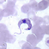
In the early stage, symptoms are typically either not present or mild, and may include fever, swollen lymph nodes , headaches, or swelling at the site of the bite. [1] After four to eight weeks, untreated individuals enter the chronic phase of disease, which in most cases does not result in further symptoms. [2] [5] Up to 45% of people with chronic infection develop heart disease 10–30 years after the initial illness, which can lead to heart failure . [2] Digestive complications, including an enlarged esophagus or an enlarged colon , may also occur in up to 21% of people, and up to 10% of people may experience nerve damage. [2] T. cruzi is commonly spread to humans and other mammals by the bite of a kissing bug. [6] The disease may also be spread through blood transfusion , organ transplantation , eating food contaminated with the parasites, and vertical transmission (from a mother to her baby). [1] Diagnosis of early disease is by finding the parasite in the blood using a microscope or detecting its DNA by polymerase chain reaction . [5] Chronic disease is diagnosed by finding antibodies for T. cruzi in the blood. [7] It affects more than 150 types of animals. [8] Prevention focuses on eliminating kissing bugs and avoiding their bites. [1] This may involve the use of insecticides or bed-nets . [9] Other preventive efforts include screening blood used for transfusions. [1] As of 2019 [update] , a vaccine has not been developed. [1] Early infections are treatable with the medications benznidazole or nifurtimox , which usually cure the disease if given shortly after the person is infected, but become less effective the longer a person has had Chagas disease. [1] When used in chronic disease, medication may delay or prevent the development of end–stage symptoms. [1] Benznidazole and nifurtimox often cause side effects, including skin disorders, digestive system irritation, and neurological symptoms, which can result in treatment being discontinued. [1] [2] As of 2019 [update] , new drugs for Chagas disease are under development, and experimental vaccines have been studied in animal models. [10] [11] It is estimated that 6.2 million people, mostly in Mexico, Central America and South America, have Chagas disease as of 2017, [1] [3] resulting in an estimated 7,900 deaths. [4] Most people with the disease are poor, [12] and most do not realize they are infected. [13] Large-scale population migrations have carried Chagas disease to new regions, which now include the United States and many European countries. [1] The disease was first described in 1909 by Brazilian physician Carlos Chagas , after whom it is named. [1] Chagas disease is classified as a neglected tropical disease . [14] Contents 1 Signs and symptoms 2 Cause 2.1 Transmission 3 Pathophysiology 4 Diagnosis 5 Prevention 6 Management 6.1 Complications 7 Epidemiology 7.1 Non-endemic countries 8 History 9 Research 9.1 Treatments 9.2 Diagnostic tests 10 See also 11 References 12 External links Signs and symptoms [ edit ] An acute Chagas disease infection with swelling of the right eye (Romaña's sign) Chagas disease occurs in two stages: an acute stage, which develops one to two weeks after the insect bite, and a chronic stage, which develops over many years. [2] [5] [15] The acute stage is often symptom-free. [2] When present, the symptoms are typically minor and not specific to any particular disease. [5] Signs and symptoms include fever, malaise , headache, and enlargement of the liver , spleen , and lymph nodes . [1] [2] [5] Rarely, people develop a swollen nodule at the site of infection, which is called "Romaña's sign" if it is on the eyelid, or a "chagoma" if it is elsewhere on the skin. [5] [16] In rare cases (less than 1–5%), infected individuals develop severe acute disease, which can cause life-threatening fluid accumulation around the heart , or inflammation of the heart or brain and surrounding tissues . [2] The acute phase typically lasts four to eight weeks and resolves without treatment. [2] Unless treated with antiparasitic drugs , individuals remain chronically infected with T. cruzi after recovering from the acute phase. [2] Most chronic infections are asymptomatic, which is referred to as indeterminate chronic Chagas disease. [2] However, over decades with chronic Chagas disease, 30–40% of people develop organ dysfunction ( determinate chronic Chagas disease), which most often affects the heart or digestive system . [2] [5] The most common manifestation is heart disease , which occurs in 14–45% of people with chronic Chagas disease. [2] People with Chagas heart disease often experience heart palpitations and sometimes fainting due to irregular heart function. [17] By electrocardiogram , people with Chagas heart disease most frequently have arrhythmias . [17] As the disease progresses, the heart's ventricles become enlarged ( dilated cardiomyopathy ), which reduces its ability to pump blood. [17] In many cases the first sign of Chagas heart disease is heart failure , thromboembolism , or chest pain associated with abnormalities in the microvasculature . [17] Also common in chronic Chagas disease is damage to the digestive system, particularly enlargement of the esophagus or colon , which affects 10–21% of people. [2] Those with enlarged esophagus often experience pain ( odynophagia ) or trouble swallowing ( dysphagia ), acid reflux , cough, and weight loss. [2] Individuals with enlarged colon often experience constipation , which can lead to severe blockage of the intestine or its blood supply . [2] Up to 10% of chronically infected individuals develop nerve damage that can result in numbness and altered reflexes or movement. [2] While chronic disease typically develops over decades, some individuals with Chagas disease (less than 10%) progress to heart damage directly after acute disease. [17] Signs and symptoms differ for people infected with T. cruzi through less common routes. ... Langgaard (1842). [52] The formal description of Chagas disease was made by Carlos Chagas in 1909 after examining a two-year-old girl with fever, swollen lymph nodes, and an enlarged spleen and liver. [52] Upon examination of her blood, Chagas saw trypanosomes identical to those he had recently identified from the hindgut of triatomine bugs and named Trypanosoma cruzi in honor of his mentor, Brazilian physician Oswaldo Cruz . [52] He sent infected triatomine bugs to Cruz in Rio de Janeiro , who showed the bite of the infected triatomine could transmit T. cruzi to marmoset monkeys as well. [52] In just two years, 1908 and 1909, Chagas published descriptions of the disease, the organism that caused it, and the insect vector required for infection. [53] [54] [55] Almost immediately thereafter, at the suggestion of Miguel Couto , then professor of the Faculdade de Medicina do Rio de Janeiro [ pt ] , the disease was widely referred to as "Chagas disease". [53] Chagas' discovery brought him national and international renown, but in highlighting the inadequacies of the Brazilian government's response to the disease, Chagas attracted criticism to himself and to the disease that bore his name, stifling research on his discovery and likely frustrating his nomination for the Nobel Prize in 1921. [53] [56] In the 1930s, Salvador Mazza rekindled Chagas disease research, describing over a thousand cases in Argentina's Chaco Province . [52] In Argentina, the disease is known as mal de Chagas-Mazza in his honor. [57] Serological tests for Chagas disease were introduced in the 1940s, demonstrating that infection with T. cruzi was widespread across Latin America. [52] This, combined with successes eliminating the malaria vector through insecticide use, spurred the creation of public health campaigns focused on treating houses with insecticides to eradicate triatomine bugs. [30] [52] The 1950s saw the discovery that treating blood with crystal violet could eradicate the parasite, leading to its widespread use in transfusion screening programs in Latin America. [52] Large-scale control programs began to take form in the 1960s, first in São Paulo , then various locations in Argentina, then national-level programs across Latin America. [58] These programs received a major boost in the 1980s with the introduction of pyrethroid insecticides, which did not leave stains or odors after application and were longer-lasting and more cost-effective. [52] [58] Regional bodies dedicated to controlling Chagas disease arose through support of the Pan American Health Organization , with the Initiative of the Southern Cone for the Elimination of Chagas Diseases launching in 1991, followed by the Initiative of the Andean countries (1997), Initiative of the Central American countries (1997), and the Initiative of the Amazon countries (2004). [30] Research [ edit ] Treatments [ edit ] Fexinidazole , an antiparasitic drug approved for treating African trypanosomiasis, has shown activity against Chagas disease in animal models. As of 2019, it is undergoing phase II clinical trials for chronic Chagas disease in Spain. [36] [59] Other drug candidates include GNF6702 , a proteasome inhibitor that is effective against Chagas disease in mice and is undergoing preliminary toxicity studies, and AN4169 , which has had promising results in animal models. [11] [60] A number of experimental vaccines have been tested in animals. ... As of 2019, vaccine research has mainly been limited to small animal models, and further testing in large animals is needed. [10] Diagnostic tests [ edit ] As of 2018, standard diagnostic tests for Chagas disease were limited in their ability to measure response to antiparasitic treatment. ... Endothelin-1 has been studied as a prognostic marker in animal models. [61] T. cruzi shed acute-phase antigen (SAPA), which can be detected in blood using ELISA or Western blot, [22] has been used as an indicator of early acute and congenital infection. [61] A novel assay for T. cruzi antigens in urine has been developed to diagnose congenital disease. [22] See also [ edit ] Drugs for Neglected Diseases Initiative Chagas: Time to Treat campaign Association for the Promotion of Independent Disease Control in Developing Countries References [ edit ] ^ a b c d e f g h i j k l m n o p q r s t u v w x y "Chagas disease (American trypanosomiasis)" . ... "Global economic burden of Chagas disease: a computational simulation model" . The Lancet Infectious Diseases . 13 (4): 342–8. doi : 10.1016/S1473-3099(13)70002-1 .CYP51A1, TNF, IL10, IFNG, MMP2, LGALS3, CCR5, PPARG, MMP9, IL2, HLA-A, HLA-DRB1, HSPA4, MTCO2P12, IL6, IL17A, MBL2, TLR4, COX2, IL18, IL1B, PTGS2, RBM45, CALR, PPP1R2C, TLR2, NFE2L2, NDUFA5, TGFB1, MYD88, CYTB, REN, IL1RN, NLRP3, ACHE, APOA1, ATM, PRL, VIP, TP53, VDR, KNG1, HP, TPI1, FN1, TLR9, CTSL, TRIM33, CYP2B6, ACE, BACE1, EPGN, NPNT, CCL4, CCL3, SPN, AMZ1, CCL2, THBS1, RRAD, TNFRSF1B, MIR208A, CCR2, SPINK1, CX3CL1, SDC4, SKIL, SLC11A1, SMPD1, SOD2, TRBV20OR9-2, KIR2DS2, TCN2, TWIST1, RO60, TRIM21, SRM, ROS1, COLEC11, PPARGC1B, PANX1, MASP2, PPARGC1A, SBNO2, SIRT2, RPIA, PLCB1, SIRT1, PTPN22, EBI3, PYCARD, HSPA14, UBAP1, GGNBP2, SBNO1, OTUB1, RABEP2, HEXIM1, KNTC1, TYK2, TNFSF11, VIM, VIPR1, VPS11, FOSL1, CDR3, DYSF, DBA2, TP63, ARHGEF10, MCU, ADAM7, EIF2S2, LMLN, MBD2, SLC25A31, TMPRSS11D, VIPR2, ABO, RAD51, CSF3, CYBB, DECR1, DHODH, DMD, EIF2S1, EIF2S3, FBN1, FCN2, FDPS, FHIT, FOXO3, GAST, G6PD, GABPA, GCK, GEM, GLUL, CTSB, CR1, HLA-G, CDKN1A, ACTB, ADA, PARP1, AGT, ALB, ALOX5, ARG1, STS, ATP2A3, ATP2B1, BCHE, BDNF, C3, CACNA1C, CAMP, CD80, CD86, HLA-DPB1, HSPD1, PPBP, COX1, NGF, NHS, NOS2, PNP, NRF1, OCA2, P2RX7, PAEP, PCNA, PDE2A, PIK3CA, PIK3CB, PIK3CD, PIK3CG, PLCB4, POLG, POMC, PPP1R12A, MIF, IGF1, MEF2D, IGHG3, IL2RA, IL4R, IL7, IL12B, IL13, IL15, ISG20, ITPR1, KIR2DL2, KIR3DL1, KRT8, LGALS9, LTA, LY6E, MAP6, MEF2A, NT5E
-
Upshaw–schulman Syndrome
Wikipedia
.; Muchitsch, E. (2012). "A new mouse model mimicking thrombotic thrombocytopenic purpura: correction of symptoms by recombinant human ADAMTS13" .ADAMTS13, THBD, F3, TFPI, VWF, CFH, HLA-DRB1, THBS1, ZFP36, TNFSF10, MAPKAPK2, ABCA1, PTPN22, TNS3, IL33, RBM45, KCNH8, RN7SL263P, SH3BP4, CCL2, STAT3, APOA1, S100B, PTGS2, PHEX, COX2, JAK2, HP, BRF1, FLT4, EPHB1, ELAVL1, SLC25A10, CASP1, CACNA1S, MTCO2P12
-
Monoclonal B-Cell Lymphocytosis
Wikipedia
Among these mutations, IVGH4-59/61 is most often mutated in low-count MBL while IGHV1-69, IGH2-5, IGHV3-23, IGH23-33, IGHV3-48 , and IGHV4-34 are most often mutated in high-count MBL and CLL/SLL. [2] [9] Finally, genetic abnormalities such as the deletion of the q arm in chromosome 13 found in low count MBL are more commonly associated with a favorable prognosis in CLL/SLL while those found in high count MBL, e.g. deletions in the q arm of chromosome 11 or p arm of chromosome 17 [13] are commonly associated with unfavorable prognoses in CLL/SLL. [9] Individuals with MBL-MZ have monoclonal B cell cells that bear complex and distinctive genomic abnormalities, such as deletions and translocations involving chromosome 7, presence of an isochromosome 17, and, rarely, mutations in the NOTCH2 and KLF2 genes. [6] Some of these genomic abnormalities are similar to those found in splenic marginal zone lymphomas and some of the MBL-MZ patients that bore these abnormalities developed this lymphoma. [2] The genetic abnormalities in atypical and non-CLL/SLL MBL have not been well-defined. [ citation needed ] The cited studies suggest that there is a step-wise accumulation of genomic abnormalities that lead to CLL/SLL MBL and MBL-MZ and then to overt malignancy. [9] It presumed that similar accumulations led to the development of atypical and non-Cll/SLL MLB and than to their respective malignancies. [6] However, given the number and diversity of these abnormalities, it is unclear which are critical determinants of these disorders. [2] A recent model based on laboratory studies of normal CD19 + B cell, monoclonal CLL/SLL MBL cells, and CLL/SLL malignant cells found that their accumulation of genomic abnormalities may be caused by progressively increasing: 1) double strand breaks in DNA , 2) activation of non-homologous end joining error-prone DNA repair mechanisms, and 3) consequential accumulation of genomic abnormalities which promote the clonal development, survival, proliferation, and ultimately malignancy of the involved B cells. [14] Infectious diseases [ edit ] Studies have identified MBL in ~30% of patients infected with the hepatitis C virus , found increased risks of CLL/SLL-MLB in patients with pneumonia, and decreased risk of CLL/CSS MBL in patients who have been vaccinated for influenza or pneumonia.NOTCH2, KRT20, MS4A1, LOC102724971, IGH, MBL2, LOC102723407, IGHV3OR16-7, IGHV3-69-1, CD19, LEF1, ITGA4, NOTCH1, IGF2BP1, IL22, MBL3P, CD274, IL21, AICDA, XPO1, HM13, CLEC12A, IMMP1L, MIR155, MIR15A, MIR21, LINC01672, COLEC10, BCL2, VIM, TP53, CXCR5, CD22, CD38, CD79B, CDK6, CCR6, IL2, IMPA1, JAK2, LAIR1, MME, NTF3, PIK3C2B, SELL, SIM1, BCL6, TNF, SPN
-
Diabetes Mellitus, Noninsulin-Dependent
OMIM
The contributions of Native American ancestors to maternal and paternal lineages were estimated as 90% and 40%, respectively. In a logistic model with higher educational status as dependent variable, the odds ratio for higher educational status associated with an increase from 0 to 1 in European admixture proportions was 9.4. ... Van Vliet-Ostaptchouk et al. (2008) genotyped 501 unrelated Dutch patients with type 2 diabetes and 920 healthy controls for 2 SNPs in strong linkage disequilibrium near the HHEX gene, rs7923837 and rs1111875, and found that for both SNPs, the risk for T2D was significantly increased in carriers of the major alleles (OR of 1.57 and p = 0.017; OR of 1.68 and p = 0.003, respectively). Assuming a dominant genetic model, the population-attributable risks for diabetes due to the at-risk alleles of rs7923837 and rs1111875 were estimated to be 33% and 36%, respectively. ... Among control subjects, the risk allele of this polymorphism was associated with impairment of insulin secretion according to the homeostasis model assessment of beta-cell function or the corrected insulin response. ... Unoki et al. (2008) detected consistent association of a SNP in KCNQ1 (rs2283228) with the disease in several independent case-control studies (additive model p = 3.1 x 10(-12); odds ratio = 1.26, 95% confidence interval = 1.18-1.34).PPP1R3A, HNF4A, PAX4, AKT2, GCK, IRS1, KCNJ11, HNF1A, HNF1B, SLC2A4, NEUROD1, PDX1, SLC2A2, IRS2, WFS1, HMGA1, ABCC8, CDKAL1, INS, INSR, ENPP1, ADCY5, CAPN10, TCF7L2, PPARG, TGFB1, LIPC, BCL2, MAPK8IP1, HP, NOS3, PPARGC1A, HMOX1, LEPR, ICAM1, UCP2, LEP, SIRT1, ADIPOQ, EDNRA, EDN1, TNF, TNFRSF1A, NOS2, KL, CPT1A, SNAP25, ATP2A2, EDNRB, RETN, GCKR, SLC30A8, GLIS3, GCGR, FTO, GLP1R, PTPN1, JAZF1, KCNQ1, IGF2BP2, MTNR1B, THADA, HMG20A, NOTCH2, PROX1, KCNK16, GPD2, UBE2E2, AP3S2, MAEA, CMIP, PEPD, GRB14, CCND2, PLEKHA1, DGKD, ZFAND3, ITGA1, PSMD6, PAM, KSR2, NFATC2, CAT, SHBG, MIR375, SLC2A1, BRAF, SLC1A2, ST6GAL1, HHEX, JADE2, IL13RA1, MIR126, SOD2, VPS26A, GSTM1, MOK, GNB3, GCG, CNKSR2, AUTS2, IGF2, GP2, INPPL1, SREBF1, SOD1, FGF21, LPL, ZNF257, FBN1, USP48, PPARA, SCTR, EPC2, IAPP, FAM234A, KLF14, CYBA, RNF6, IDE, ETS1, GIPR, CASP3, TNFRSF1B, SFRP4, OGG1, ND1, ADAMTS9, GPX1, AR, NFKB1, MIR27A, EGFR, MIR192, PCSK2, COX2, PRKCB, MIR144, HBA1, TIMP1, C2CD4A, ATF3, SLC22A3, MIR221, TMEM18, C3, C2CD4B, MIR483, MIR222, MIR423, RELA, FAS, CISD2, CBS, HK1, ATP2A3, MIR130B, HLA-DRB5, MIR203A, MIR142, CCDC92, MIR140, MIR200A, MIR98, MIR204, PAX6, ECE1, MIR214, ITLN1, MIR10B, MIR30A, MIR377, MIR1260A, CDO1, RARRES2, CCR5, SMAD5, MAT1A, NUS1, MIR1249, MIR1296, MAPK8, MIR1226, MIR1306, MRAS, MIR1228, MIR744, MIR939, CD36, MIR885, MIR1307, MIR151A, GPT, RBP4, IGF1, ID1, MIR127, MIR215, HSD11B1, MIR205, HPX, MIR195, MTOR, MIR141, GIP, HIF1A, MIR181C, GH1, UCP3, SOCS3, MIR17HG, MIR125B1, MIR432, MIR92B, MIR628, MIR532, MIR487B, BCL2L11, CYP1A2, S100A6, IL6, MIR485, MIR409, MIRLET7D, CCL2, MIR339, MIR335, MIR1301, MIR33B, MIR1908, MIR3173, MIR4482, BCL2L1, MIR4516, MIR8061, MIR6741, PRKAA1, PCK1, PCSK1, NFE2L2, BHMT, MIR7704, PPARD, MIR1260B, NUCB2, NKX6-1, ADRA2A, SERPINF1, AKT1, ZC3HC1, AGER, SERPINE1, MIR6803, KCNU1, AVP, MIR2116, MAPK1, TMEM155, MMP9, CASP8, PTGS2, G6PC, NR3C1, GSK3B, IGF1R, CCN2, REG3A, PIK3R1, CALCA, MAPK3, MAFA, HMGCR, MMP3, NPPB, SERPINA12, CRTC2, PC, ABCG2, CD38, BECN1, BAX, MMP1, PRKCZ, PTGDS, PRKN, MADD, HTR2C, AGTR2, TRPC1, ARRB2, LNPEP, AOC3, TRPC6, DCX, DEFB1, SMAD4, MIF, DNMT1, SLC9A1, CARTPT, NOG, PLAT, PLN, MTNR1A, UGT1A1, NFE2L1, DNM1L, CYP2E1, PPP1R3C, FOXM1, UGT1A6, SRD5A1, NR1D1, NOS1, THBD, AGRP, GJA1, PRKCE, CYB5R4, MAPK9, TGFBR2, CCKAR, CDKN2B, MAPK14, ACOT2, CYTB, MKI67, CTF1, SCN1B, DAG1, F7, ANKRD23, COL3A1, OXCT1, FEM1B, SRC, CALM1, CYP11A1, HSD3B1, KLF11, MAP1LC3A, KCNMB1, ARX, NCOA6, HSD17B3, TSPAN8, PNPLA3, GPT2, CASP7, BAD, ABCC2, ASIP, CASP12, PHOX2A, EPAS1, ATP2A1, PPP2CA, AK1, GADD45GIP1, AANAT, ATP5F1B, SLC16A11, STAR, BEST1, MPST, MTR, ALDOB, PRKCI, CCHCR1, MC4R, LMNA, CRHR1, EIF3F, SDF2L1, HLA-DQB1, SLC12A3, ATF6, CDKN2B-AS1, CAMK1D, NEUROG3, CTNNB1, ALMS1, ARAP1, ATM, UMOD, CDC123, ANK1, GRK5, DUSP9, NOX4, ABO, CYP19A1, ARL15, ELN, SRR, TP53INP1, CASR, TM6SF2, PTPN22, KIF11, CUBN, MAP2K7, TMEM163, GPR55, SCGN, TFAP2B, AGMO, HMGA2, DNER, ALDH7A1, RBMS1, HECTD4, RFC2, LAMA1, AHR, PRPF31, SLC26A9, PROM1, CTBP1-DT, ARG1, UBE2Z, DGKB, POU5F1, AHI1, ZMIZ1, RP9, INS-IGF2, ZRANB3, COBLL1, FAF1, TCF19, PRC1, ACSL1, TH, ND5, PTPRD, RASGRP1, GLP2R, RBPJ, TUB, GATAD2A, BAZ1B, LINGO2, NRL, ALK, PBX4, SSR1, NDUFAF6, PRKAG2, CLIP2, WRN, POC5, DHDDS, OAS1, SNHG17, ABCA4, SGCD, SLC16A13, CAMTA1, CTRB1, ADGRL3, EHMT2, SPPL3, STUB1, CAMK2G, TRDN, MACF1, TSBP1, STX8, TTLL6, PTPRN2, CAMKK2, RREB1, TRPM1, BCL9, LIG4, ATXN1, CTBP1, SUGP1, ROM1, TPCN2, MPHOSPH9, MCM6, SH2B3, POMGNT1, MASP1, FBXW7, RBPJL, SGCG, LIMK1, PPP2R2C, PDE6B, CERKL, FAM161A, RBP3, HIVEP2, HLA-DRB9, HLA-DOB, PRIM2, GPR119, MAGEC3, ASCC2, HK2, PRPH2, PEX10, HLA-DRB1, HGSNAT, SSTR4, NPY, SUMO4, ARL6, MNX1, GUCA1B, LCN2, UNC5D, MOG, OSBPL7, PCSK9, TRNL1, NECTIN2, PRKAA2, RTN4RL1, TCF7, TTC8, MAK, PYY, CYB5D2, TTC39C, MAP6, DLEU7, MFAP1, CFAP77, RELN, MBNL1, RFLNA, EDARADD, RDH12, MCC, LINC02694, LINC01344, RPSAP52, PSMD9, PCARE, TNFRSF11B, NTRK2, PRKAB1, MMP2, PDE6G, NRG4, ST6GALNAC3, APOA5, LPA, CTTNBP2, NTRK3, LRMDA, PDE6A, ZSWIM3, LTA, HFE, PTH1R, PTH, PTGFRN, PCNT, REG1A, RHO, OSBPL1A, SLC5A2, ZNF513, SELE, CCDC149, C8orf37, CCNQ, PRRX1, PLCD1, IFNG, SLC5A1, IDH3B, IMPDH1, LINC01010, PLCB3, IL17A, IDH3A, IL18, SLC9B2, C17orf58, ND6, PPARGC1B, SELP, ZNF775, IL10, PLG, TRNQ, TRNS1, TRNS2, TRNK, SI, TRNH, TRNW, IL1RN, TRNF, IL4, IL1B, TRNE, IL1A, ST3GAL4, ST3GAL3, PPIL6, PDILT, CXCL8, ARL9, NEK2, ND4, REN, RP1, EYS, KCNQ3, PIK3CD, SPP1, HORMAD2, CHCHD6, RP2, PIK3CB, PIK3CA, NLRP3, MTHFR, SLCO4C1, SDHAF4, MSH3, RLBP1, KIF9-AS1, RGS13, RGS7, RGR, RFC1, NFKBIL1, RPGR, RPE65, MRPL12, CNDP1, CDHR1, COX3, REEP6, COX1, FFAR4, GPBAR1, SCD, ATXN7, ATXN2, SLC22A1, SAG, HNF1A-AS1, ZNF101, GOLGA6A, PON1, PIK3CG, TTLL8, CHMP4B, IL17REL, EHMT1, LINC01122, KCNQ1OT1, INTS8, RAB3GAP1, ENPP7P10, RALY, NLGN1, CPEB3, MGLL, MINDY1, SPATA7, CD59, CD81, UBAP2, ARHGAP15, CDK5, NUDT6, POLR3A, KIZ, CDKN2A, TRIOBP, SNRNP200, PALLD, ZHX3, LINC00844, ABCB9, ABCB10, CRB1, PRC1-AS1, BRS3, ARHGEF18, RPGRIP1L, ADAMTS9-AS2, LINC01426, CAD, UBE2E2-AS1, PROX1-AS1, CA4, OSER1-DT, SLC30A10, FBXL7, CEP68, ZZEF1, ANGPTL8, APOM, ARPIN-AP3S2, LMCD1-AS1, CNR1, ZBED3-AS1, ACKR3, MERTK, PDSS2, ATP8B2, DLEU1, LRFN2, RAMP2, CALCOCO2, CPA1, GPR158, TOPORS, STARD9, NAMPT, FRY, CRP, CRX, PITPNM2, CNGA1, CNGB1, GTF2I, NFAT5, PNPLA6, CETP, KLHL7, TEX14, ANKH, LINC00271, RAI1, HMGA2-AS1, CXCR6, PRPF8, SUMO2P17, CHUK, YKT6, ZGLP1, TXNIP, CLCNKB, C1GALT1, PRCD, ARL2BP, BLM, NR2E3, LOC102723407, AGT, AGTR1, LRP12, LINC02030, AHSG, PURG, LINC02484, ALB, LINC02841, PELO, AKR1B1, EXOC6, HPGDS, PALD1, FOXP1, CCND2-AS1, NSG1, LINC02576, SND1, LRP1B, FSCN3, ADRB3, ADIPOR1, ABCA1, ANKFY1, SNX7, PEX5L, GLRX5, HSD17B12, ASB3, ANGPTL4, ADA, ADRB2, ADARB1, SOST, IMPG2, GHRL, SCAPER, LINC01611, ADRA1A, ADRA2B, TBL2, ANPEP, RCBTB1, VPS33B, HNF4A-AS1, OSBPL3, SCAANT1, TXNL4B, PTPN23, MIR5094, ABI3BP, PINX1, SAMM50, FSCN2, BBS2, PRPF6, SRBD1, DIANPH, BDNF, DARS2, BGLAP, SBNO1, AMACR, LINC00824, ASCL2, LINC02010, APP, GIN1, APOA1, APOB, APOC3, HORMAD2-AS1, IFT172, APOE, CNTLN, APRT, POLDIP2, ETS1-AS1, PABPC4-AS1, LINC01339, TSBP1-AS1, C5orf67, ARL3, PKN2-AS1, GPSM1, TSPAN3, PCSK6, CST3, AGBL5, PABPC4, PEA15, EXT2, EYA2, OASL, FABP4, GDAP1L1, FABP2, OFD1, GCC1, ADIPOR2, NPRL3, IFT88, FGF23, FGF14, ZNF408, BEST3, MIR29A, ESR1, ALDH1A2, PRPF3, PIM3, NRXN1, NRXN3, PIEZO2, LRAT, LPAR2, XYLT1, PRPF4, APLN, KIF9, SEMA4A, SELENBP1, CLN8, MGAM, MTMR3, AKTIP, ZSCAN20, ZIC1, THSD4, TLR4, TULP1, TNKS2, ZNF34, GOT2, TP53, TNFAIP6, SLC2A10, FFAR1, UCP1, C16orf74, GPR42, TGFBR3, MLX, LINC02245, GSTP1, GSTT1, SPHKAP, MIR146A, FOXO1, GABPA, XRCC4, SCD5, CPED1, PCNX2, VWF, VSNL1, BICC1, GABRA4, USH2A, GABRG3, GAD1, GAD2, VEGFA, VDR, VCAM1, CLRN1, MED23, YTHDC2, DPP4, UBE3C, GTF2IRD1, DSPP, RHOBTB1, ACE, G6PC2, DECR1, MAGI2, DBP, SLC7A14, CLOCK, FCHSD2, ACE2, SINHCAF, USP3, CELSR1, RBM19, FGF19, MIR4435-2HG, RAPGEF5, GDF15, MTSS1, UVSSA, SFI1, CYP2C9, WSCD2, DEPDC5, TRIB3, DHX38, IFT140, SENP2, CYP21A2, KIAA1549, DOCK4, DARS1, MRPS35, BCHE, LIPE, AIMP2, RNF19A, LDLR, USF1, ARNTL, NOS1AP, AHSA1, GSTK1, PTEN, RBM45, RENBP, CTLA4, IGFBP1, CRK, GRAP2, DMD, GYS1, PON2, CCL5, POMC, IGFBP3, MME, STAT3, LGALS3, CYP2C19, SLCO6A1, LPIN1, F3, STAP2, APOA2, AQP7, FOXA2, SST, SLC17A5, MET, HMGB1, GPR151, NIDDM1, SELENOP, NIDDM2, GPRC6A, PCK2, GGT1, OR10A4, DIO2, HSPA5, GC, CYP3A4, LINC01672, MPO, GGTLC5P, SIRT6, TLR2, GLO1, ELMO1, BCL11A, MLXIPL, ARID4B, NRF1, P2RX7, MIR155, MSTN, CYP2B6, ACR, GGTLC3, MRGPRX1, ALDH2, PDR, GGT2, MRGPRX3, APOL1, HSPA1A, SIRT3, GPR166P, VN1R17P, OXER1, SLC2A11, FZD4, FNDC5, SNCA, MRGPRX4, MIR21, HSPA4, ABCG1, HSPA1B, LPAR3, CHI3L1, NR3C2, FGF2, CD14, ISL1, HAMP, LGR6, ARNT, ZBTB7C, CAV1, GGTLC4P, SGK1, PSEN1, NR1H4, CISD1, SELENOS, CD163, TMBIM4, VIP, CCN4, MFN2, ROCK1, IL22, GGTLC1, VIM, SLC22A2, SOD3, PLIN1, OGA, CNBP, PLA2G2A, SAT1, HAVCR1, FETUB, SFRP5, NAT2, EPO, IARS1, GLUL, LBP, MIR27B, STS, APOA4, ACACB, KNG1, MIR29B1, GAS6, IL15, CD44, HLA-DQA1, ATN1, FXN, CD68, ADM, MIR29B2, LOC102724197, MEFV, CRY2, HGF, FGF1, MTTP, DDIT3, ANGPTL3, BCL2A1, SIRT2, DKK1, INSRR, KCNJ4, IL33, FADS2, CASP1, IL6R, IL37, CCK, SORT1, TBC1D4, MT1A, HEBP1, PRL, MBL2, PARP1, GRK2, ARHGEF11, ROS1, TRPM5, PTX3, FADS1, PYCARD, IL2RA, KRT16, IL5, XPR1, IL2, BMP4, HIRA, LPAL2, FABP1, PADI4, CHDH, FANCD2, PTK2B, BRCA2, SOCS1, FN1, XBP1, SLC47A1, FOXC2, SLBP, PLA2G7, FGB, GHSR, DGAT1, LIPG, EPHB2, IGFBP2, MIR34A, ELANE, MT2A, HSPG2, SOAT1, DPPA3, SULT1E1, TPD52, SH2B1, HHIP, HDLBP, NR1I2, AZU1, MIR122, BTC, CPE, MAP4K4, PDHX, STAM2, NCAM1, ACP1, CMKLR1, NGF, NHS, PKD1, UTS2, MYH9, PGC, MTCO2P12, IFNL3, RAPGEF1, SORCS1, KIR3DL1, GPX3, KDR, P2RY12, TIMP3, SNAP23, FLVCR1, GRN, HDAC9, AMY2A, TNFSF14, COMT, RPL29, GPD1, PDE3B, RAPSN, DACT1, SPX, TRAF6, PVT1, CYP2D6, AKR1A1, KEAP1, MIR223, ALOX12, HDAC3, MARCHF1, ISG20, RNU1-1, ANGPT1, C1QTNF9, TNMD, GAS5, IL13, CXCL12, SPARC, ENO1, CCL16, GPR142, CXCL10, ENHO, STRA6, ST13, TFRC, NPC1, CACNA1E, STK11, STX1A, CCR2, PDK4, RUNX2, NRG1, ERBB2, S100A9, SQSTM1, C1QL3, BCAR1, CLPS, LRG1, TOMM40, GEM, ACACA, DDAH2, FKBP5, CISH, CDK4, LGR5, METRN, MUSK, CREB1, MIR17, FLAD1, TAS2R13, CD40LG, PSMA6, MIR143, SLC39A7, CLU, POU2F1, NR1H3, PTGES2, FOXP3, OLR1, PCNA, LRP5, GAPDH, SLC2A9, ABCB6, PRKCA, NR0B2, XRCC1, ENSA, CXCR4, F5, PLA2G10, NR4A1, PLA2G6, ABCG5, ACHE, FOXO3, FBRS, STAT5A, P2RX3, CACNA1D, FSD1, HPSE, NPPA, FGFR1, KLK3, ARSA, PRMT1, IGF2R, CHGA, KLF4, PPP1R3B, DHX40, ANXA1, PNPLA2, ELAVL2, ERP44, CIDEC, PRDM16, FBXO8, BMP7, VPS13C, STEAP4, NNMT, CCNL1, MCIDAS, HSPA2, APOD, KHDRBS1, APOH, DST, MIR30D, ADAM17, FCGR3B, FCGR3A, ISYNA1, POU2F3, OSM, MIR145, SMUG1, B2M, NR1H2, NTS, MIR152, TNFSF10, WNK1, UTS2R, TM7SF2, GGCT, WNT5B, TNFRSF12A, NUP62, DDAH1, COL18A1, CMA1, IMPACT, HEMGN, GFAP, TNFSF11, F2, SP6, F2R, OGN, FAAH, PDE5A, BTF3P11, IL18R1, PBX1, ERBB4, NBEAL2, SIRT4, TCF4, SIRT5, HCCS, ELOVL6, GTF2H1, TTR, CILP2, TEK, FSD1L, TERF1, TFAM, PHLPP1, BACE2, TRPV1, MIR182, G6PD, ESR2, VEGFC, TGFBI, GRB10, TGM2, BMP2, OPA1, PARL, LINC01194, SLC6A4, ENTPD1, PRKG1, CENPJ, DRD2, LOX, KCNJ1, DCTN4, MC3R, PPIG, TUBB4B, JAK2, JAK1, S100A8, CERS6, ITGAM, NOD1, SPRY2, RNR2, JPH3, ITGA2B, LAD1, TMEM154, LITAF, STK38, PTPN2, RPS6KB1, KLB, PLF, MBOAT4, UCN3, KCNMA1, ALOX5, KCNJ15, TIMM8A, SGSM3, BRD2, PLTP, CEACAM5, RRAD, PRTN3, KCNJ9, H6PD, RXRA, MUTYH, PSAT1, PROS1, IRAK1, ITGA2, ANGPTL2, PGR, AMD1P2, EBI3, LRP2, C1QTNF5, CREBRF, PPIA, ADCY3, PHOSPHO1, ABCB1, RAC1, LRP1, SFTPD, EGR1, CYP2J2, FOXD3, CAPN1, RN7SL263P, BMS1, COPA, RAD1, TFB1M, TMPRSS6, SERPINA1, C1QTNF1, AMD1, NTN1, C5AR2, CDK9, CD33, MMP8, CASQ1, METRNL, ALX4, CYP2C18, NPC1L1, TLR9, SCARB1, INTU, SELL, LARS2, CYBB, ARID1B, KLF7, MIR495, SLCO1B1, CX3CR1, DOK5, CYP2C8, CYP1A1, P2RY1, NCOA5, SLC27A4, MAGEE1, FST, GFPT2, DUSP12, LRPPRC, CDKN1B, CELA3A, RNLS, FAT1, FBN2, SLC52A2, FCGR2B, ATP6AP2, CDKN1C, PREX1, KMT2D, SEMA6A, KLHL42, CHRM3, MICA, CPT2, CDKN1A, CP, CRYZ, CDC42, COX4I1, NR1I3, FCN3, CTSD, FABP6, MIR486-1, COX8A, PDLIM5, PTPRU, CDK2, IVNS1ABP, ABCC5, POSTN, OGT, CSF2, HOTAIR, NISCH, AGXT2, BSG, P2RX2, DEFB103A, XYLT2, COL9A3, DYRK1A, MIR30C2, P2RX6, CIB1, EP300, RCAN1, FAM3A, MIR30C1, ABCG8, ATG7, MIR29C, SLC30A6, CAV3, EPHX2, ARID3A, CEBPB, GGA3, CEL, DNAJB3, MIR33A, MIR34C, MIR320A, CELA1, CAMP, CPB2, CALR, EGF, IL32, CAPN2, ADAM28, CES1, LILRB1, SLC25A20, CAPN3, SULF2, NOD2, HSPB3, EPOR, ARHGEF12, VASH1, ESRRA, TRIM72, SERPING1, ZNF410, COMP, MIR20B, PINK1, ICOSLG, CD5L, CHPT1, DAPK3, ABHD6, CYP17A1, CYP11B2, KLF5, MEG3, PDAP1, CYP3A5, SUCLA2, TXNRD2, CNDP2, ARHGAP22, TBC1D1, MEPE, KLRK1, SEMA3A, NPEPPS, AAA1, SPHK1, ATF7IP, PER2, DNASE1, FOXO6, SORBS1, TSC22D1, SARDH, NQO1, DHCR7, SOCS2, GDF11, P2RY2, NR4A3, SLAMF1, LDHA, LECT2, LGALS1, LIF, C1QTNF3, FAM3B, LMX1A, DUOX1, PVALB, ICOS, PTPRN, PTPN9, RMC1, CYP2R1, APLNR, AFP, AFM, KIF6, SMAD7, KCNIP1, ADORA1, MAPT, DUOX2, MBP, IL20, ADH1B, PLIN2, MDM2, MEF2A, NANS, FRMD3, TERF2IP, ALOX5AP, IL1R1, IL6ST, SLC39A1, IL12B, SDC2, SDC1, CXCL5, SRL, ALOX15, SPINK4, ITGAX, MSMO1, TSPAN31, S100B, COPD, SFXN1, GHS, RXRG, KCNJ6, DESI1, RRAS, PRRT2, DLL1, RORC, NPAS4, KISS1, KLK1, KRT18, PRSS55, PRKCD, LARP1BP2, HDAC7, PIK3C2G, TAAR1, NM, SLC25A3, PHB, ATP6V1H, PGF, PFKM, NOTCH1, NT5C3A, PEG3, NPHS1, BFAR, IL23A, ERFE, TRPM6, ACAT1, PDCD1, PWAR1, PCYT1A, OCA2, R3HDML, PCBD1, OPRM1, P2RX1, P2RX4, P2RX5, PAK1, ACLY, NFKBIA, PRKAR1A, POR, PRKAB2, MMP7, MMP12, MMP13, SRGN, PTPA, MMP14, MRC1, RNU6-392P, GPRC5B, GAL, MT1E, FITM2, MT1X, NF2, MTHFD1, METTL9, PLXNA2, SLC35G1, PLK1, GNPDA2, PLD1, PLCG1, PLAGL1, PLAG1, PLA2G1B, PKNOX1, PKLR, IKBKB, CHIT1, CRISPLD2, AZGP1, GSN, KLRC4-KLRK1, GSR, UGT2B15, TF, TERT, GATA6, SERPINA3, ANGPTL6, TERC, TXN2, VIPR1, UCHL1, TMEM132A, TAPBP, CFH, VLDLR, ERVK-18, C1QTNF12, HLA-DMA, ATP5PF, GAST, VTN, STX4, BID, GFER, STAT5B, TLR3, TRH, ACAD10, CRISP2, GLA, CEP55, CD2AP, CBLIF, P2RX5-TAX1BP3, MIR107, BCR, CIMT, GPR39, THBS1, TTN, GHR, FFAR2, TLE1, TKT, TXN, GCA, GPS2, KLF10, SESN2, GFPT1, ARID5B, GORASP2, MIR199A1, FGL1, SPINK1, AQP9, SOX4, SORD, SNRNP70, FKBP4, AOC2, ANXA6, SPZ1, HTN1, SLC30A1, APPL1, HTR2A, SLC16A1, TRPM7, ANGPT2, GPC5, SLC6A2, ST8SIA4, SLC2A5, ANKRD55, IGFALS, QRSL1, FHL1, MIR199A2, MIR20A, STAT4, LINC-ROR, PGR-AS1, SSTR5, WNT5A, MIR130A, ONECUT1, FLT1, SREBF2, NAT10, C4B_2, MYDGF, LOC107832851, DMAP1, LARP6, LY75-CD302, RASD1, RBM14-RBM4, LSR, NBAS, DEFB4B, MCS+9.7, HMGA1P8, LOC110806263, MMP26, RHOT1, CCL28, ALG1, METTL3, LIN7C, LINC00994, DIP, RPS10-NUDT3, GNG12-AS1, RAB14, WASHC1, CYRIB, VIM-AS1, NDUFA12, SLC50A1, NEIL3, LOC102723996, SIRT1-AS, THRIL, MIR1910, SLC38A2, ALL2, NLGN3, CASZ1, ENAH, ZFAND6, OPN1MW3, VPS35, EXOSC4, RNPC3, UGT1A10, UGT1A8, UGT1A7, ERRFI1, UGT1A5, UGT1A9, MIR3188, TEMPS, UGT1A4, BNC2, TRPM4, TET2, FBLIM1, SALRNA1, CBSL, LOC102723971, UGT1A3, TREM2, MIR3939, APPL2, MOCOS, ZC4H2, LGR4, DEFB103B, COMMD3-BMI1, MIR4756, USE1, ASAH2, ENOX1, BOP, CAMK2N1, SLC6A20, SLC52A1, CST12P, SAGE1, PLUT, WWOX, LRP1-AS, ADA2, MIR3666, IL17D, SHC3, CERNA3, HDL3, FXYD5, MIR4463, PERCC1, SOX6, UGGT2, ROBO4, DUSP26, SUCNR1, IGSF21, CISD3, TIMD4, TRIM47, KCTD1, MIF-AS1, TTC28-AS1, EXOC3L2, SLC9C1, UCN2, CYP4V2, WNT3A, GALP, SLC22A16, LY86-AS1, KRT90P, DSEL, HCCAT5, GORAB, BIRC8, AD7, RNU12, AQP11, H19, TXNRD3, SLC46A1, OPN4, PRDM6, CGB8, CGB5, MUC19, LINC00523, PGP, TRMT10A, ABCC11, BPIFA4P, HS6ST3, MMAB, PHF6, ZACN, SLC27A1, LINC-PINT, MALAT1, PPP1R1B, KAT8, CHKB-CPT1B, SLC41A2, HORMAD1, IMMP2L, LEKR1, SAMD12, MIXL1, GPR61, LINC01193, PWAR4, C7orf50, GLYR1, UBASH3B, ORAI1, PTPN5, GPIHBP1, MFSD9, HCAR2, ZNF469, ZBED3, ZGPAT, BRINP3, ZFP69, HSD17B13, ACTBL2, L3MBTL3, TIRAP, TAS2R12P, TAS1R3, NRK, CLDN19, SIK1, UPP2, PPM1K, SLC2A12, CBLL2, FAM3D, ACVR1C, DQX1, SGMS2, NLRP6, ARID2, PIFO, MLKL, TET3, PDIK1L, MTPN, LRGUK, ROMO1, OR2AG1, PIWIL4, SESN3, FGFBP3, GSC, MT1P3, HT, GPAT4, BEAN1, CD300LG, PTPRVP, SLC30A7, IL23R, FAM78B, CENPX, KLHDC7A, SGMS1, MED19, CADM2, ANGPTL5, DCD, THEM4, TADA1, AKNAD1, SLC41A1, OLIG1, SLC22A12, CASC2, ANKK1, OSBPL8, PTF1A, WDR72, NEGR1, HCG27, IL27, SPESP1, MARCHF8, FOLH1B, TPRG1L, LYPLAL1, UNC5B, TCHHL1, SKA1, MSI2, GGTA2P, JMJD1C, SLC39A12, TPPP2, SPIC, RFX6, SLC29A4, SETDB2, INHBE, MICC, MT1IP, CCAR2, MIR323B, MARK4, MIR146B, MIR202, SPTBN4, MIR496, MIR499A, MIER1, USP37, NPS, CHD8, POTEM, TSHZ3, ECSCR, HNP1, MIR424, MIR382, SLC25A19, EIF2AK4, IGAN1, NIF3L1, ELOVL5, BACH2, EXOC4, RNASEK, MIR378A, PLIN5, POTEKP, PGBP, UBL5, CXCL16, NEUROD4, POU5F1P3, CHCHD2P9, TNFSF12-TNFSF13, USP36, KMT5AP1, MUC5B, OPN1MW2, CPA6, HYMAI, CDK11A, SLC12A9, ARNTL2, CA10, SMARCAD1, OCTN3, CELF4, SPHK2, MIR708, C20orf181, MIR661, GOPC, PELI1, KIDINS220, POU5F1P4, CXADRP1, NCF1, CFAP97, WASH6P, XPO5, MIR590, MIR657, MIR593, PLEKHG5, MRS2, GJD2, ATP10A, SLC39A10, PROK2, MIR99A, TOMM5, MUL1, ASRGL1, MIR15A, MIR15B, MIR18A, MIR187, STN1, MIR200B, ADM2, PRR5L, BBS10, LIN28A, NLRX1, NEIL1, MIR206, TNFAIP8L2, MIR154, MOGAT2, MIR147A, MIR106B, MFRP, MED25, TLR10, NIPA2, MOB2, COLEC12, SETD7, ALPK1, TAS1R2, MIR125A, MIR128-1, TSEN2, MIR132, COASY, MIR210, IRX3, MIR95, MBOAT7, WDR13, MIR296, PDIA2, GORASP1, DCLRE1C, GMCL2, GMCL1, NFKBIZ, NSD1, MIR30E, FN3K, DMRTA1, MIR31, SLC17A9, UBE2O, FNDC4, GNPNAT1, RFX7, GRAMD2B, MIR217, LY6G6E, MIR219A1, KCTD15, MIR22, VKORC1, MIR23A, SLC30A5, MIR23B, BRD9, WNK4, MIR26B, FRTS1, ACD, SCLY, UTRN, LARS1, HACD3, IK, IGH, IGFBP5, IGFBP4, IFNA13, IFNA1, ICAM3, HTR4, HTR2B, HTC2, HSPD1, HSP90AA1, HSPB2, HSPB1, HSPA9, HSD11B2, HES1, HRC, HRAS, HOXA5, HNRNPK, HNRNPF, HNRNPA1, FOXA3, HMBS, IL3, IL4R, IL7, KCNJ10, L1CAM, KRT31, KRT19, KRT7, KPNA1, KIR3DL2, KIR2DS1, KEL, KCNN3, KCNN2, KCNK3, KCNJ3, CXCR1, KCND3, KCNC4, KCNB1, ITGB2, INSIG1, IMPA1, ILK, TNFRSF9, IL16, IL15RA, IL12A, HLF, HLA-DRA, HLA-DQB2, GDF1, GNAO1, GLRX, GLS, GCLC, GLB1, GK, GJA8, GJA4, GH2, GDNF, GDF2, OPN1MW, SFN, GCH1, GBP2, GATA4, GATA3, GAP43, GALNT2, GABRG2, GABPB1, FUT6, FUT2, NR5A1, GOLGB1, GPI, HLA-DQA2, HSD17B10, HLA-DPB1, HLA-DOA, HLA-DMB, HLA-A, CFHR2, HCRT, HBA2, HAS3, HARS1, HADH, HADHA, GZMB, GPM6A, GYG1, GTF3A, GSTA4, GSTA1, GRB2, FFAR3, GPR35, GPER1, GPR21, CXCR3, GPR1, LAMB3, LAMP1, LAMC2, CCN3, OAS3, NR4A2, NUCB1, NTRK1, YBX1, SLC11A2, NPY2R, NPR3, NPR2, NPR1, NPM1, NME1, P4HB, NGFR, NFYC, NFYB, NFYA, NFATC1, NEFL, NDUFS4, NAGLU, MYO5A, MYLK, MYL2, OMG, PAEP, MYF5, PDK2, PIN1, PIK3C3, PIK3C2A, SERPINE2, SERPINB6, PGM1, PFKFB3, PFKFB2, PFDN2, PER1, PECAM1, PDHB, PRDX1, PDGFRB, PDGFB, PDE4D, PDC, PDB1, PCOLCE, SERPINA5, PCDH8, PAWR, PAPPA, SERPINB2, MYH6, MYD88, LAMP2, SH2D1A, MAX, MAS1, MAOA, MAN2A1, MAN1A1, MAFD2, SMAD3, SMAD2, SMAD1, MXD1, LYZ, LY75, MC5R, LY9, LTF, LTBR, CYP4F3, LRP6, LPP, LOXL2, FADS3, LIPA, LCN1, STMN1, MB, SMCP, MUC1, MST1, MTRR, RNR1, ATP8, MT1L, MT1M, MT1JP, MT1H, MT1G, MT1F, MT1B, MSX2, MSH5, ADAM11, MSH2, MPI, ALDH6A1, KMT2A, MGST3, MGP, MGAT2, MFAP2, MAP3K5, MEF2C, ME1, FOS, FOLR2, FOLR1, CA5A, CCND3, CCKBR, CBR3, CBR1, RUNX3, RUNX1, CAV2, CAMK2A, CALCR, CACNB3, CACNA1C, CA2, CD4, VPS51, C5, CAPN5, C4B, C4A, TSPO, BRCA1, BPI, BNIP3, BMP6, BMI1, CD247, CD86, BLK, CGA, AP3S1, CKM, CKB, CIDEA, CHRNB4, CHRNA4, CHN2, CHM, CEACAM7, CEACAM3, CGB3, CFTR, CD34, CTSC, CENPA, CECR, CEBPA, CDX2, CDKN3, CDK6, CDH13, CDH2, CD48, ENTPD3, BLVRA, BGN, CLCN3, AIF1, AMY1C, AMY1B, AMY1A, BIN1, AMPD1, AMH, ALPP, ALPL, ALDH3B1, ALCAM, ALAS1, AIC, ANXA5, ADORA3, ADORA2B, ADH1C, ADD1, ADCYAP1, ACVR2B, ACTG2, ACTG1, ACTB, ACAA1, AAVS1, ANK2, APCS, CFB, ATIC, BCS1L, OPN1SW, HCN2, BCAT1, AVPR2, ATP5PO, ATP7A, ATP5MC1, ATP4A, ATP2B1, ATP12A, ATHS, BIRC2, SERPINC1, ARSL, ARR3, RND3, RHOA, AQP5, AQP2, FASLG, APOC4, APOC2, BIRC3, CLCN2, CLCN5, FOLH1, EFNB3, EPHA4, ENO2, MARK2, ELK3, ELAVL1, EIF4G1, EIF4EBP1, EIF4A2, EGR3, EGR2, CELSR2, EFNB2, EPHX1, LPAR1, TYMP, EBM, E2F1, DUSP6, DRD3, DPYS, DPYD, DPT, DNTT, DMP1, EPHB1, ERBB3, DLD, FASN, FMO3, FLNA, FLII, FHIT, FGFR4, FGF11, GPC4, FDXR, FCP1, FCGRT, FBP1, FAP, ERG, FABP5, FABP3, F2RL2, F2RL1, EXT1, EVC, ETV3, ETFA, ESRRG, ESRRB, FBL, DMBT1, SEPTIN1, CLK2, COL17A1, CS, CRMP1, CREM, CREBBP, CRABP2, CPT1B, COX10, COX7A1, COX5B, MAP3K8, CORT, COL9A2, CSHL1, COL9A1, COL8A1, COL6A3, COL4A5, COL4A2, COL2A1, COL1A1, CNTF, CNR2, CMM, CCR1, CSF3, CST2, DIAPH2, BRINP1, CFD, DES, DEFB4A, DEFA3, DEFA1, DDX3X, DDT, AKR1C2, DCN, DBI, DBH, CYP27B1, CTAA1, CYP27A1, CYP8B1, CYP7A1, CYP2A6, CYP1B1, CYB5A, CXADR, CUX1, CTSL, CTSH, CTRL, PKM, PLA2G4A, PLEK, RBM14, SPTLC1, AGPAT1, PROCR, HYOU1, CARM1, UNC13B, CAP1, TIMM44, FAM3C, IFI30, LYPLA1, SPON1, SLC19A2, SPON2, NSA2, PEMT, ATP8A1, DLC1, CITED2, KLF2, WARS2, TCIRG1, SIGMAR1, FSTL3, SLC35A1, OLFM4, OLIG2, CCL27, BTN2A1, ADAMTS13, ESM1, LIAS, METAP2, EHD1, MALT1, FGL2, NMU, CD300C, CYP46A1, PPP1R13L, NPC2, CPLX1, WASF3, STARD10, SLC17A3, CAPN9, EBP, CD226, CTCF, IGF2BP3, IGF2BP1, USP16, ZMPSTE24, FLOT1, TREH, AIM2, GDF3, CXCL14, STXBP5L, GAL3ST1, ADAMTS2, ATG5, AKAP6, CHST3, FHL5, HOMER1, EIF2AK3, ITM2B, KIF20B, NCR1, TJP2, PEX16, TMPRSS11D, TGFBRAP1, ADGRG1, PIWIL1, ITGB1BP1, MSC, LARGE1, HACD1, SOX13, WTAP, TRIM13, MAFB, NUTF2, MPHOSPH10, TSHZ1, RBM6, HIPK3, PREB, AKT3, SCO2, KCNE2, THRAP3, EXOG, RNF10, NCOR1, SGSM2, HEPH, IP6K1, TOX, HDAC4, SART3, ECE2, AQR, EIF5B, LPIN2, NCOR2, PTPRT, MAP4K5, SLC16A7, DHDH, MCTS1, RGCC, SLC27A6, REM1, TRBV7-8, IGHD1-14, SLCO1B3, PCLO, MCAT, EIF3K, PDLIM3, SESN1, KLF15, SIT1, SERP1, IL17B, DISC1, DKK4, SIGLEC7, ACAD8, GREM1, BSCL2, PCOLCE2, CNNM1, DBNL, TBK1, HSPB8, MBL3P, CSAD, WAC, BPIFA1, TLR7, DNAJC27, IGF2-AS, GP6, IPO11, HSPA14, IRAK4, SLC35B3, G0S2, MYLIP, SLC40A1, TBX21, ERO1A, SLC2A8, SLC39A3, RHOD, PADI1, GPR132, HOOK2, CD274, CTNNA3, NUPR1, MYCBP, CHEK2, FAIM2, PSD3, SYNE1, TTC28, COBL, ARC, SIK2, FBXO28, SYT11, TBC1D9, CAND2, KDM1A, NT5C2, DICER1, RAB18, SACM1L, SBNO2, CARD8, MLXIP, COG2, ACOT7, MGAT4A, SLCO2B1, PTGDR2, AKAP13, CRTC1, NCS1, KLHL3, PANX1, PITPNC1, KIFBP, ATRNL1, TENM4, TKFC, PART1, MGAT4C, ARIH1, KLK5, TAFA5, SLC39A6, LY96, ZFPM2, BACE1, ZMYND8, DAPK2, FAM215A, SEC14L2, PES1, BRD4, TRAM1, NPTXR, TRS-AGA2-3, MLYCD, SLC33A1, ZBED1, SERPINF2, XCL1, SLC2A3, SLC1A7, SLC1A5, SKP2, SKIL, SIX3, SHC1, SRSF5, SELPLG, SDHA, CX3CL1, CCL22, SLC6A8, CCL20, CCL11, CCL8, CCL7, CCL4, CCL3, SCO1, SCN8A, SCN7A, SCN2A, SAA2, SLC5A5, SLC8A1, S100A12, SRI, TADA2A, TACR3, TAC3, TAC1, SYT4, SYT1, STXBP3, STX5, STIM1, STC1, SSTR2, SRD5A2, SLC9A3, SPRR2A, SPG7, SOX5, SOX2, SORL1, SIGLEC1, SMPD1, SLPI, SLCO1A2, SLC18A2, SLC10A2, SAA1, S100A4, TAL1, DNAJC3, PTMAP4, PTMA, PTGER3, PTBP1, PTAFR, PSMD3, PSMD2, PSMB6, PSMA7, PSG2, TMPRSS15, EIF2AK2, PTPN6, MAPK7, PRKD1, PPP1R2, POU3F1, POU2F2, PODXL, SEPTIN5, PNLIP, PMM2, PML, PMAIP1, PTPN4, PTPN11, RYR2, REG1B, RTN1, CLIP1, RPS19, RPS6KA3, RPS6KA2, RPN2, RPE, RORB, RNH1, RIT2, REV3L, REL, PTPRB, RCN2, RARB, RANGAP1, RAG1, RAB27A, RAB5B, RAB4A, PYGB, PTPRS, PTPRF, PTPRC, TAF1, TCP1, HTR3B, API5, ADAM19, MBTPS1, TNFRSF25, PAGE1, VAMP4, VAMP8, HSD17B6, JRK, KHSRP, DENR, PIAS1, CST7, TNFRSF14, DGKZ, IRS4, DOC2B, DYRK3, LOH19CR1, BRAP, PICALM, ZNF239, SLC14A2, AAAS, FOSL1, TNFSF12, RAB11A, MAFK, NOL3, PCSK7, HGS, MTMR7, USP2, USP14, PAPSS2, RPL14, CCRL2, TAF1C, F2RL3, KALRN, TRPA1, RIPK2, EIF2B5, ZPR1, FUBP1, PER3, TMEM11, BANF1, GALR2, TNFRSF11A, DLK1, TNFRSF18, SIGLEC5, ECB2, HBFQTL2, PRDX2, TLL1, HSP90B1, NR2C2, TPO, TPM4, TPM1, TOP2A, TOP1, TNFAIP3, NR2E1, TLR5, TLR1, TLE3, TRPM2, TJP1, TIMP2, TIAM1, THBS4, THBS2, TGFB2, TGFA, TFPI, TFAP2A, TERF2, TEP1, TRAF3, TST, TFEB, NSD2, LAP, PRRC2A, MANF, SLMAP, ZNF236, ZFP36, SF1, YWHAG, YY1, XDH, XBP1P1, WARS1, TNFRSF4, VPREB1, VEGFB, VDAC1, UPK2, UGT1A, UGCG, SCGB1A1, UBE2B, TYROBP, TYK2, TXNRD1, LINC02605

-
Gastrointestinal Stromal Tumor
Wikipedia
"Gastrointestinal stromal tumors (GIST): a model for molecule-based diagnosis and treatment of solid tumors".KIT, PDGFRA, SDHB, SDHC, TULP3, NME7, SDHA, SUFU, IFT52, SMO, CDON, TGFBR2, HIPK2, RB1, CD3E, IFT88, DYRK2, PTCH2, SALL3, KIF3A, STK36, ZBTB11, C2CD3, IFT172, RPL24, PTCH1, B9D1, RAB23, HIPK1, CTNNA1, DMD, TMEM17, BOC, FOXF1, DISP1, GLI1, GLI2, GLI3, NDST1, IHH, GLIS2, NPC1, TMEM231, HHIP, CC2D2A, HHAT, ELP4, CDKN2A, H3P10, PIK3CB, PIK3CG, PIK3CD, PIK3CA, PAX3, MIR4683, HOTAIR, MIR34C, MIRLET7C, CD34, DPP4, E2F1, MTOR, MEN1, TPX2, RASSF1, BANK1, TESC, SLC2A4, SLC12A3, SNAI1, TP53, TSC1, REC8, AKT1

-
Second-Impact Syndrome
Wikipedia
"Implementation of Neuropsychological Testing Models for the High School, Collegiate, and Professional Sport Settings" .


