-
Disfigurement
Wikipedia
In various religious and spiritual contexts, disfigurement has been variously described as being a punishment from the divine for sin (such as Yahweh 's defacement of Cain for Abel 's murder in Judaism), as being (such as Paul of the New Testament 's arguments about Christ 's sufferings) caused by supernatural forces of hate and evil against the good and just, which will be later atoned for, or as being without explanation per se with people just having to endure. ... Find sources: "Disfigurement" – news · newspapers · books · scholar · JSTOR ( July 2016 ) ( Learn how and when to remove this template message ) Lon Chaney 's version of " Erik " in the 1925 film The Phantom of the Opera had pervasive facial disfigurements, including jagged teeth and sunken-in eyes. ... A common origin of his skin and hair colors revolve around chemical burns as the result of the Joker character either falling into, jumping into, or being thrown into a vat of noxious chemicals. In Tim Burton 's 1989 film adaptation of Batman , the Joker character, in this version a criminal originally known as "Jack Napier", receives his distinct rictus grin as the result of a botched plastic surgery that he received after a ricocheted bullet that Napier intended to harm Batman badly injured the Joker's face. ... Perhaps the Punisher's most iconic nemesis, he was played in 2008's Punisher: War Zone by Dominic West . [10] Nick Cave and the Bad Seeds ' popular alternative rock song " Red Right Hand ", first released in 1994's Let Love In , describes a nightmarish figure with a blood-red, disfigured hand (as referred to in the title [11] ).

-
Febrile Infection-Related Epilepsy Syndrome
Wikipedia
PMID 21883180 . ^ van Baalen, A; Häusler, M; Plecko-Startinig, B; Strautmanis, J; Vlaho, S; Gebhardt, B; Rohr, A; Abicht, A; Kluger, G; Stephani, U; Probst, C; Vincent, A; Bien, CG (August 2012). ... Neuropediatrics . 43 (4): 209–16. doi : 10.1055/s-0032-1323848 . PMID 22911482 . ^ Fox, Kristy; Wells, Mary Ellen; Tennison, Michael; Vaughn, Bradley (July 11, 2017). ... Lancet Neurol . 10 : 99–108. ^ Carabello, RH; Reyes, G; Avaria, MF; Buompadre, MC; Gonzalez, M; Fortini, S; Cersosimo, R (2013). "Febrile infection related epilepsy syndrome: a study of 12 patients". ... Retrieved 7 October 2014 . ^ Appenzeller, S; Helbig, I; Stephani, U; Haeusler, M; Kluger, G; Bungeroth, M; Müller, S; Kuhlenbäumer, G; Van Baalen, A (2012). ... Epilepsy Res . 69 : 67–79. ^ Nabbout, R; Mazzuca, M; Hubert, P; Peudennier, S; Allaire, C; Flurin, V; Aberastury, M; Silva, W; Dulac, O (2010).
-
Papillary Fibroelastoma
Wikipedia
. ^ Matsumoto N, Sato Y, Kusama J, Matsuo S, Kinukawa N, Kunimasa T, Ichiyama I, Takahashi H, Kimura S, Orime Y, Saito S (2007). ... PMID 24839649 . ^ Takada A, Saito K, Ro A, Tokudome S, Murai T (2000). "Papillary fibroelastoma of the aortic valve: a sudden death case of coronary embolism with myocardial infarction". ... PMID 18191538 . ^ Gopaldas, R. R.; Atluri, P. V.; Blaustein, A. S.; Bakaeen, F. G.; Huh, J.; Chu, D. (2009). ... Thorac Cardiovasc Surg . 55 (3): 204–7. doi : 10.1055/s-2006-924439 . PMID 17410513 . v t e Cancers from and involving the heart Primary Papillary fibroelastoma Rhabdomyoma Angiosarcoma Teratoma Cystic tumour of the atrioventricular nodal region Other Myxoma Atrial Lipoma Secondary

-
Factor Xiii Deficiency
Wikipedia
FXIII concentrate is only available under the Federal Drug Administration's Investigational New Drug (IND) Program, or through clinical trial. [4] Recombinant factor XIII [ edit ] Recombinant factor XIII (rFXIII) is the only drug alternative to receiving blood transfusions, the traditional treatment for factor XIII deficiency. Novo Nordisk ’s rFXIII, catridecacog , was approved by the US Food and Drug Administration in 2014. ... One of the biggest fears in developing rFXIII was that the body would mount an immune-response to the protein; however, several safety and pharmacokinetics studies have reported no immunogenic response to rFXIII or associated yeast products. [2] See also [ edit ] Factor XIII References [ edit ] ^ Dorgalaleh A, Naderi M, Hosseini MS, Alizadeh S, Hosseini S, Tabibian S, et al. (2015). ... Cite journal requires |journal= ( help ) ^ a b c Lovejoy A, Reynolds T, Visich J, Butine M, Young G, Belvedere M, Blain R, Pederson S, Ishak L, Nugent D (2006). "Safety and pharmacokinetics of recombinant factor XIII-A2 administration in patients with congenital factor XIII deficiency" . ... External links [ edit ] Classification D ICD - 10 : D68.2 ICD - 9-CM : 286.3 OMIM : 134570 134580 MeSH : D005177 DiseasesDB : 31412 SNOMED CT : 18604004 External resources eMedicine : ped/3040 v t e Disorders of bleeding and clotting Coagulation · coagulopathy · Bleeding diathesis Clotting By cause Clotting factors Antithrombin III deficiency Protein C deficiency Activated protein C resistance Protein S deficiency Factor V Leiden Prothrombin G20210A Platelets Sticky platelet syndrome Thrombocytosis Essential thrombocythemia DIC Purpura fulminans Antiphospholipid syndrome Clots Thrombophilia Thrombus Thrombosis Virchow's triad Trousseau sign of malignancy By site Deep vein thrombosis Bancroft's sign Homans sign Lisker's sign Louvel's sign Lowenberg's sign Peabody's sign Pratt's sign Rose's sign Pulmonary embolism Renal vein thrombosis Bleeding By cause Thrombocytopenia Thrombocytopenic purpura : ITP Evans syndrome TM TTP Upshaw–Schulman syndrome Heparin-induced thrombocytopenia May–Hegglin anomaly Platelet function adhesion Bernard–Soulier syndrome aggregation Glanzmann's thrombasthenia platelet storage pool deficiency Hermansky–Pudlak syndrome Gray platelet syndrome Clotting factor Hemophilia A/VIII B/IX C/XI von Willebrand disease Hypoprothrombinemia/II Factor VII deficiency Factor X deficiency Factor XII deficiency Factor XIII deficiency Dysfibrinogenemia Congenital afibrinogenemia Signs and symptoms Bleeding Bruise Hematoma Petechia Purpura Nonthrombocytopenic purpura By site head Epistaxis Hemoptysis Intracranial hemorrhage Hyphema Subconjunctival hemorrhage torso Hemothorax Hemopericardium Pulmonary hematoma abdomen Gastrointestinal bleeding Hemobilia Hemoperitoneum Hematocele Hematosalpinx joint Hemarthrosis
-
Neurocysticercosis
Wikipedia
Neurocysticercosis / ˈ nj ʊər oʊ ˌ s ɪ s t i ˌ s ɜːr ˈ k oʊ s ɪ s / is a specific form of the infectious parasitic disease cysticercosis that is caused by the infection with Taenia solium , a tapeworm found in pigs. ... PMID 20045747 . ^ Abba K, Ramaratnam S, Ranganathan LN (17 March 2010). Abba K (ed.).TLR4, IL10, GSTT1, SLCO6A1, HPGDS, COX1, TACSTD2, ICAM1, GSTK1, GSTM1, GSTM3, CYTB, CD14, EPC1, FOXP3, CD38, VEGFA, TRAF1, TNF, CTSB, TAC1, SLC12A3, FGF2, GSTP1, MMP9, FOLH1, ISG20, FAS, IL2RA, IFNG, GFAP, HPRT1, HLA-A, ALK

-
Cerebrovascular Disease
Wikipedia
Benjamin; Hoang, Jenny; Hunt, Christopher H; Kennedy, Tabassum A; Khalessi, Alexander A; Mack, William; Patel, Nandini D; Perlmutter, Joel S; Policeni, Bruno; Schroeder, Jason W; Setzen, Gavin; Whitehead, Matthew T; Cornelius, Rebecca S; Corey, Amanda S; Corey, A. S (2017). "ACR Appropriateness Criteria ® Cerebrovascular Disease" . ... C; Fisher, M; Furie, K. L; Heck, D. V; Johnston, S. C; Kasner, S. E; Kittner, S. J; Mitchell, P. ... T; Hoh, B. L; Janis, L. S; Kase, C. S; Kleindorfer, D. O; Lee, J. ... Donald; Saver, Jeffrey L.; Albers, Gregory W.; Alberts, Mark J.; Chaturvedi, Seemant; Feldmann, Edward; Hatsukami, Thomas S.; Higashida, Randall T.; Johnston, S.ACE, TNF, ITGB3, ICAM1, ITGA2B, NOS2, EDNRA, COL4A1, HDAC9, F2, F5, CST3, MTHFR, NOTCH3, LPA, APP, ZFHX3, CASZ1, ABO, SH2B3, ALB, ALOX5AP, NOS3, PLA2G7, IL1B, GLA, PTGS2, IL1RN, IL6, PDE3A, KCNK3, LRCH1, CDK6, PLAT, EDN1, WNT2B, APOA1, VKORC1, UCP2, PITX2, JAK2, SOD1, TGFB1, MMP12, FGA, RGS7, NKX2-5, COL3A1, FOXF2, CXCR3, FOXC1, NR3C1, TSPAN2, PLAU, IL10, VEGFA, AGER, CCL2, PRKAA2, MMP9, MMP3, ITGAV, IRF1, HTR4, PRPF8, ZCCHC14, TBX3, PAOX, PDE4D, IGF1, CASP3, ITM2B, ANK2, SMOX, SH3PXD2A, NGF, MMP2, ANGPT1, S100B, ADM, KDR, HDAC2, LIF, AGTR2, ANGPT2, MIF, NOS1, PDE5A, ESR2, HGF, IL18, TIMP1, EPHX2, APOD, ASCL1, HDAC1, NEDD4L, CASP2, NDUFC2, PDGFRA, MAPK8, GFRA1, CFH, TGFA, HDAC4, CXCL1, CD44, SRC, CAST, NPY1R, NDFIP1, PPP1R15A, NOSTRIN, NPPA, ERBB4, ACAN, HMGCR, CSPG4, PRKCD, BMP4, CBS, PRKCH, LDLR, ALDH2, CDKN2B-AS1, PIK3CA, ADA2, ENG, TREX1, ABCC6, TCF7L2, PROCR, KCNQ1, HBB, PRKAG2, MPL, GTF2I, FTO, TNXB, PARK7, HTRA1, NAA25, ELN, GUCY1A1, SMARCA4, PHACTR1, LIPC, FLNA, APOE, NFE2L2, PHF14, MICAL2, PHF20L1, COX7A2L, AGXT, ALOX5, HIF1A, ASB3, GTF2IRD1, GDF15, CCHCR1, MYD88, POLK, ZC3HC1, CDKAL1, AQP4, TET2, MYH11, DPP4, PTPRG, GYS1, PTPRF, FADS2, ADIPOQ, SEMA5B, CELSR2, SNAP29, RNF43, GSDME, ZGLP1, FADS1, AGTR1, GRHL1, PMF1-BGLAP, GCG, SERPINE1, SLC19A2, PLCG1, F10, TRIM29, FASTK, ABHD2, LIPC-AS1, ADAMTS13, TSPAN15, PMF1, SIK3, LINC02828, SERPINA5, PCNT, GCKR, TSPOAP1-AS1, ESR1, CERT1, AGT, GNAQ, GLP1R, BAZ1B, JAG1, ACAD9, C20orf181, LINC00624, PRG3, TBL2, PPARG, EPO, PON1, PLG, GFAP, SIGMAR1, CYP11B1, CHDH, HMGB1, SLC5A2, MECP2, INPPL1, TTR, CHD3, TULP2, IMPA2, SHBG, FUNDC2, UBE2Z, ILF3, F11-AS1, TSPAN33, BDNF, TCF21, SELP, COL4A2, MIR100HG, TPP2, EHMT1, XYLT1, SLC5A4, FMNL2, NLRP3, LIMK1, CEACAM20, SPSB4, ARL6IP6, SLC7A13, TGFBI, LEP, LAMC2, NAGS, TLR4, CCM2, PCSK9, SPINK2, COPD, ITPK1, XYLT2, PKD1L3, VHL, CSF3, CMAHP, ATXN2, SLC39A8, RFC2, REN, TRNL1, PLPP3, HSD11B2, WDR12, ALDH1A2, CARMIL1, ZPR1, SELENBP1, ZCCHC8, CYP2C19, MAML3, MMUT, SLC44A2, CCDC102B, CSF2, WDFY4, CLIP2, PAPPA2, VWF, ZNF880, CXCL12, SCN5A, ZAP70, SUGP1, CRP, SLC30A3, MPO, ATP2B1, CAD, FGB, NHS, SPP1, F3, LGALS3, LPL, DBP, ACTB, SLC6A4, PROC, PCYT1A, NINJ2, VCAM1, GABPA, ACSM3, SGCA, AVP, RETN, APOA5, SIRT1, CERNA3, ADD1, APOB, IL17A, PSG2, P2RY12, CEACAM5, GDNF, NPPB, ENO2, CEACAM7, INSRR, REST, CEACAM3, DLG4, DECR1, PARP1, ADRB2, CTNND1, NGB, KL, RCBTB1, FABP4, IL1A, ABCA1, THBD, CXCR4, MIR126, DSPP, TMX2-CTNND1, TP53, CYP4F2, SELE, GOLGA6A, ABCB1, BRCA1, ITGB2, CSE1L, PTGS1, GH1, HSPB3, PLA2G15, PAEP, ACSS2, ACCS, F2R, CPB2, HSPA4, SLC8A1, G6PD, PLA2G2A, HMOX1, CD14, APC, BCAR1, CDKN2A, HABP2, CLDN5, STAT3, TRPM4, ITGA2, NRG1, CYP11B2, CHM, CYBB, CX3CR1, XRCC1, HSPB1, HSPB2, TP63, CD36, FGF23, TEK, PLA2G6, KLK3, AR, RBP4, TNFRSF11B, ACTA2, NR3C2, F11, HPSE, ACHE, TSPO, PLA2G1B, PIK3CG, SIRT3, CAV1, FGFR1, MAG, PIK3CD, PIK3CB, CST12P, OLR1, PREP, COX8A, CXCL8, OCLN, ACE2, SERPINB2, MANF, NLN, MAPK3, CCR2, CXCL10, ALOX15, ANXA1, TLR2, LTA, TAC1, CD59, KCNJ13, ABCC8, CDK5, SOD2, C3AR1, C3, SLC25A1, CCL11, CETP, CIMT, CD200, IRF4, MIR146A, RAC1, CHI3L1, LCN2, MIR221, FGG, CDKN2B, HP, CYP2C9, MTOR, SAMHD1, GJA1, SETD2, CYBA, GAL3ST1, DLD, SLC35A1, PTPN22, TREM2, PROZ, GSK3B, GPR17, NTN1, GAS6, WDHD1, ABCB6, GP6, GRIN2B, GNB3, DCX, NOX4, PVR, CASP6, ABCD2, MIR363, IL33, CD200R1, PYCARD, TGM2, GP1BA, GSTM1, NRGN, RPSA, ENTPD1, LAD1, THBS1, BCL2, MIR21, PWAR1, GRIN1, MAP3K5, SGSM3, MIR223, BMP7, EFNA5, NEFL, SLC12A2, APOC3, GRIA2, SMN1, SMN2, IL37, APEX1, NPY, IS1, VPS51, SERPINA3, EGR1, KLK4, MMP1, ANGPTL4, ADAM17, PTX3, TIMP2, TBXAS1, NOTCH1, AHR, OPA1, CBSL, TCFL5, PC, UCHL1, CNR1, ARG1, IKZF5, F13A1, COX2, NR1I2, PDE4A, PADI4, ZMYM2, IL2, RNF213, TNFSF10, MTCO2P12, AOC3, HTR2A, FN1, IGFBP3, IGFALS, ENPP1, IFNG, ARRB2, MIR149, MMRN1, CCR5, SLC2A1, GAP43, SLC52A2, HDAC6, TM7SF2, TSPAN31, NANS, REG1A, PON2, POLG, TNFRSF1A, TNNI3, DHX40, DAPK1, HFE, F8, MARCHF1, TRPM2, ADH1B, HLA-DRB1, CYP4A11, CHAT, SH2B2, RELA, MIR210, REM1, SRA1, PVALB, MIR15A, MIR200B, PTPRC, ABCD3, SERPINB6, PTH, POU2F3, ATF6, TBC1D9, RBPMS, BTG3, UPK3B, NES, PPARA, PANX1, DEFB4B, ATG7, PPIA, TPSG1, CAP2, MAPK1, PSMD4, RENBP, MIR503, MAP2K1, PRL, MIR424, POLDIP2, MIR133B, CDNF, MIR17HG, PROS1, PSD, GDF11, CEBPZ, MIR155, TIGAR, FLVCR1, RTEL1, LRRFIP1, TRAF6, MSC, SACM1L, TREM1, MAP9, TMSB4X, PDGFD, SESN2, SYS1, GDE1, GSTO1, TIMP3, ORAI1, THY1, EIF2AK3, TH, KRT90P, CLDN1, F2RL3, TNFSF4, KALRN, SPHK2, CSRP3, MRS2, ST8SIA4, APOL1, LRRC8A, NDRG2, CFLAR, HDAC3, CDK5R1, CPAT1, PKNOX2, TRPV1, GORASP1, UGCG, WNK1, TYMS, WNT3A, TNFRSF12A, RNASE3, MALAT1, UBQLN1, SHH, SGK1, RGMB, GADL1, MFN2, SFPQ, PWAR4, RBM8A, CDCA5, CCS, MIR107, MIR122, SCN2A, MIR132, MIR134, S100A9, RYR1, OR10A4, NPAS4, RGS5, APH1A, PTGES, AZIN2, TCN2, ZEB1, MARCHF10, CENPV, TBL1X, CKLF, TAL1, SYT1, PRDX6, TRIM21, SRF, KIF6, SOX9, ARHGEF10, SOAT1, AKR1A1, A2M, GPER1, DHCR24, GPT, GPX1, MC4R, MBL2, GPX3, GRN, NQO1, DEFB4A, COX1, MAS1, CALCA, GRM2, MAP6, MAOB, CALR, CAPN1, C4B, BTF3P11, BSG, SEPTIN1, GCHFR, ARNTL, ARSA, MSTN, MSR1, STS, ALDH7A1, GDF10, MNAT1, RCAN1, MMP10, MFAP1, BCKDHA, GCLC, IFNGR2, CYP19A1, CYP3A5, CYP2J2, IRF5, INS, CD68, IL13, CLDN7, CPB1, IL12A, TNC, CXCR2, IL9, COL17A1, IL6R, IL4R, IL4, CHRM3, IFNB1, INPP5D, CD40LG, CYP1A1, CD40, LTC4S, LTA4H, CASP1, CTSL, HBA1, HBA2, CASP9, CASQ1, CTNNB1, CSF1R, KCNK2, HPX, ITGAM, HSPA1A, ITGA4, APRT, IGFBP7, ADCYAP1, NDUFAB1, ENPEP, F7, MYH6, MYO5A, EZH2, AKT1, P4HB, ADH1C, FCN2, FES, NFKB1, ELAVL2, OGG1, NOP2, APLNR, ODC1, ERCC2, NPR3, NR4A2, FGF2, GRK2, NT5E, PAWR, NTF3, ACR, PDGFRB, FABP2, ELANE, MEPE, PCDHGA3, KANK2, LTB4R2, CTLA4, CTAA1, ANKS1B, SPIRE1, IL25, FAM20C, AGXT2, ACKR3, VCAN, RTN4, CSF3R, PELI1, JPH3, ERN1, ALG1, MOAP1, MAPK14, AMACR, NSUN5, CLU, CCDC88A, DDAH1, CUX1, SELENOS, CCR6, HDAC8, SS18L1, ZC4H2, NKRF, MARCKSL1, TSKU, RTN4R, SMUG1, MYDGF, CHMP2B, NBEAL1, SLC12A5, RNF19A, GCA, NLRC4, COMT, SNHG1, SCAF1, IL21, CRH, HPSE2, SH3BP4, IGAN1, CPOX, F9, F12, NEUROG2, LRRC4, TINAGL1, CP, FABP1, CXCL16, SPDEF, CRYGD, CRHBP, LTB4R, COL1A2, ALPK3, SH3RF1, MPP5, CRYAB, CHD8, COL11A2, CRK, ROBO3, CHD7, GBA3, ESD, MARK4, BACE1, CRIP1, HAMP, EGF, DRAM1, STAP2, FXYD5, TLR7, MARK2, DYRK1A, UBR5, ANGPT4, DVL1, HBEGF, LAMTOR2, ENSA, DCDC2, DTNA, SCLY, SIRT6, IL23A, LSR, HTRA2, ATP6V1H, SF3B6, CMPK1, DRD1, DPYSL2, CLEC1B, MZB1, MRPL18, RMC1, PSAT1, NT5C, DUOX2, EEF1A2, IL20, IL22, NEUROG3, AK3, F11R, TRAT1, ECE1, MRPL4, SH3GLB1, EDNRB, EGR2, EDN3, EDN2, S1PR1, EGFL7, EDA, BCL11A, EPHA4, CXADR, DUOX1, ANKRD1, SIGLEC7, TRPM7, NPTN, EPHB4, DEFB1, DCN, SLC17A5, TTC19, MKS1, SERGEF, SARS2, TUG1, LRIT1, MOCOS, ERCC5, RHOT1, RNLS, SLC40A1, IMPACT, CYP2C8, CACYBP, TMED9, INTU, ANGPTL3, DNMT3B, DNASE1, DMRT1, RABGEF1, TERF2IP, DMD, XRN1, TOLLIP, KRT20, DIAPH2, CROT, EPB41L4B, HPGDS, CRCP, SERP1, IL17B, DLL4, UGT1A1, EPHB2, AGO2, PIK3C2B, COL18A1, CKMT2, FASLG, MIR141, MIR143, MIR145, ARR3, AREG, MIR17, MIR181C, MIR195, MIR200A, AQP9, MIR206, FAS, SERPINC1, MIR211, APOH, MIR24-1, MIR29B1, MIR29B2, MIR34A, MIR93, MIR98, ANG, TNFSF12-TNFSF13, AMH, MIR140, MIR130A, PPP1R42, BBS2, SERPINA9, RAB7B, CEACAM1, BGLAP, BCL2A1, MCIDAS, BCHE, IRF2BP2, ASPG, SLC26A5, ENHO, THEMIS, ATM, RTL1, CCL4L1, SMIM10L2A, AZGP1, AXL, CRIP3, KIF1A, MIR103A1, ATRX, MIR124-1, MIR125A, ZFAS1, AMBP, MIR371A, OPN1MW3, ENDO1, ADRB1, MIR1247, FAS-AS1, MTRNR2L8, APELA, ADRA2B, ADORA2A, RBM14-RBM4, PTCSC3, PGR-AS1, EMSLR, AKR1B1, ADCY9, LOC102724197, ADAM10, ACVRL1, ACOX1, CREST2, ASIC1, RN7SL263P, ACACA, LOC110806262, STIN2-VNTR, SOD2-OT1, AGRP, MIR874, MIR374B, DEFB104B, ALCAM, MIR451A, MIR410, MIR146B, MIR494, MIR181D, MIR499A, MT1IP, SMIM10L2B, CXADRP1, SPAG11A, SFTPA1, GGTLC5P, SCARNA6, MIR574, MIR638, GGTLC3, GGT2, OPN1MW2, AIF1, GGTLC4P, AHSG, CELIAC2, BHMT, CHRNA4, TSLP, PPP1R1B, SLA2, MAML2, SPIRE2, SPZ1, ATP5MD, ZNF566, SNHG12, ABCC11, CD38, DCLK3, CD34, CD47, KCNK17, RHOT2, CD28, LRSAM1, MS4A1, CREB3L1, TRIM47, NLRP12, TIMD4, GGTLC1, INTS4, TSPAN10, RNF146, BID, CDKN1B, MMEL1, NOX5, CGA, TNFAIP8L2, CECR, MYH14, SHCBP1, CEBPD, CEBPB, AGBL2, RABEP2, MINDY3, TUBB1, CDC42, ZC3H12A, ARMC9, RAB11FIP1, CDC25A, COASY, PDCD1LG2, MPIG6B, CD74, FANCC, INTS5, MTG1, SERPINH1, SPECC1, CACNA1C, C1orf52, HJV, PDIK1L, KCTD7, CALM1, CALD1, PTCRA, MLKL, MCEMP1, TET3, HTR3D, PRSS55, MUC16, CACNA1A, CA8, MSRB3, CALHM1, CA3, HSPA12A, TAS2R50, C5, HECTD4, BTBD8, STPG4, TICAM1, LYPD4, APCDD1, WIPF2, TP53INP1, SERPINA6, CAT, TMEM54, CASR, LRRC3B, CASP8, HSPA12B, LRG1, ANTXR2, GSTO2, FOPNL, TSACC, RBM45, CARS1, DEFB104A, TRIM69, TRPM6, CACUL1, TTC7B, CAPN2, IL34, UNC45B, DDAH2, MGLL, PSD4, NNT, MAT1A, MARS1, SOX10, SPG7, MAPT, MAP2, SREBF1, MAOA, SSB, ST2, STAT1, MAL, STAT5A, STAT5B, STAT6, STC1, SULT1E1, STIM1, STK11, SMAD4, SMAD3, SMAD2, SMAD1, MAP3K7, TAT, MATN2, SUMO2, SUMO3, SLC6A13, MET, SHMT1, SI, SLC1A2, MEN1, SLC4A1, ME1, MDM4, SLC6A1, MDM2, SLC6A11, SLC8A2, SMS, SMCP, SLC8A3, CD46, SLC12A3, SLC19A1, MCL1, SLPI, SMARCA1, MBP, SMO, SMPD1, TBX1, LRP6, LRP4, TRAF2, JAK3, ITPR2, TNFRSF1B, ITGAL, TNNT2, TNR, ITGA5, IRF6, CRISP2, NR2C2, TRAF1, HSP90B2P, TLR5, TRPC1, IRF3, IDO1, TXN, FOXK2, TYRP1, UBE2V1, IL15, UCN, IL12B, UMOD, KCNJ11, KCNMA1, TCF3, LGALS3BP, LRP1, LOXL1, LOX, LNPEP, TRA, PRDX2, TEAD1, TMBIM6, LIFR, TFPI, LGALS9, LGALS2, KIR3DL1, TGFBR1, TGFBR3, LGALS1, LDHC, LBP, KTN1, KRT7, KNG1, KLKB1, KLK1, TIMP4, SHC1, MFGE8, MAP3K11, PSMB7, NRAS, SLC11A2, NPC1, PRNP, NOTCH4, KLK6, NME1, NM, PSEN1, NINJ1, PSMA6, PSMD9, PRKAR1A, PTEN, PTGER1, PTGER2, PTGER4, NEDD9, NCAM1, PTPRB, NBN, NUBP1, MYO6, GADD45B, NRDC, PRH2, MTRR, SERPINF2, PFN1, PFKFB3, SERPINF1, PIN1, PKM, PDGFB, PDC, PLAG1, PAM, PAK3, PRDX1, PLIN1, PRH1, FXYD1, PLXNA2, PLXNB1, PMAIP1, POMC, P2RX7, P2RX4, PON3, OSM, OMG, PTPA, MUC2, MOK, SRSF3, CCL4, S100A12, MT1E, MT1B, SARS1, MT1A, SCD, MS, MRC1, SCT, MOS, CCL3, CCL7, MT1F, MOG, CCL19, CCL25, CX3CL1, SDC4, MMP14, SELL, SELPLG, SELENOP, SETMAR, MMP8, S100A10, S100A8, RAG1, MT1M, RAP1A, RARRES2, RASA1, RASGRF2, MTR, OPN1LW, MTNR1A, MT2A, MT1X, MT1L, RFC1, TRIM27, S100A1, MT1JP, RHO, RNASE2, MT1H, ROCK1, ROS1, RPS6KB1, RRAD, RTN1, MT1G, RYR3, UNG, UROD, USF1, OPN1MW, APBB3, KLF2, CITED2, SEMA3A, TUBA1B, IRF9, NOD1, SPAG11B, RAPGEF3, RBM14, EMG1, CAP1, FSTL3, SEMA4D, CHERP, GBE1, GATM, GATA4, SORBS1, TXNRD2, GATA3, AHSA1, GART, PAICS, GEM, RAMP1, TXNIP, GJA4, IQCB1, NOS1AP, SEMA3E, GPR37, RAPGEF5, KEAP1, GP9, FGF19, NR1I3, GOLGB1, GLO1, MAMLD1, RAMP2, GIPR, DNM1L, NR1H3, GIP, GHR, NAMPT, GGT1, FARP1, TRIM13, GGCX, CDK2AP2, PDLIM5, POSTN, GRIN2A, MLC1, NTNG1, CARD8, FHL1, FGFR4, SIRT2, PDCD11, MYH15, FAIM2, KIF1B, FGF1, CYFIP1, DMXL2, VEGFD, MAN2B2, FCN1, FCGR1A, SIRT5, FBN2, FAP, TARDBP, TRS-AGA2-3, NPTXR, BRD4, TNFRSF13B, PHB, FOXJ1, YME1L1, STIP1, GALNT3, GALNT2, FUT1, PPARGC1A, LYVE1, BRD8, FUS, SUB1, SPIN1, PRSS21, FSHMD1A, COPS5, USP18, FXN, FPR2, TPPP, FOLH1, ADAMTS5, NUP42, RPP14, PSIP1, FOXO3, PDCD10, FOXM1, GPX4, CELSR1, VASP, HTR1D, AKAP1, MADCAM1, BRAP, IDUA, IDS, KLF11, SEMA7A, IDE, IAPP, KHSRP, HTR2B, RTCA, IFNAR2, HTR1A, HTC2, VAMP8, BECN1, TNFRSF25, MBTPS1, RNGTT, TNFSF14, TNFSF12, HSPA2, TNFRSF10A, IFNAR1, CUBN, SYNGAP1, GET1, IL11, VGF, VIM, BEST1, VSNL1, VTN, CXCR1, WAS, IL6ST, WNT3, WNT5A, XBP1, NR4A3, XPNPEP1, ZFP36, MZF1, ZBTB16, IL2RB, IL1R1, IGSF1, PRRC2A, AIMP2, GHS, TFPI2, NRP1, HSPA1L, ABCG1, ADAMTS2, GZMB, GYPC, ABCG2, GSTZ1, GSTT1, AIM2, GSTM2, HOMER2, HOMER1, ROCK2, MAPK8IP1, ADAMTS1, COX5A, GSTA1, GSR, TBPL1, NPEPPS, GSN, CXCL14, CCL4L2, CXCL3, CLOCK, GRINA, CARTPT, GRAP2, ZFYVE9, HSPA1B, HLA-DPA1, PROM1, HRES1, HRH1, PER2, TLX2, MGAM, HNRNPU, HMOX2, HLA-DRB3, HLA-DPB1, SOCS3, SEMA5A, H1-3, ARTN, ANGPTL1, HLA-B, AIFM1, CBFA2T2, HLA-A, SLC33A1, HACD1, UBE2K, NOG, HCLS1, H3P8

-
Neonatal Stroke
Wikipedia
. ^ Derugin, N., Ferriero, D. M., Vexler, Z. S. (1998) Neonatal reversible focal cerebral ischemia: a new model. Neuroscience Research 32, 349-353. ^ a b c d e f g h i j k l m n Rees, S., Harding, R., Walker, D. (2011). ... International Journal of Developmental Neuroscience, 29, 551-563. ^ a b c d e f g h i j Chauvier, D., Renolleau, S., Holifanjaniaina, S., Ankri, S., Bezault, M., Schwendimann, L., et al. (2011). ... Stroke, 37, 2678-2683. doi : 10.1161/01.STR.0000244810.91105.c9 . ^ a b Alberi, L., Chi, Z., Kadam, S. D, Mulholland, J. D., Dawson, V. ... Seminars in Fetal and Neonatal Medicine, 14(5), 323-328 ^ Reeves, S., A., Gibbs, R., S., Clark, S., L. (2011).
-
Ovarian Pregnancy
Wikipedia
P.; Bhatt, S; Dogra, V. S. (2008). "Diagnostic clues to ectopic pregnancy" . ... New York: MacMillan. p. 255ff. ^ a b c d e f g h Helde, M. D.; Campbell, J. S.; Himaya, A.; Nuyens, J. J.; Cowley, F. ... European Journal of Obstetrics & Gynecology and Reproductive Biology . 114 (1): 92–96. doi : 10.1016/j.ejogrb.2003.09.038 . PMID 15099878 . ^ Priya, S.; Kamala, S.; Gunjan, S. (2009). "Two interesting cases of ovarian pregnancy after in vitro fertilization-embryo transfer and its successful laparoscopic management". ... PMID 16749658 . ^ Manjula, N. V.; Sundar, G.; Shetty, S.; Sujani, B. K.; Mamatha. "A rare case of a ruptured ovarian pregnancy" . ... PMID 17000523 . ^ Kudo, M.; Tanaka, T.; Fujimoto, S. (1988). "A successful treatment of left ovarian pregnancy with methotrexate".
-
Anti-Aqp4 Disease
Wikipedia
Early data suggested that then-practiced forms of HSCT were very effective only in the short term. [24] However later study data had most patients thriving, with no relapses within 5 years. [25] References [ edit ] ^ a b Wingerchuk DM, Banwell B, Bennett JL, Cabre P, Carroll W, Chitnis T, de Seze J, Fujihara K, Greenberg B, Jacob A, Jarius S, Lana-Peixoto M, Levy M, Simon JH, Tenembaum S, Traboulsee AL, Waters P, Wellik KE, Weinshenker BG (July 2015). ... S2CID 6371457 . ^ Masaki K, Suzuki SO, Matsushita T, Matsuoka T, Imamura S, Yamasaki R, Suzuki M, Suenaga T, Iwaki T, Kira J (2013). ... S2CID 3808843 . ^ Bergamaschi R, Tonietti S, Franciotta D, Candeloro E, Tavazzi E, Piccolo G, Romani A, Cosi V (February 2004). ... S2CID 3443914 . ^ Arru G, Sechi E, Mariotto S, Farinazzo A, Mancinelli C, Alberti D, Ferrari S, Gajofatto A, Capra R, Monaco S, Deiana GA, Caggiu E, Mameli G, Sechi LA, Sechi GP (2017). ... PMID 28017256 . S2CID 206291987 . ^ Watanabe S, Misu T, Miyazawa I, Nakashima I, Shiga Y, Fujihara K, Itoyama Y (September 2007).
-
Distal Spinal Muscular Atrophy Type 1
Wikipedia
M.; Guenther, U. -P.; Rudnik-Schoneborn, S.; Varon, R.; Zerres, K.; Schuelke, M.; Hubner, C.; Von Au, K. (2007). ... PMID 19019300 . ^ a b Wang, C. H.; Finkel, R. S.; Bertini, E. S.; Schroth, M.; Simonds, A.; Wong, B.; Aloysius, A.; Morrison, L.; Main, M.; Crawford, T. ... PMID 17761659 . ^ Nizzardo, M; Simone, C; Rizzo, F; Salani, S; Dametti, S; Rinchetti, P; Del Bo, R; Foust, K; Kaspar, B. ... M.; Guenther, U. -P.; Rudnik-Schoneborn, S.; Varon, R.; Zerres, K.; Schuelke, M.; Hubner, C.; Von Au, K. (2007). ... PMID 18263757 . Messina, M. F.; Messina, S.; Gaeta, M.; Rodolico, C.; Salpietro Damiano, A.

-
Glutathione Synthetase Deficiency Of Erythrocytes, Hemolytic Anemia Due To
OMIM
Their red cells lacked GSH and were severely deficient in GSH-S. No neurologic abnormalities or 5-oxoprolinuria were present. A concurrent glutathione-S-transferase (GST; see 138350) deficiency was also found in red cells. The GSH-S activity was one-half normal in the parents, but GST was normal, indicating that GSH-S deficiency is the primary defect.
-
Paresis
Wikipedia
In medicine, paresis ( / p ə ˈ r iː s ɪ s , ˈ p æ r ə s ɪ s / ) is a condition typified by a weakness of voluntary movement, or by partial loss of voluntary movement or by impaired movement.BCHE, PLAT, ATP1A2, COL4A1, SCN1A, SLC2A1, ALAD, STAT4, KLRC4, TTR, TSC2, TSC1, TPP2, TLR4, TGFBR3, SLC1A3, SMARCB1, CFHR3, RASA1, PRNP, PDGFB, OPA1, NOTCH1, DNM1L, TREX1, PDCD10, DOCK6, TUBB2B, CYP26C1, UBAC2, EOGT, WDR62, IL23R, ANGPTL6, IRF2BPL, ARHGAP31, NF2, TBC1D24, DLL4, ADA2, ERAP1, SUFU, PNPLA8, TBK1, RHOBTB2, NID1, TRNW, FAS, IL12A, TRNV, SP110, HLA-DQB1, HLA-B, CFHR1, CFH, GNAQ, GBE1, GALC, MTOR, FGFR1, ENG, COL3A1, CCR1, CACNA1A, C4A, ATP1A3, IL10, RBPJ, KRAS, ND5, TRNS2, TRNS1, TRNQ, TRNL1, TRNK, MEFV, ND6, TRNF, ND1, CYTB, COX3, COX2, COX1, TRNC, IL12A-AS1, FAP, ENPEP, MMP9, BDNF, NOTCH3, CIMT
-
Adult-Onset Immunodeficiency Syndrome
Wikipedia
You can help by adding to it . ( September 2017 ) Society and culture [ edit ] The swash.com website uses AIDS 2.0 as the moniker for maybe another, apparently highly contagious AIDS-like condition described by The Epoch Times . [4] The Daily Beast has described this disease emphatically as not AIDS 2.0. [5] References [ edit ] ^ Browne, S. K.; Burbelo, P. D.; Chetchotisakd, P.; Suputtamongkol, Y.; Kiertiburanakul, S.; Shaw, P. ... L.; Jutivorakool, K.; Zaman, R.; Ding, L.; Hsu, A. P.; Patel, S. Y.; Olivier, K. N.; Lulitanond, V.; Mootsikapun, P.; Anunnatsiri, S.; Angkasekwinai, N.; Sathapatayavongs, B.; Hsueh, P.
-
Metastrongylosis
Wikipedia
External links [ edit ] Classification D ICD - 10 : B83.8 v t e Parasitic disease caused by helminthiases Flatworm/ platyhelminth infection Fluke/trematode ( Trematode infection ) Blood fluke Schistosoma mansoni / S. japonicum / S. mekongi / S. haematobium / S. intercalatum Schistosomiasis Trichobilharzia regenti Swimmer's itch Liver fluke Clonorchis sinensis Clonorchiasis Dicrocoelium dendriticum / D. hospes Dicrocoeliasis Fasciola hepatica / F. gigantica Fasciolosis Opisthorchis viverrini / O. felineus Opisthorchiasis Lung fluke Paragonimus westermani / P. kellicotti Paragonimiasis Intestinal fluke Fasciolopsis buski Fasciolopsiasis Metagonimus yokogawai Metagonimiasis Heterophyes heterophyes Heterophyiasis Cestoda ( Tapeworm infection ) Cyclophyllidea Echinococcus granulosus / E. multilocularis Echinococcosis Taenia saginata / T. asiatica / T. solium (pork) Taeniasis / Cysticercosis Hymenolepis nana / H. diminuta Hymenolepiasis Pseudophyllidea Diphyllobothrium latum Diphyllobothriasis Spirometra erinaceieuropaei Sparganosis Diphyllobothrium mansonoides Sparganosis Roundworm/ Nematode infection Secernentea Spiruria Camallanida Dracunculus medinensis Dracunculiasis Spirurida Filarioidea ( Filariasis ) Onchocerca volvulus Onchocerciasis Loa loa Loa loa filariasis Mansonella Mansonelliasis Dirofilaria repens D. immitis Dirofilariasis Wuchereria bancrofti / Brugia malayi / | B. timori Lymphatic filariasis Thelazioidea Gnathostoma spinigerum / G. hispidum Gnathostomiasis Thelazia Thelaziasis Spiruroidea Gongylonema Strongylida ( hookworm ) Hookworm infection Ancylostoma duodenale / A. braziliense Ancylostomiasis / Cutaneous larva migrans Necator americanus Necatoriasis Angiostrongylus cantonensis Angiostrongyliasis Metastrongylus Metastrongylosis Ascaridida Ascaris lumbricoides Ascariasis Anisakis Anisakiasis Toxocara canis / T. cati Visceral larva migrans / Toxocariasis Baylisascaris Dioctophyme renale Dioctophymosis Parascaris equorum Rhabditida Strongyloides stercoralis Strongyloidiasis Trichostrongylus spp.
-
Papuloerythroderma Of Ofuji
Wikipedia
Skin biopsies reveal a dense lymphohistiocytic infiltrate, eosinophils in the papillary dermis, and increased Langerhans cells (S-100 positive). Systemic steroids are the treatment of choice and may result in long-term remissions. [1] : 57 [2] It was characterized in 1984. [3] [4] Use of PUVA in treatment has been described. [5] See also [ edit ] Pruritus List of cutaneous conditions Erythroderma References [ edit ] ^ James, William; Berger, Timothy; Elston, Dirk (2005). ... ISBN 978-1-4160-2999-1 . ^ Torchia D, Miteva M, Hu S, Cohen C, Romanelli P (March 2010). ... Dermatology . 220 (4): 311–320. doi : 10.1159/000301915 . PMID 20339287 . ^ Ofuji S, Furukawa F, Miyachi Y, Ohno S (1984).
-
Temperature Sensitivity Complementation, Cell Cycle Specific, H142
OMIM
The ts mutant Syrian hamster cell line AF8 (116950) shows arrest in G1, 8.6 hours before the S phase; the complementing human gene is on chromosome 3. ... Arrest at nonpermissive temperatures occurs at the early S phase.
-
Cloacal Exstrophy
Wikipedia
It is associated with a defect of the ventral body wall and can be caused by inhibited mesodermal migration. [4] The defect can often be comorbid with spinal bifida and kidney abnormalities. [5] See also [ edit ] Bladder exstrophy References [ edit ] ^ Ben‐Neriah, Z., Withers, S., Thomas, M., Toi, A., Chong, K., Pai, A., Velscher, L., Vero, S., Keating, S., Taylor, G. and Chitayat, D. (2007), OEIS complex: prenatal ultrasound and autopsy findings.
-
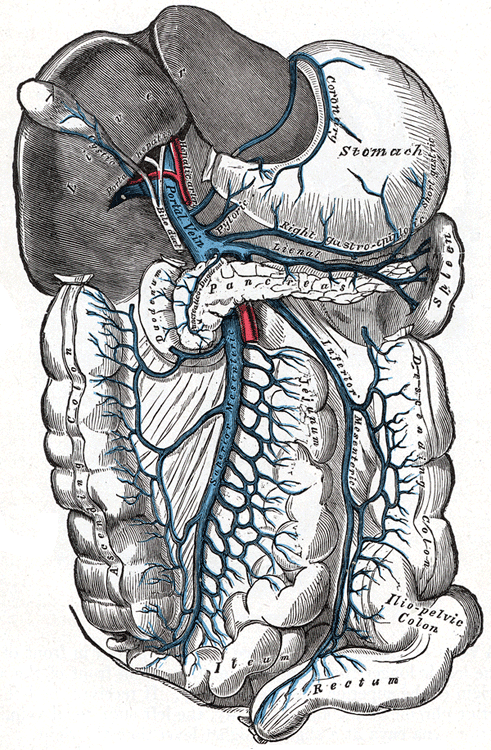
Pylephlebitis
Wikipedia
.; Visvalingam, V.; Indaram, A.; Bank, S. (2001). "Pylephlebitis--diagnosis and management". ... Kumar, V.; Abbas, A. K.; Fausto, N.; Robbins, S. L.; Cotran, R. S. (2015). Robbins and Cotran Pathologic Basis of Disease , 9th ed.; Philadelphia: Elsevier Saunders.
-
Complicated Grief Disorder
Wikipedia
PMID 12416919 . ^ DeVaul RA, Zisook S (May 1976). "Psychiatry: unresolved grief. ... Primary Care . 6 (2): 391–402. PMID 258819 . ^ Zisook S, Shuchter S, Schuckit M (June 1985). ... PMID 6823793 . ^ a b c d e f Shear MK, Simon N, Wall M, Zisook S, Neimeyer R, Duan N, et al. (February 2011). ... Presentation World Congress of Behavioral and Cognitive Therapies, Boston, MA; 2010. ^ Nakajima S, Shirai A, Maki S, et al. Mental health of the families of crime victims and factors related to their recovery. ... Further reading [ edit ] Tafà M, Cerniglia L, Cimino S, Ballarotto G, Marzilli E, Tambelli R (2018).
-
Arterial Stiffness
Wikipedia
PMID 16461838 . ^ Willum-Hansen T, Staessen JA, Torp-Pedersen C, Rasmussen S, Thijs L, Ibsen H, Jeppesen J (February 2006). ... S2CID 23321317 . ^ Fernandez-Fresnedo, G.; Rodrigo, E.; de Francisco, A. L. M.; de Castro, S. S.; Castaneda, O.; Arias, M. (2006). ... ISSN 1046-6673 . PMID 17130269 . ^ Cheng, S.; Vasan, R. S. (2011). "Advances in the Epidemiology of Heart Failure and Left Ventricular Remodeling" . ... PMC 3621875 . PMID 22083151 . ^ Whelton, S. P.; Blankstein, R.; Al-Mallah, M. ... A.; Hundley, W. G.; Polak, J. F.; Blumenthal, R. S.; Nasir, K.; Blaha, M. J. (2013).


