-
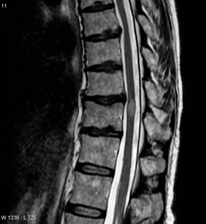
Transverse Myelitis
Wikipedia
Therefore, the signs and symptoms depend on the area of the spine involved. [5] Back pain can occur at the level of any inflamed segment of the spinal cord. [1] If the upper cervical segment of the spinal cord is involved, all four limbs may be affected and there is risk of respiratory failure – the phrenic nerve which is formed by the cervical spinal nerves C3 , C4 , and C5 innervates the main muscle of respiration , the diaphragm . [ citation needed ] Lesions of the lower cervical region (C5–T1) will cause a combination of upper and lower motor neuron signs in the upper limbs, and exclusively upper motor neuron signs in the lower limbs.
-
Low Blood Pressure (Hypotension)
Mayo_clinic
What's considered low blood pressure for one person might be OK for someone else. ... Sometimes, low blood pressure can be life-threatening. ... Certain health conditions and use of medications may cause low blood pressure. Conditions that can cause low blood pressure Medical conditions that can cause low blood pressure include: Pregnancy. ... A heart attack, heart failure, heart valve disease and an extremely low heart rate (bradycardia) can cause low blood pressure. ... Tests Other tests may be done to determine the cause of low blood pressure. Blood tests. Blood tests can help diagnose low blood sugar (hypoglycemia), high blood sugar (hyperglycemia or diabetes) or a low red blood cell count (anemia), all of which can lower blood pressure.
-
Papillary Urothelial Neoplasm Of Low Malignant Potential
Wikipedia
PUNLMP (Papillary Urothelial Neoplasm of Low Malignant Potential) Micrograph of a PUNLMP. ... They cannot be reliably differentiated from low grade papillary urothelial carcinomas using cytology, [1] and their diagnosis (vis-a-vis low grade papillary urothelial carcinoma) has a poor inter-rater reliability . [2] Pathologic grading and staging tumors are: graded by the degree of cellular atypia (G1->G3), and staged: papilloma papillary tumor of low malignant potential (PTLMP) papillary urothelial carcinomas low grade papillary urothelial carcinomas high grade. Differential diagnosis [ edit ] Papilloma. Low grade papillary urothelial carcinoma. Treatment [ edit ] PUNLMPs are treated like non-invasive low grade papillary urothelial carcinomas, [1] excision and regular follow-up cystoscopies. There is a rare occurrence of a pelvic recurrence of a low-grade superficial TCC after cystectomy.

-
Low-Grade Fibromyxoid Sarcoma
Wikipedia
Low-grade fibromyxoid sarcoma Micrograph of a low-grade fibromyxoid sarcoma. H&E stain . Specialty Pathology Low-grade fibromyxoid sarcoma ( LGFMS ) is a rare type of low grade sarcoma first described by Harry Evans in 1987. ... Unlike many other types of cancer, low grade fibromyxoid sarcoma can metastasize after many years, sometimes decades after the initial presentation of the tumor. [1] References [ edit ] ^ Evans, Harry L (2011). "Low-Grade Fibromyxoid Sarcoma: A Clinicopathologic Study of 33 Cases With Long-Term Follow-Up".

-
Polymorphous Low-Grade Adenocarcinoma
Wikipedia
Polymorphous low-grade adenocarcinoma Low magnification micrograph of a polymorphous low-grade adenocarcinoma, showing the typical variation of architectural arrangement. ... It is non-aggressive when compared to other oral cavity tumors, i.e. it is a low-grade tumor. [1] It forms glands, i.e. it is an adenocarcinoma . ... Micrograph of a polymorphous low-grade adenocarcinoma. H&E stain . ... References [ edit ] ^ a b Arathi N, Bage AM (2009). "Polymorphous low-grade adenocarcinoma of parotid gland: a rare occurrence" . ... PMID 19136798 . ^ a b Paleri V, Robinson M, Bradley P (April 2008). "Polymorphous low-grade adenocarcinoma of the head and neck".PRKD1, S100A1, S100B, MIB1, ACKR3, SPTLC3, ANO1, PRKD2, PRKD3, BMS1, CXCR4, ACACA, CD34, NFIB, MYB, MKI67, KRT7, KIT, ETV6, ERBB2, DDX3X, CDKN2A, H3P10

-
Immunoglobulin D Level In Plasma, Low
Omim
Dunnette et al. (1978) suggested autosomal recessive inheritance of low IgD level in plasma. They had previously shown that the distribution of the log of IgD levels in a population sample is not unimodal (13-14% of persons have a low level). A family study was then done on persons with low IgD and the above conclusion arrived at from analysis of the data. Immunology - Low plasma IgD Inheritance - Autosomal recessive ▲ Close
-
Low Pressure Hydrocephalus
Wikipedia
Please help to improve this article by introducing more precise citations. ( April 2011 ) ( Learn how and when to remove this template message ) Low pressure hydrocephalus Ventricles position Specialty Neurology Low-pressure hydrocephalus (LPH) is a condition whereby ventricles are enlarged and the individual experiences severe dementia , inability to walk, and incontinence – despite very low intracranial pressure (ICP). Low pressure hydrocephalus appears to be a more acute form of normal pressure hydrocephalus . If not diagnosed in a timely fashion, the individual runs the risk of remaining in the low pressure hydrocephalic state or LPHS. ... References [ edit ] Further reading [ edit ] Pang, Dachling; Altschuler, Eric (1994). "Low-Pressure Hydrocephalic State and Viscoelastic Alterations in the Brain". ... Owler, B.K.; Jacobson, E.E.; Johnston, I.H. (2001). "Low pressure hydrocephalus: Issues of diagnosis and treatment in five cases".CFAP43, NPHP1, CSF2, TUBB3, LAMC2, PMPCA, ERCC6, ERCC8, NXPH1, ACE, NPHP4, NEK8, MUC1, AQP4, ANKS3, APOE, UMOD, INVS, ALDH3A2, MLYCD, DNAJC13, ALB, NPHP3, CPVL, GLIS2, ANKS6, MAPKBP1, TYRP1, TBPL1, PER2, PROM1, VIM, TRPC1, TNF, TAZ, PSMD10, PSEN1, PRNP, PRKD1, PAX2, EPO, C3, BCL2, MIR4274

-
Lowe Syndrome
Medlineplus
Lowe syndrome is a condition that primarily affects the eyes, brain, and kidneys. ... Progressive kidney problems in older children and adults with Lowe syndrome can lead to life-threatening renal failure and end-stage renal disease (ESRD). Frequency Lowe syndrome is an uncommon condition. ... Causes Mutations in the OCRL gene cause Lowe syndrome. The OCRL gene provides instructions for making an enzyme that helps modify fat (lipid) molecules called membrane phospholipids. ... Learn more about the gene associated with Lowe syndrome OCRL Inheritance Pattern This condition is inherited in an X-linked pattern.OCRL, INPP5B, CLCN5, ACTB, ARHGAP1, PHETA1, INPP5E, INPP5K, INPP5D, SCRN1, SLC22A8, APPL1, ABCC4, SLC22A6, SIX2, PALLD, NR1H4, NBAS, BFAR, SNX9, SHH, MCOLN1, PTBP2, SESN2, SCRN2, PIFO, PHETA2, ASPM, SMARCA1, PTEN, SCN7A, RAB5A, AFP, ANXA2, CES1, CETP, ABCC2, CTSD, DCX, G6PD, GH1, GRB2, HOXD13, HPRT1, HTC2, INPP5A, MTM1, NAGLU, PAFAH1B1, PIP, PLEK, PTBP1, ADRB2, ARSH
-
Low-Set Ears
Wikipedia
Clinical sign of congenital conditions Low-set ears Specialty Medical genetics Low-set ears are a clinical feature in which the ears are positioned lower on the head than usual. They are present in many congenital conditions. Specifically, low-set ears are defined as outer ears positioned two or more standard deviations lower than the population average. [1] Low-set ears can be associated with conditions such as: Down syndrome [2] Turner syndrome Noonan syndrome [3] Patau syndrome [4] DiGeorge syndrome [5] Cri du chat syndrome Edwards syndrome Fragile X syndrome Okamoto syndrome It is usually bilateral, but it can be unilateral in Goldenhar syndrome . [6] See also [ edit ] LEOPARD syndrome References [ edit ] ^ Sivan Y, Merlob P, Reisner SH (June 1983). "Assessment of ear length and low set ears in newborn infants" . J. ... Retrieved 27 October 2010 . ^ "ear (low set)" . GPnotebook . External links [ edit ] Classification D ICD - 10 : Q17.4 ICD - 9-CM : 744.29 ( CDC/BPA 744.245) External resources MedlinePlus : 003303 v t e Congenital malformations and deformations of ears Size Macrotia Microtia Anotia Position Low-set ears Other Accessory auricle Mondini dysplasia This medical sign article is a stub .ARID1A, IFT52, PAM16, MRPS16, KCNK4, EFEMP2, DONSON, PSAT1, SLC25A24, POMT2, PYCR2, ANKRD11, IFT81, DCPS, CCDC22, PCLO, AHDC1, GPKOW, INTU, FOXP1, B9D1, FGF20, VPS33B, B3GAT3, KIFBP, TCTN3, AUTS2, SETBP1, C2CD3, GMNN, ACOX1, SAMHD1, ZDHHC9, OSGEP, CHD7, POMGNT1, ACER3, ASXL2, SETD5, PHIP, PPP2R3C, TMEM70, MKS1, BCOR, QRICH1, AHI1, GTPBP2, MAGEL2, PUS7, TMCO1, WNT4, RIPK4, BCL11A, RAB23, OTUD6B, DYNC2LI1, PIGT, LARP7, MBTPS2, TMEM216, MMACHC, NIPBL, PACS1, ORC6, EBP, STAMBP, POMT1, COLEC10, RNASEH2A, ZBTB18, COG5, PIBF1, TUBB3, RXYLT1, KLHL41, CWC27, ZMPSTE24, IRX5, SF3B4, CD96, GPC6, HUWE1, GNE, HDAC6, SLC12A6, MED12, WASHC5, FIG4, RUSC2, ZBTB24, KIAA0586, STAG2, SEPTIN9, MAPRE2, KIAA0556, PIGN, ATP6V0A2, KAT6B, TGDS, EXOSC2, ADNP, MED13L, SPECC1L, CLASP1, RPGRIP1L, SATB2, CAMTA1, MAPK8IP3, B4GAT1, POGZ, TAB2, SPART, CNOT1, DHX30, CILK1, CHSY1, MRAS, B4GALT7, TREX1, MAN1B1, POLR3A, PEX26, FRMD4A, TMEM94, B3GLCT, KLHL40, DIS3L2, SYT2, TWIST2, MYSM1, C12orf57, CEP41, PGAP3, MARS2, COG7, ATPAF2, TMEM67, HS6ST2, RSPRY1, UBE3B, WDR73, POMGNT2, GPT2, MYO18B, TMEM107, TRAF7, POMK, ANTXR1, KIAA1109, RNASEH2C, CDCA7, ITCH, AMER1, CCBE1, VAC14, B3GALNT2, OCLN, RNU4ATAC, CRPPA, AGRN, KIF7, CTU2, RSPO2, SH3PXD2B, UNC80, KCTD1, CHAMP1, NALCN, PTF1A, EBF3, STAC3, PHACTR1, JMJD1C, HYLS1, TUBB, TAPT1, ARL13B, ASXL1, ARX, GLIS3, BMPER, ESCO2, CEP120, PLVAP, CDT1, ARMC9, FBXO11, VIPAS39, ALX4, SLC5A7, EPG5, DOCK6, CC2D2A, WDR35, ARID1B, PRR12, TBC1D24, GATAD2B, ADGRG6, KIF15, FAM20C, MRPS22, INPP5E, LRRC8A, LMOD3, SMG9, KLHL7, ZC4H2, TMEM165, CENPJ, LMBRD1, MCTP2, MBD5, TENM3, PIEZO2, MRPS14, SMOC1, TCTN1, FRAS1, GREB1L, ALG13, TCTN2, CSPP1, EHMT1, ALG9, TXNDC15, SRD5A3, FAT4, EFL1, RNASEH2B, TMEM231, SLC39A8, FKRP, ALG8, COLEC11, CPLANE1, UPF3B, TMEM237, PORCN, NXN, NSD1, STRA6, IFIH1, XYLT2, EIF4A3, CEP104, ACP5, MEGF8, GP1BB, GNS, GNAI3, GLUL, GLE1, GK, GJB2, GJA1, GDNF, GBA, GATA4, FZD2, MTOR, FLNB, FLNA, FHL1, FGFR2, FGFR3, FGFR1, GPC4, FKTN, FBN1, FANCB, EYA1, EVC, ERCC5, ERCC4, BRF1, H3-3A, HELLS, LMNA, MYCN, MVK, ATP6, KMT2A, MEIS2, MEF2C, MECP2, MCM5, MAF, SMAD4, MAB21L1, LRP2, LIFR, HNRNPK, LBR, KRAS, KIF22, KCNJ2, IREB2, INSR, IGF2, IGF1R, IGBP1, HSPG2, HSD17B4, HRAS, EP300, EEF1A2, MYO9A, EDNRA, CHD3, CHAT, CENPF, CDKN1C, CDH11, CDC42, CDC6, CBL, CAMK2G, BUB1B, BRAF, BMPR1A, BMP2, ATRX, ATR, ATP6V1E1, ATP6V1A, ATIC, ASCL1, ARVCF, ARL3, ALX3, AKT1, ADSL, ADAR, ACTB, ACTA1, CHD4, CHRNA1, CHRND, DDX3X, EDN3, DVL3, DVL1, SLC26A2, ATN1, DPH1, DNMT3B, DLX4, DHPS, DHODH, DHCR24, DHCR7, DAG1, CHRNG, CTSD, CTNNB1, CSNK2A1, CRMP1, CREBBP, CPT2, COMT, COL13A1, COL11A2, COL11A1, COL4A1, COL2A1, MYH3, NEB, CEP57, SOX4, USP9X, LZTR1, SHOC2, KAT6A, PCGF2, YWHAE, WNT7A, WNT5A, WNT3, VARS1, UFD1, TWIST1, HIRA, TPM3, TPM2, TFAP2B, TFAP2A, TCF20, TBX15, TBX2, TBCE, TBX1, TALDO1, TAF1, VAMP1, SOX11, SOX9, RBM10, SMC1A, NAA10, PHOX2B, PUM1, TTC37, SEC24C, ADAMTS3, CHST3, PLAA, EFTUD2, CRLF1, LARGE1, PLOD3, AP3D1, KYNU, HERC1, TRRAP, CCNK, EED, CDK13, CDK10, AP3B1, ITGA8, CNTNAP1, PEX3, PPM1D, OFD1, PLA2G6, CDC45, SOX5, SON, NF1, SNRPB, PSMD12, MASP1, PRPS1, PPP2R1A, PPP1CB, POR, POLE, EXOSC9, PMM2, PLCB4, PEX12, PEX10, PCNT, PBX1, PAX1, PRDX1, PAFAH1B1, OTX2, ORC4, ORC1, NUP88, NRAS, NPR2, NOTCH3, NOTCH2, NFIX, NFIA, PTCH1, PTEN, PTH1R, RPS23, SNAP25, SMS, SMARCD2, SLC25A1, SLC18A3, SLC6A9, SLC6A8, SKI, SIM1, SCN4A, SCN1A, RREB1, RPS6KA3, PTPN11, RPL35A, RIT1, RET, DPF2, RB1, RARB, RAF1, RAC1, QARS1, ALDH18A1, PEX5, PEX2, SCN1A-AS1
-
Low Back Pain
Wikipedia
Low back pain Other names Lower back pain, lumbago Low back pain is a common and costly complaint. ... These exercises only work if they are limiting low back pain. [52] Exercise programs that incorporate stretching only are not recommended for low back pain. ... "Diagnosis and treatment of acute low back pain". American Family Physician . 85 (4): 343–50. ... "How do we define the condition 'recurrent low back pain'? A systematic review" . ... "Clinical decision support and acute low back pain: evidence-based order sets".PLAT, SPARC, NAB2, SPAST, HSPB8, HTRA1, ALDH18A1, STAT6, FAS, TBX6, ABCD1, NGF, IL1B, LBP, TNF, IL6, CALCA, CXCL8, COMT, IL1A, LEP, CCL2, NUBP1, EMSLR, TP63, CASP9, COASY, BDNF, CRP, CISD3, SEMA3A, SLCO6A1, COPD, RGS6, GDF15, NLRP3, MIR204, ADAMTS3, ADAMTS4, ZFYVE9, MSC, FST, ITLN1, ADAMTS5, RTN4, MCF2L2, SIRT1, PES1, SPECC1, TSKU, LCS1, F11R, DNAJC5, PER2, MARCHF1, PACC1, SPEN, ACR, GDF5, ZNF35, ACAN, AMH, BMP3, TSPO, CASP1, CAT, CD6, DNMT3B, NR3C1, IL1RN, IL6ST, MMP1, MMP9, OPRM1, FURIN, PPARG, PTGS2, RARRES2, RASA1, RHAG, RTN1, CCL4, ACP5, TAC1, TLR4, TP53, VDR, SMS

-
Hypochloremia
Wikipedia
Hypochloremia Chlorine Specialty Endocrinology Hypochloremia (or Hypochloraemia ) is an electrolyte disturbance in which there is an abnormally low level of the chloride ion in the blood . ... External links [ edit ] Classification D ICD - 10 : E87.8 ICD - 9-CM : 276.9 v t e Electrolyte imbalances Sodium High Salt poisoning Low Hypotonic Isotonic Cerebral salt-wasting syndrome Potassium High Low Chloride High Low Calcium High Low Symptoms and signs Chvostek sign Trousseau sign Milk-alkali syndrome Disorders of calcium metabolism Calcinosis ( Calciphylaxis , Calcinosis cutis ) Calcification ( Metastatic calcification , Dystrophic calcification ) Familial hypocalciuric hypercalcemia Phosphate High Low Magnesium High Low This medical treatment –related article is a stub .
-
Proteinuria, Low Molecular Weight, With Hypercalciuria And Nephrocalcinosis
Omim
A number sign (#) is used with this entry because low molecular weight proteinuria is caused by mutation in the chloride channel-5 gene (CLCN5; 300008) on chromosome Xp11.22. Description Low molecular weight proteinuria with hypercalciuria and nephrocalcinosis is a form of X-linked hypercalciuric nephrocalcinosis, a group of disorders characterized by proximal renal tubular reabsorptive failure, hypercalciuria, nephrocalcinosis, and renal insufficiency. ... More than 50% of their urinary proteins were those with a molecular weight of less than 40 kD, defined by Suzuki et al. (1985) as low molecular weight (LMW) proteins, including lysozyme (153450) and beta-2-microglobulin (109700). ... Igarashi et al. (1995) found that patients with low molecular weight proteinuria tended to have hypercalciuric nephrocalcinosis without rickets or renal failure. ... Nakazato et al. (1997) identified mutations in the CLCN5 gene in affected members of 2 Japanese families with low molecular weight proteinuria. Akuta et al. (1997) identified mutations in the CLCN5 gene in 7 of 10 unrelated Japanese patients with low molecular weight proteinuria, hypercalciuria, and nephrocalcinosis.
-
Cerebral Salt-Wasting Syndrome
Wikipedia
Cerebral salt-wasting syndrome Other names CSWS Specialty Endocrinology Cerebral salt-wasting syndrome ( CSWS ) is a rare endocrine condition featuring a low blood sodium concentration and dehydration in response to injury (trauma) or the presence of tumors in or surrounding the brain . In this condition, the kidney is functioning normally but excreting excessive sodium. [1] The condition was initially described in 1950. [2] Its cause and management remain controversial. [3] [4] Contents 1 Signs and symptoms 2 Causes 3 Diagnosis 4 Treatment 5 References 6 External links Signs and symptoms [ edit ] Signs and symptoms of CSWS include large amounts of urination (polyuria, defined as over three liters of urine output over 24 hours in an adult), high amounts of sodium in the urine, low blood sodium concentration, [1] excessive thirst (polydipsia), extreme salt cravings, dysfunction of the autonomic nervous system (dysautonomia), and dehydration . ... Advanced symptoms include muscle cramps , lightheadedness, dizziness or vertigo , feelings of anxiety or panic, increased heart rate or slowed heart rate , low blood pressure and orthostatic hypotension which can result in fainting . [5] Other symptoms frequently associated with dysautonomia include headaches , pallor , malaise , facial flushing, constipation or diarrhea , nausea , acid reflux , visual disturbances, numbness, nerve pain, trouble breathing , chest pain, loss of consciousness, and seizures . [5] Causes [ edit ] CSWS is usually caused by brain injury/trauma or cerebral lesion, tumor, or hematoma. [ citation needed ] Diagnosis [ edit ] CSWS is a diagnosis of exclusion and may be difficult to distinguish from the syndrome of inappropriate antidiuretic hormone (SIADH), which develops under similar circumstances and also presents with hyponatremia. [1] The main clinical difference is that of total fluid status of the patient: CSWS leads to a relative or overt low blood volume [3] whereas SIADH is consistent with a normal or high blood volume (due to water reabsorption via the V2 receptor). [1] If blood-sodium levels increase when fluids are restricted, SIADH is more likely. [6] Additionally, urine output is classically low in SIADH and elevated in CSWS. [1] Treatment [ edit ] While CSWS usually appears within the first week after brain injury and spontaneously resolves in 2–4 weeks, it can sometimes last for months or years. In contrast to the use of fluid restriction to treat SIADH, CSWS is treated by replacing the urinary losses of water and sodium with hydration and sodium replacement. [1] The mineralocorticoid medication fludrocortisone can also improve the low sodium level. [1] [7] References [ edit ] ^ a b c d e f g Yee AH, Burns JD, Wijdicks EF (April 2010). ... External links [ edit ] Classification D DiseasesDB : 32234 External resources eMedicine : ped/354 v t e Electrolyte imbalances Sodium High Salt poisoning Low Hypotonic Isotonic Cerebral salt-wasting syndrome Potassium High Low Chloride High Low Calcium High Low Symptoms and signs Chvostek sign Trousseau sign Milk-alkali syndrome Disorders of calcium metabolism Calcinosis ( Calciphylaxis , Calcinosis cutis ) Calcification ( Metastatic calcification , Dystrophic calcification ) Familial hypocalciuric hypercalcemia Phosphate High Low Magnesium High Low
-
Electroencephalogram, Low-Voltage
Omim
Inheritance An autosomal dominant mode of inheritance was established for low-voltage EEG by Vogel (see review by Vogel and Motulsky, 1986). ... However, epileptic seizures had not been observed in the families of Steinlein et al. (1992), and no low-voltage patterns were reported in families with benign neonatal convulsions. ... The linkage between BFNC and D20S19 was confirmed by Malafosse et al. (1990, 1992) and Ryan et al. (1991). Low voltage EEG, benign neonatal convulsions, and Fanconi anemia (FA; 227650) are all linked to the VNTR marker CMM6 (D20S19). ... Clusters of rare cutter sites around CMM6 pointed to at least 4 closely related CpG islands. Lab - Low-voltage EEG - Absent EEG alpha waves Inheritance - Autosomal dominant (20q) ▲ Close
-
Nail Low-Sulfur Protein
Omim
See 161070. Nails - Low-sulfur nail proteins Inheritance - Autosomal dominant ▲ Close
-
Trousseau Sign Of Latent Tetany
Wikipedia
Note the flexed wrist and MCPs , and the extended DIPs and PIPs Differential diagnosis low calcium Trousseau sign of latent tetany is a medical sign observed in patients with low calcium . [1] From 1 to 4 percent of normal patients will test positive for Trousseau's sign of latent tetany. [2] This sign may be positive before other manifestations of hypocalcemia such as hyperreflexia and tetany , as such it is generally believed to be more sensitive (94%) than the Chvostek sign (29%) for hypocalcemia. [3] [4] To elicit the sign, a blood pressure cuff is placed around the arm and inflated to a pressure greater than the systolic blood pressure and held in place for 3 minutes. ... Volume 2: 112-114. v t e Electrolyte imbalances Sodium High Salt poisoning Low Hypotonic Isotonic Cerebral salt-wasting syndrome Potassium High Low Chloride High Low Calcium High Low Symptoms and signs Chvostek sign Trousseau sign Milk-alkali syndrome Disorders of calcium metabolism Calcinosis ( Calciphylaxis , Calcinosis cutis ) Calcification ( Metastatic calcification , Dystrophic calcification ) Familial hypocalciuric hypercalcemia Phosphate High Low Magnesium High Low This medical sign article is a stub .

- Low Phospholipid-Associated Cholelithiasis Orphanet
-
Liddle Syndrome 2
Omim
Hiltunen et al. (2002) reported a Finnish man who developed hypertension associated with low serum aldosterone, suppressed renin activity, and persistent hypokalemia at 25 years of age. His mother had been hypertensive since age 40 years and had a low-normal serum potassium with low plasma renin activity. ... He had hypokalemic metabolic alkalosis and low plasma renin activity; serum aldosterone was in the low-normal range. ... However, the N530S variant was detected in 1 of 291 healthy Finnish blood donors as well as in 1 of 175 control Finnish men, aged 50 to 69 years, who had low-normal blood pressure; neither individual was available for further evaluation. ... INHERITANCE - Autosomal dominant CARDIOVASCULAR Vascular - Hypertension METABOLIC FEATURES - Metabolic alkalosis - Hypokalemia ENDOCRINE FEATURES - Low plasma renin activity - Low plasma aldosterone level MISCELLANEOUS - Phenotype ameliorated by low-salt diet and antagonists of the epithelial channel of the distal nephron - No improvement with antagonists of the mineralocorticoid receptor MOLECULAR BASIS - Caused by mutation in the sodium channel, nonvoltage-gated 1, gamma gene (SCNN1G, 600761.0001 ) ▲ CloseSCNN1B, SCNN1G, SCNN1A, REN, NEDD4L, NEDD4, CYP11B2, NR3C2, CFTR, AGT, MUL1, ASIC5, HGS, PRKN, HSD11B2, HMBS, UBE2K, CYP11B1, CBLL2

-
Apolipoprotein A-I Deficiency
Orphanet
Clinical description Although extremely low plasma HDL cholesterol may be detected in any patient fortuitously from birth, the age of symptom onset and the clinical presentation varies greatly. ... Diagnostic methods Diagnosis is based on biochemical analysis of plasma Apo A-I and HDL cholesterol levels showing extremely low HDL cholesterol levels and very low to undetectable Apo A-I (inferior to 5 mg/dL). Low HDL cholesterol levels are associated with normal VLDL and LDL cholesterol levels, and normal or decreased triglyceride levels. ... In case of carotid atherosclerosis or cardiovascular complications, a low-fat diet balanced in anti-oxidants (e.g. ... Regular cardiovascular monitoring should be offered to Apo AI deficient patients with extremely low HDL cholesterol (<20 mg/dL) because of the increased risk (Odds Ratio x2-3) of coronary artery disease.
-
Hypocholesterolemia
Wikipedia
Hypocholesterolemia Other names Low cholesterol Formula structure of cholesterol Causes Statins, hyperthyroidism, adrenal insufficiency, malabsorption, malnutrition, etc. Hypocholesterolemia is the presence of abnormally low ( hypo- ) levels of cholesterol in the blood ( -emia ). ... PMID 1355411 . ^ a b Criqui MH. (1994). Very Low Cholesterol and Cholesterol Lowering. ... "Relations of trait depression and anxiety to low lipid and lipoprotein concentrations in healthy young adult women" . ... "A Randomized Trial of Alerting to Hypocholesterolemia Results of the Low Indexes of Metabolism Intervention Trial-C (LIMIT-C)" .PCSK9, SAR1B, ABCA1, PEX12, FLCN, UBE3B, HSD3B7, CCT5, MSMO1, DHCR7, PMM2, APOB, CAV1, PIK3CD, PIK3CA, PIK3CG, LDLR, CD44, PIK3CB, MTTP, ACTB, IL6, CXCR4, HMGCR, CNBP, XBP1, AHSA1, GRAP2, XPR1, TM4SF5, TRPA1, AIMP2, CAV2, SACM1L, VEGFA, UCP3, UCP1, TNF, SREBF2, SREBF1, SOD1, SLC6A3, SLC2A4, TRPV1, TBC1D9, ANXA6, ROS1, ADAM10, ANTXR2, NLRP3, PRRT2, AGT, AGTR1, PRDM16, CFAP97, NGLY1, TRPV6, HDL3, RXFP3, AKT1, TAS2R14, NPC1L1, ANGPTL3, POLDIP2, RNF19A, BACE1, APOA4, RGS2, RHO, CASP1, KCNK3, ITIH4, CXCL10, CXCL8, HTR3A, HSD11B1, FDFT1, FASN, F3, ERN1, EGF, EFNB2, DSC2, CAT, CYBB, CYBA, MAPK14, CRK, CP, LCAT, MAP3K5, CDC42, MFAP1, PTK2, MAPK8, MAPK3, MAPK1, PRKCB, PRKCA, APP, APRT, AQP2, ACP3, AQP4, SERPINI1, ABCB1, BRAF, P2RX7, OSBP, NOS3, MVD, MUC5AC, MIR214


