-
Silent Sinus Syndrome
Wikipedia
Silent sinus syndrome Other names Imploding antrum syndrome Specialty ENT surgery Silent sinus syndrome is a spontaneous, asymptomatic collapse of the maxillary sinus and orbital floor associated with negative sinus pressures. ... Usually the diagnosis is suspected clinically, and it can be confirmed radiologically by characteristic imaging features that include maxillary sinus outlet obstruction, sinus opacification, and sinus volume loss caused by inward retraction of the sinus walls. ... References [ edit ] Illner A, Davidson HC, Harnsberger HR, Hoffman J (2002). "The silent sinus syndrome: clinical and radiographic findings". ... Full text Numa WA, Desai U, Gold DR, Heher KL, Annino DJ (2005). "Silent sinus syndrome: a case presentation and comprehensive review of all 84 reported cases". ... "Queen Meresankh III – the oldest case of bilateral Silent Sinus Syndrome (c. 2620/10 - 2570 BC)?".
-
Sinus Node Dysfunction
Wikipedia
Sinus node dysfunction (SND) Other names Sick Sinus Syndrome or Sinoatrial node disease Telemetry strip of a 44-year-old female with asymptomatic sinus pause found while admitted for mildly symptomatic COVID-19 pneumonia. ... This ECG from the same patient shows atrial fibrillation at around 126 beats per minute. The most common complication of sinus node dysfunction is the development of tachycardia-bradycardia syndrome with abnormal atrial rhythms such as atrial tachycardia , atrial fibrillation , and flutter . [5] [3] These rhythms increases the risk of clot formation in the atrium, embolization, and stroke . [5] Developing sinus arrest, sinus node exit block, sinus bradycardia , atrioventricular block , and other types of abnormal rhythms are also common complications. [5] [6] Sinus node dysfunction has a close association with the presence of atrial fibrillation due to their shared etiology of remodeling. [6] Causes [ edit ] Sinus node dysfunction can be caused by intrinsic and extrinsic factors that affect the normal functioning of the sinus node. ... Electrolyte abnormalities such as hyperkalemia , hypokalemia , and hypocalcemia can also alter normal sinus node functioning. Hypothyroidism , hypoxia , hypothermia , and various toxins have also been associated with sinus node dysfunctions. [5] [6] Diagnosis [ edit ] Electrocardiogram (ECG) of a 64-year-old female with sinus bradycardia . ... Electrocardiogram [ edit ] The primary 12-lead electrocardiogram (ECG) finding in sinus node dysfunction is inappropriate sinus bradycardia . [7] Sinus node dysfunction can also present with sudden sinus arrest with or without junctional escape , sinoatrial block , prolonged asystolic period followed by tachycardias, or tachycardia-bradycardia syndrome presenting as various atrial arrhythmias such as atrial fibrillation , flutter , tachycardia , or paroxysmal supraventricular tachycardia . [7] [5] Clinical Diagnosis [ edit ] Diagnosing sinus node dysfunction requires clinical symptoms as well as ECG abnormalities. ... "Incidence of and Risk Factors for Sick Sinus Syndrome in the General Population" .SCN3B, SCN5A, HCN4, KCNJ5, MYH6, CACNA1D, CACNA2D1, CACNB2, GPD1L, SCN10A, SCN2B, SCN1B, POMC, KCND3, PKP2, KCNJ8, ANK2, KCNJ3, GJC1, TBX3, MIR17HG, MIR106B, NAA50, DECR1, DNAH8, EMD, KCNE2, TGFB1, SLC8A1, KCNQ1, GJA1, GJA5, GNB2, TLX2, IL10, KCNJ1, PITX2, APRT, MFAP1, MIR423

-
Sick Sinus Syndrome 1
OMIM
Description The term 'sick sinus syndrome' encompasses a variety of conditions caused by sinus node dysfunction. ... Electrocardiogram typically shows sinus bradycardia, sinus arrest, and/or sinoatrial block. Episodes of atrial tachycardias coexisting with sinus bradycardia ('tachycardia-bradycardia syndrome') are also common in this disorder. ... Susceptibility to sick sinus syndrome (SSS3; 614090) is influenced by variation in the MYH6 gene (160710). ... Inheritance The inheritance pattern of sick sinus syndrome in the families reported by Benson et al. (2003) was autosomal recessive.MYH6, SCN5A, HCN4, POMC, CACNA1D, KCNJ5, HCN1, SCN10A, CACNA1C, KCNE5, GPD1L, GNB5, AKAP9, PKP2, RANGRF, KCNE3, SLMAP, TRPM4, SCN3B, SCN1B, SGO1, ABCC9, SCN2B, CACNA2D1, KCNJ8, CACNB2, CALM2, KCND3, MECP2, PDCD1, ICAM1, TRPM7, COL1A2, TXN2, CCN2, FBN1, FGFR3, FN1, IL6, IGF1, MEF2C, HDAC4, TBX18, VEGFA, VCAM1, TTN, THBS1, TGFB1, CCL2, LMNA, AGT
-
Sick Sinus Syndrome
MedlinePlus
Sick sinus syndrome (also known as sinus node dysfunction) is a group of related heart conditions that can affect how the heart beats . "Sick sinus" refers to the sino-atrial (SA) node, which is an area of specialized cells in the heart that functions as a natural pacemaker. ... These changes lead to abnormal heartbeats and the other symptoms of sick sinus syndrome. A particular variation in another gene, MYH6 , appears to increase the risk of developing sick sinus syndrome. ... In older adults, sick sinus syndrome is often associated with age-related changes in the heart. ... Learn more about the genes associated with Sick sinus syndrome HCN4 MYH6 SCN5A Inheritance Pattern Most cases of sick sinus syndrome are not inherited.
-
Sinus Tachycardia
Wikipedia
Please help to improve this article by introducing more precise citations. ( April 2012 ) ( Learn how and when to remove this template message ) Sinus tachycardia ECG of a 29-year-old female with sinus tachycardia with a heart rate of 125 bpm Specialty Cardiology Sinus tachycardia (also colloquially known as sinus tach or sinus tachy ) is an elevated sinus rhythm characterized by an increase in the rate of electrical impulses arising from the sinoatrial node . ... Thus, sinus tachycardia is a medical finding that can be either physiological or pathological . ... Treatment [ edit ] Treatment for physiologic sinus tachycardia involves treating the underlying causes of the tachycardia response. ... Patients who are unresponsive to such treatment can undergo catheter ablation to potentially repair the sinus node. [2] Acute myocardial infarction [ edit ] Sinus tachycardia can present in more than a third of the patients with AMI but this usually decreases over time. ... ISBN 978-1-4496-1589-5 . ^ a b c d e Allison Henning, Conrad Krawiec (2020). "Sinus Tachycardia". StatPearls . PMID 31985921 .CYP1A1, GCG, CPT2, TPM3, MYPN, EHMT1, TMEM127, SDHAF2, KIF1B, ZMPSTE24, TMEM94, KMT2B, BANF1, SLC25A11, VHL, SDHD, TPM2, SDHC, SDHB, SDHA, RET, PEX5, MDH2, MAX, LMNA, FH, DLST, CRP, TLX1NB

-
Inappropriate Sinus Tachycardia
Wikipedia
Inappropriate sinus tachycardia Other names IST Specialty Cardiology Inappropriate sinus tachycardia is a rare type of cardiac arrhythmia within the category of supraventricular tachycardia (SVT). ... Invasive treatments include forms of catheter ablation such as sinus node modification [7] (selective ablation of the sinus node), complete sinus node ablation (with associated implantation of a permanent artificial pacemaker ), and AV node ablation in very resistant cases (creation of iatrogenic complete heart block , necessitating implantation of a permanent artificial pacemaker). ... "Radiofrequency Catheter Modification of the Sinus Node for "Inappropriate" Sinus Tachycardia". ... Avaliação dos resultados imediatos e tardios" [Sinus node modification by catheter using radiofrequency current in a patient with inappropriate sinus tachycardia. ... "Radiofrequency catheter modification of sinus node for inappropriate sinus tachycardia: A case report".
-
Sinus Bradycardia
Wikipedia
Sinus bradycardia is a sinus rhythm with a rate that is lower than normal This article needs additional citations for verification . ... Unsourced material may be challenged and removed. Find sources: "Sinus bradycardia" – news · newspapers · books · scholar · JSTOR ( August 2014 ) ( Learn how and when to remove this template message ) Sinus bradycardia ECG of Sinus bradycardia with a heart rate of 43 bpm. Specialty Cardiology Symptoms lightheadedness dizziness hypotension vertigo syncope Diagnostic method electrocardiogram Sinus bradycardia is a sinus node dysfunction with a rate that is lower than normal. ... Generally, smaller species have higher heart rates while larger species have lower rates. [5] Pathophysiology & etiology [ edit ] Rhythm strip of sinus bradycardia at 50 bpm Sinus bradycardia is commonly seen in normal healthy persons and athletes in the absence of pathophysiological diseases or conditions. [1] Different factors or etiologies could lead to the dysfunction of the sinus node, causing a malformation or prolongation of the impulse. ... "Sinus Bradycardia". StatPearls . Treasure Island (FL): StatPearls Publishing.ANK2, CALM1, TNNI3K, BVES, TRDN, AKAP9, HCN4, KCNE2, NOS1AP, SNTA1, SLC20A2, SCN10A, SCN5A, SCN4B, KCNQ1, KCNJ5, KCNH2, KCNE1, COL2A1, CAV3, CALM2, PRKAG2

-
Aneurysm Of Sinus Of Valsalva
Wikipedia
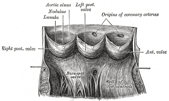
Aneurysm of sinus of Valsalva Other names Aortic sinus aneurysm Aorta laid open to show the semilunar valves . (Aortic sinus, also known as "sinus of Valsalva", is labeled at upper left.) ... Aortic sinus aneurysms can burst or rupture into adjacent cardiac chambers, which can lead to heart failure if untreated. ... The manifestations appear depending on the site where the sinus has ruptured. For example, if the sinus ruptures in a low pressure area like the right atrium or right ventricle then a continuous type of murmur is heard. ... It is also accompanied by a superficial thrill. A ruptured Sinus of Valsalva abscess represents a surgical emergency.
-
Cavernous Sinus Thrombosis
Wikipedia
Cavernous sinus thrombosis Oblique section through the cavernous sinus. Specialty Neurology Cavernous sinus thrombosis ( CST ) is the formation of a blood clot within the cavernous sinus , a cavity at the base of the brain which drains deoxygenated blood from the brain back to the heart. ... References [ edit ] ^ a b c "Guidelines Cavernous sinus thrombosis" (PDF) . ^ "Cavernous sinus thrombosis - NHS Choices" . www.nhs.uk . ... Retrieved 27 May 2016 . ^ a b "Cavernous sinus thrombosis: MedlinePlus Medical Encyclopedia" . www.nlm.nih.gov . Retrieved 27 May 2016 . ^ a b "Cavernous Sinus Thrombosis - Eye Disorders" . ^ Zhang J, Stringer MD (July 2010).

-
Congenital Dermal Sinus
Wikipedia
Congenital dermal sinus Other names Spinal congenital dermal sinus Vertebral column Congenital dermal sinus is an uncommon form of cranial or spinal dysraphism . [1] [2] It occurs in 1 in 2500 live births. [2] It occurs as a dermal indentation, found along the midline of the neuraxis and often presents alongside infection and neurological deficit. [1] Congenital dermal sinus form due to a focal failure of dysjunction between the cutaneous ectoderm and neuroectoderm during the third to eight week of gestation . [1] [2] [3] [4] Typically observed in the lumbar and lumbosacral region, congenital dermal sinus can occur from the nasion and occiput region down. [1] [2] [3] Early diagnosis and treatment is crucial for cases of congenital dermal sinus. ... Contents 1 Embryogenesis 2 Diagnosis 2.1 Clinical features 2.1.1 Cutaneous abnormalities 2.1.2 Infection 2.1.3 Neurological deficit 2.2 Imaging 3 Treatment 4 Historically 5 References Embryogenesis [ edit ] During normal development, cutaneous ectoderm separates from neuroectoderm to allow for the insertion of mesoderm . [2] That is, the skin separates from the tissue of the spinal cord to allow proper formation of the vertebral column . [2] In cases of congenital dermal sinus there is a failure in this process, resulting in formation of a persistent connection between the skin and neural tissue. [2] This manifests as a tract extending from the surface of the skin to the spinal cord lined with stratified squamous epithelium , surrounded by dermal and neurological tissue. [2] [4] The tract may terminate in the deep fascia , or even make contact with neural elements. [1] Congenital dermal sinus may form at any point along the midline of the neuraxis, however, the majority form in the lumbar and lumbosacral region (41% and 35% of cases respectively). [2] Diagnosis [ edit ] Congenital dermal sinus is often diagnosed in infants and children . [1] Early diagnosis is important in congenital dermal sinus, so that treatment can be provided early, to prevent progression of associated complications. [1] [2] [3] [4] Clinical features [ edit ] There three key hallmarks of congenital dermal sinus: cutaneous abnormalities, infection , and neurological deficits. [ citation needed ] Cutaneous abnormalities [ edit ] Congenital dermal sinus is a tract from the surface layer of the skin, through the deeper tissues into the cranial or spinal cavity. [1] The skin findings of this tract can include: Pit along neuraxis [3] Flat capillary hemangioma [3] Hypertrichosis [1] [2] [3] Skin tag [2] Abnormal pigmentation [1] [2] Subcutaneous lipoma [1] [2] [3] Signs of local infection [1] [2] [3] Infection [ edit ] The stratified squamous epithelium of the congenital dermal sinus tract can extend to the spinal fascia of the dura mater or all the way to the spinal cord . [3] [4] Thus, the congenital dermal sinus forms a point of entry for infection, this can allow for the formation of an abscess . [2] [4] Infection can then travel up the spinal cord to result in meningitis , which can be fatal if left untreated. [1] [4] Neurological deficit [ edit ] Congenital dermal sinus is often also associated with spinal fluid drainage, intradural cysts and spinal cord tethering; conveying neurological deficit. [3] Neurological deficit can occur due to spinal cord compression from intradural dermoid cyst growth in the epidermis and dermis . [3] Tethered spinal cord can result in gait difficulties and sphincter dysfunction, as well as compressing the spine. [4] Neurological deficits are more likely to occur where diagnosis has not been timely, allowing cysts and or infection. [2] [3] Imaging [ edit ] Magnetic Resonance Imaging (MRI) is the preferred tool for diagnostic and preoperative imaging of congenital dermal sinus. [1] [2] [3] [4] MRI allows the neural structures to be observed, visualizing the tract and its anomalies and lesions. [1] [2] [3] [4] For example, exposing tethered cord, inclusion tumors or spinal cord malformations. [2] Observation by X-ray is limited in diagnosis, especially due to immature calcification of infants less than 18 months. [4] X-ray may be used in conjunction with MRI or sonogram images to assist preoperatively. [2] Treatment [ edit ] Treatment of congenital dermal sinus involves complete resection of the tract as well as intradural exploration. [3] Prophylactic surgical removal of the congenital dermal sinus tract is beneficial for the patient, allowing neurological and bladder function to be maintained. [1] Early surgical intervention results decreases the risk of infection and/or tumour progression – factors typically associated with delayed presentation of congenital dermal sinus. [2] ] Intradural exploration is necessary as excision of the entire tract, as well as any of its intradural connections, reduces need for further surgical intervention. [3] The surgical technique involves ‘removing the cutaneous lesion in ellipse’. [3] The tract of the congenital dermal sinus must then be explored and excised, with intradural lesions dissected. [3] If not all epithelial tissue is removed, there is a possibility for the dermoid cyst to reoccur and require further operation. [3] Further operations are limited by postoperative and post-infection scarring. [3] Historically [ edit ] Prior to pervasive use and availability of advanced methods of neuroimaging, it is possible that the rate of incidence of congenital dermal sinus has been supplemented by the incidence of coccygeal pits. [2] [3] Coccygeal pits are distinct from congenital dermal sinus as they are found within the gluteal cleft , rather than above the gluteal cleft. [2] [3] The caudally orientated coccygeal pits are not associated with intradural pathology and do not need to be excised, unlike the cephalically oriented tracts of the congenital dermal sinus which confer great intradural pathology and require surgical intervention. [2] [3] While coccygeal pits occur in 4% of neonate population, congenital dermal sinus is only found in 1 in 2500 live births. [2] [3] References [ edit ] ^ a b c d e f g h i j k l m n o p Wang, YM; Chuang, MJ; Cheng, MH (September 2011). "Infected spinal dermal sinus tract with meningitis: a case report" (PDF) . ... PMID 12949296 . ^ a b c d e f g h i j k l m n o p q r s t u v w x Elton, S; Oakes, JW (January 2001). "Dermal sinus tracts of the spine" . Neurosurgical Focus . 10 (1): 1–4. doi : 10.3171/foc.2001.10.1.5 . ^ a b c d e f g h i j Jindal, A; Mahapatra, AK (September 2001). "Spinal congenital dermal sinus: an experience of 23 cases over 7 years" .
-
Paranasal Sinus And Nasal Cavity Cancer
Wikipedia
The talk page may contain suggestions. ( December 2020 ) Paranasal sinus and nasal cavity cancer Bone of nasal cavity Specialty Oncology Symptoms Nose bleeds , Headaches , blocked sinus, diplopia , [1] etc. ... See also [ edit ] Head and neck cancer Paranasal sinus Nasal cavity References [ edit ] ^ a b c d e f g h i j k l m National Cancer Institute (2019). Paranasal Sinus and Nasal Cavity Cancer Treatment (Adult) (PDQ®)–Patient Version. ... Retrieved 2019-05-12 . ^ a b c d e f National Cancer Institute (2019). Paranasal Sinus and Nasal Cavity Cancer Treatment (Adult) (PDQ®)–Patient Version. ... Treatment of nasal cavity and paranasal sinus cancer with modern radiotherapy techniques in the postoperative setting—the MSKCC experience.

-
Coronary Sinus Stenosis
Orphanet
A rare congenital anomaly of the coronary sinus characterized by its stenosis at the ostium, lumen, or origin, typically leading to dilation of the vessel. ... The malformation may be associated with other cardiac anomalies, such as coronary artery-coronary sinus fistula, unroofed coronary sinus, atrial septal defect, coronary sinus-left atrium fistula, total anomalous pulmonary venous connection, and ventricular septal defect.
-
Sinus Venosus Atrial Septal Defect
Wikipedia
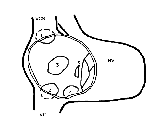
Sinus venosus atrial septal defect ASD locations. (1: upper sinus venosus defect; 2: lower sinus venosus defect.) Specialty Cardiac surgery A sinus venosus atrial septal defect is a type of atrial septal defect primarily associated with the sinus venosus . They represent 5% of atrial septal defects. [1] They can occur near the superior vena cava or inferior vena cava , but the former are more common. [2] They can be associated with anomalous pulmonary venous connection . [3] References [ edit ] ^ Robbins and Cotran Pathologic Basis of Disease 8th Edition ^ "Yale: Congenital Heart Disease: Sinus Venosus ASD" . Retrieved 2009-01-09 . ^ Attenhofer Jost CH, Connolly HM, Danielson GK, et al. (September 2005). "Sinus venosus atrial septal defect: long-term postoperative outcome for 115 patients" . ... External links [ edit ] Classification D ICD - 10 : Q21.1 ICD - 9-CM : 745.8 MeSH : C548009 C548009, C548009 v t e Congenital heart defects Heart septal defect Aortopulmonary septal defect Double outlet right ventricle Taussig–Bing syndrome Transposition of the great vessels dextro levo Persistent truncus arteriosus Aortopulmonary window Atrial septal defect Sinus venosus atrial septal defect Lutembacher's syndrome Ventricular septal defect Tetralogy of Fallot Atrioventricular septal defect Ostium primum Consequences Cardiac shunt Cyanotic heart disease Eisenmenger syndrome Valvular heart disease Right pulmonary valves stenosis insufficiency absence tricuspid valves stenosis atresia Ebstein's anomaly Left aortic valves stenosis insufficiency bicuspid mitral valves stenosis regurgitation Other Underdeveloped heart chambers right left Uhl anomaly Dextrocardia Levocardia Cor triatriatum Crisscross heart Brugada syndrome Coronary artery anomaly Anomalous aortic origin of a coronary artery Ventricular inversion
-
Dural Arteriovenous Fistula
Wikipedia
Type I [ edit ] Type I dural arteriovenous fistulas are supplied by meningeal arteries and drain into a meningeal vein or dural venous sinus. The flow within the draining vein or venous sinus is anterograde. ... Type II [ edit ] The high pressure within a Type II dural AV fistula causes blood to flow in a retrograde fashion into subarachnoid veins which normally drain into the sinus. Typically this is because the sinus has outflow obstruction. ... Type II IIa - confined to sinus with reflux (retrograde) into sinus but not cortical veins. IIb - drains into sinus with reflux (retrograde) into cortical veins (10-20% hemorrhage). Type III Drains direct into cortical veins (not into sinus) drainage (40% hemorrhage). Type IV Drains direct into cortical veins (not into sinus) drainage with venous ectasia (65% hemorrhage).CERNA3, PCYT1A, VEGFA, NDC1, TSPAN2, COIL, TGFBR2, PTEN, ACVRL1, CD38, SERPINE1, NFE2L2, MMP2, GABPA, F5, ENG, SERPINB2

-
Preauricular Sinus And Cyst
Wikipedia
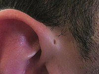
Preauricular sinus and cyst Other names Congenital auricular fistula , Geswein Hole , Congenital preauricular fistula , Ear pit , [1] : 782 or a Preauricular cyst [2] Preauricular sinus on right ear Specialty Otorhinolaryngology A preauricular sinus is a common congenital malformation characterized by a nodule, dent or dimple located anywhere adjacent to the external ear . [3] Frequency of preauricular sinus differs depending the population: 0.1–0.9% in the US, 0.9% in the UK, and 4–10% in Asia and parts of Africa. [4] Preauricular sinuses are inherited features, and most often appear unilaterally . ... "Surgical treatment of recurring preauricular sinus: supra-auricular approach" . Acta Otorhinolaryngologica Italica . 28 (6): 302–5. PMC 2689545 . PMID 19205595 . ^ "Preauricular Sinus" . Stedman's Medical Spellchecker . 2006 . ... "Surgical treatment of recurring preauricular sinus: supra-auricular approach" . Acta Otorhinolaryngologica Italica . 28 (6): 302–5. ... Further reading [ edit ] Tan T, Constantinides H, Mitchell TE (November 2005). "The preauricular sinus: A review of its aetiology, clinical presentation and management".
-
Phocomelia-Ectrodactyly-Deafness-Sinus Arrhythmia Syndrome
Orphanet
Phocomelia-ectrodactyly-deafness-sinus arrhythmia syndrome is characterised by phocomelia (involving arms more severely), ectrodactyly, ear anomalies (bilateral anomalies of the pinnae), conductive deafness, dysmorphism (long and prominent philtrum, mild maxillary hypoplasia) and sinus arrhythmia.
-
Hypertelorism-Preauricular Sinus-Punctual Pits-Deafness Syndrome
Orphanet
Hypertelorism-preauricular sinus-punctual pits-deafness syndrome is a rare developmental defect during embryogenesis syndrome characterized by hypertelorism, bilateral preauricular sinus, bilateral punctal pits, lacrimal duct obstruction, hearing loss, abnormal palmar flexion creases and bilateral distal axial triradii.
-
Sinus Pericranii
Wikipedia
Sinus pericranii Specialty Vascular surgery Sinus pericranii ( SP ) is a rare disorder characterized by a congenital (or occasionally, acquired) epicranial venous malformation of the scalp. [1] Sinus pericranii is an abnormal communication between the intracranial and extracranial venous drainage pathways. ... Contents 1 Signs and symptoms 2 Cause 3 Mechanism 4 Diagnosis 5 Treatment 6 See also 7 References Signs and symptoms [ edit ] Sinus pericranii typically present as soft palpable masses along midline skull, which may fluctuate in size depending on body positioning. ... The hypothesis of a spontaneous origin in the current case of SP is supported by no evidence of associated anomalies, such as cerebral aneurysmal venous malformations, systemic angiomas, venous angioma dural malformation, internal cerebral vein aneurysm, and cavernous hemangiomas. Mechanism [ edit ] Sinus pericranii is a venous anomaly where a communication between the intracranial dural sinuses and dilated epicranial venous structures exists.
-
Umbilical-Urachal Sinus
Wikipedia
Umbilical-urachal sinus Other names Urachal sinus Specialty Urology Umbilical-urachal sinus is a congenital disorder of the urinary bladder caused by failure of obliteration of proximal or distal part of the allantois , and the presentation of this anomaly is more common in children and rarer in adults. [1] [2] [3] It is thought have been first described by Cabriolus in 1550. [4] Complications [ edit ] Infection , with possible abscess formation. ... DiSantis DJ, Siegel MJ, Katz ME. 11 (1): 59–66. doi : 10.1148/radiographics.11.1.1996398 . PMID 1996398 . ^ "Urachal sinus presenting with abscess formation" . ... Cite journal requires |journal= ( help ) v t e Congenital malformations and deformations of urinary system Abdominal Kidney Renal agenesis / Potter sequence , Papillorenal syndrome cystic Polycystic kidney disease Meckel syndrome Multicystic dysplastic kidney Medullary sponge kidney Horseshoe kidney Renal ectopia Nephronophthisis Supernumerary kidney Pelvic kidney Dent's disease Alport syndrome Ureter Ectopic ureter Megaureter Duplicated ureter Pelvic Bladder Bladder exstrophy Urethra Epispadias Hypospadias Posterior urethral valves Penoscrotal transposition Vestigial Urachus Urachal cyst Urachal fistula Urachal sinus Medicine portal This article related to pathology is a stub .
-
Pilonidal Disease
Wikipedia
Signs and symptoms may include: [6] Intermittent pain/discomfort or swelling above the anus or near the tailbone Opaque yellow (purulent) or bloody discharge from the tailbone area Unexpected moisture in the tailbone region Discomfort sitting on the tailbone, doing sit-ups or riding a bicycle—any activities that roll over the tailbone area Some people with a pilonidal cyst will be asymptomatic . [7] Pilonidal sinus [ edit ] Pilonidal sinus (PNS): is a sinus tract, or small channel, that may originate from the source of infection and open to the surface of the skin. [8] Material from the cyst drains through the pilonidal sinus. ... "Penis: an unusual site for pilonidal sinus". International Urology and Nephrology . 38 (3–4): 607–8. doi : 10.1007/s11255-005-4761-5 . ... "Surgical interventions for the treatment of sacrococcygeal pilonidal sinus disease in children: A systematic review and meta-analysis". ... PMID 31754976 . ^ Lanigan M (September 27, 2012). "Pilonidal Cyst and Sinus" . Medscape . WebMD . Retrieved February 8, 2013 . ^ Hodges RM (1880). ... External links [ edit ] Classification D ICD - 10 : L05 ICD - 9-CM : 685 MeSH : D010864 DiseasesDB : 31128 External resources eMedicine : emerg/771 Wikimedia Commons has media related to Sinus pilonidalis . NHS Choices for pilonidal sinus treatment


