Load FindZebra Summary
Disclaimer:
FindZebra Search conducts a search using our specialized medical search engine.
FindZebra Summary uses the text completions API
(subject to OpenAI’s API data usage policies)
to summarize and reason about the search results.
The search is conducted in publicly available information on the Internet that we present “as is”.
You should be aware that FindZebra is not supplying any of the content in the search results.
FindZebra Summary is loading...
-
Organophosphate Poisoning
Wikipedia
The effects of OP exposure on infants and children are at this time currently being researched to come to a conclusive finding. [12] [13] Evidence of OP exposure in pregnant mothers are linked to several health effects in the fetus. ... These syndromes result after acute and chronic exposure to OP pesticides. Cholinergic syndrome occurs in acute poisonings with OP pesticides and is directly related to levels of AChE activity. ... Paraoxonase ( PON1 ) is a key enzyme involved in OP toxicity and has been found to be critical in determining an organism's sensitivity to OP exposure. PON1 can inactivate some OPs through hydrolysis. PON1 hydrolyzes the active metabolites in several OP insecticides such as chlorpyrifos oxon, and diazoxon, as well as, nerve agents such as soman, sarin, and VX. ... This test has been shown to be just as effective as a regular laboratory test and because of this, the portable ChE field test is frequently used by people who work with pesticides on a daily basis. [28] Treatment [ edit ] Current antidotes for OP poisoning consist of a pretreatment with carbamates to protect AChE from inhibition by OP compounds and post-exposure treatments with anti-cholinergic drugs.

-
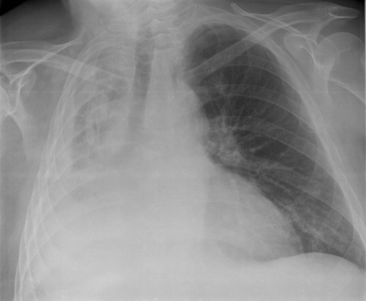
Cryptogenic Organizing Pneumonia
Orphanet
Cryptogenic organizing pneumonia (COP) is a form of idiopathic interstitial pneumonia characterized pathologically by organizing pneumonia (OP) that presents with non-specific flu-like symptoms, as well as cough and dyspnea and where no etiological agent is found. ... Diagnostic methods Diagnosis is based on clinical picture, imaging, histopathology of the lung and the exclusion of all causes of secondary OP. High resolution computed tomographic scan reveals 3 main imaging patterns: multiple patchy alveolar opacities that may migrate (in the majority of cases; typical COP), a solitary focal nodule or mass (focal COP), or diffuse infiltrative opacities (infiltrative or progressive fibrotic COP). Video-assisted thoracoscopy is the method of choice in obtaining lung tissue but transbronchial biopsies, core needle biopsies, and recently transbronchial cryobiopsies are alternate methods. Hallmark histological findings of OP are patchy filling of the lung alveoli and respiratory bronchioles by buds of granulation tissue composed of fibroblasts and myofibroblasts (Masson bodies). ... Differential diagnosis The main differential diagnosis is idiopathic chronic eosinophilic pneumonia. Secondary OP must also be excluded by elimination of its possible causes, including primary biliary cirrhosis, ulcerative colitis, Crohn disease, Sweet syndrome, sarcoidosis, Behçet disease, primary pulmonary lymphoma, drugs, infections, and all connective tissue diseases, especially rheumatoid arthritis and idiopathic inflammatory myopathies.UNC119, CD68, CRP, NR3C1, HDAC2, CXCL8, TNF, TNFRSF1A, TNFRSF1B, CD163, ING4, COP1, HT, TOMM5, WG, SFTPA1

-
Osteoporosis-Pseudoglioma Syndrome
OMIM
Frontali and Dallapiccola (1986) likewise concluded that Beighton's ocular osteogenesis imperfecta is osteoporosis-pseudoglioma syndrome, and Beighton (1986) acknowledged the diagnosis. Teebi et al. (1988) described OPS in 2 brothers and a sister whose parents were phenotypically normal first cousins. ... Population Genetics From a review of published cases, Frontali et al. (1985) suggested that OPS may be more frequent in Mediterranean countries. Mapping Gong et al. (1996) analyzed 16 DNA samples (7 affected individuals) from 3 different consanguineous kindreds with OPS. Using a combination of traditional linkage analysis and homozygosity mapping, they assigned the OPS locus to 11q12-q13. ... Gong et al. (1996) found that the most likely location of the OPS gene is in a 3-cM region between GSTP1 (134660) and D11S1296.
-
Idiopathic Interstitial Pneumonia
Wikipedia
There are seven recognized distinct subtypes of IIP. [2] Contents 1 Diagnosis 2 Development 3 References 4 External links Diagnosis [ edit ] Classification can be complex, [3] and the combined efforts of clinicians , radiologists , and pathologists can help in the generation of a more specific diagnosis. [4] [5] Idiopathic interstitial pneumonia can be subclassified based on histologic appearance into the following patterns: [6] [7] Histology Clinical Correlates Desquamative interstitial pneumonia (DIP) DIP Diffuse alveolar damage (DAD) ARDS , AIP , TRALI Nonspecific interstitial pneumonia (NSIP) NSIP Respiratory bronchiolitis RB-ILD Usual interstitial pneumonia (UIP) CVD , IPF , drug toxicity , pneumoconiosis Organizing pneumonia Cryptogenic organizing pneumonia Lymphoid interstitial pneumonia (LIP) LIP Usual interstitial pneumonia is the most common type. [8] Development [ edit ] Table 1: Development of the (histologic) idiopathic interstitial pneumonia classification Leibow et al. (1969) Katzenstein (1998) [9] ATS/ERS (2002) [7] UIP UIP UIP DAD DAD NSIP NSIP DIP DIP/RB DIP RB BIP OP OP LIP ( LPD ) LIP GIP (HMF) (HMF) UIP=usual interstitial pneumonia; DAD=diffuse alveolar damage; NSIP=non-specific interstitial pneumonia; DIP=desquamative interstitial pneumonia; RB=respiratory bronchiolitis; BIP=bronchiolitis obliterans interstitial pneumonia; OP=organizing pneumonia; LIP=lymphoid interstitial pneumonia; LPD= lymphoproliferative disease (not considered a diffuse lung disease); GIP= giant cell interstitial pneumonia ; HMF=heavy metal fibrosis, no longer grouped with diffuse lung disease Lymphoid interstitial pneumonia was originally included in this category, then excluded, then included again. [10] References [ edit ] ^ Richard K.IL13, IL4, MUC5B, MUC1, SFTPC, HNF4A, SFTPD, FAM13A, CD163, RECK, CXCR4, NKX2-1, USF2, TIAL1, THBD, TGFB1, TERT, TERC, CXCL13, ANGPT2, POSTN, SELE, SRRM2, RBMS3, FOXP3, PI15, RTEL1, TLR9, PNO1, MAK16, RTKN2, MUC21, SFTPA1, PSIP1, SDC4, CXCL12, IL13RA2, CD34, CHI3L1, CRP, VCAN, CCN2, DCN, DSG3, EGFR, ELANE, HNRNPD, IFNG, IL4R, MPO, BMP3, NCL, OVGP1, SERPINE1, PARN, PCBP2, PPARG, PTBP1, PTEN, PTPRC, CCL2, CCL7, CCL18, SFTPA2

-
X-Linked Congenital Stationary Night Blindness
GeneReviews
Electroretinogram Findings in Complete and Incomplete X-Linked Congenital Stationary Night Blindness View in own window ERG Finding Complete ( NYX X-linked CSNB) Incomplete ( CACNA1F X-linked CSNB) Scotopic rod b-wave Severely reduced or absent Reduced Mixed scotopic a-wave Normal Slightly reduced Mixed scotopic b-wave Reduced Reduced Scotopic OP Absent Slightly reduced Photopic a-wave Normal, slightly reduced, sawtooth (square) shaped Reduced Photopic b-wave Slightly reduced Reduced Photopic OP Lost, except for OP4 All OPs are lost. 30-Hz flicker Normal / slightly reduced Reduced w/double peak OP = oscillatory potential Note: Pupillary responses have been described in the literature and in textbooks as "paradoxic" (i.e., miosis of pupils when lights are turned off, as opposed to dilation). ... X-linked CSNB should be suspected in a female proband with the following ERG findings (observed in some heterozygous females): Reduced oscillatory potentials (OPs) associated with rod activity [Rigaudière et al 2003] Reduced b-wave amplitudes (with unaffected OPs) in one heterozygous female [Rigaudière et al 2003] Establishing the Diagnosis Male proband.
-
Transsexual
Wikipedia
Michael Bailey , and Martin Lalumiere , who she says "have completely failed to appreciate the implications of alternative ways of framing sexual orientation." [46] The terms androphilia and gynephilia to describe a person's sexual orientation without reference to their gender identity were proposed and popularized by psychologist Ron Langevin in the 1980s. [47] The similar specifiers attracted to men , attracted to women , attracted to both or attracted to neither were used in the DSM-IV . [48] Many transsexual people choose the language of how they refer to their sexual orientation based on their gender identity, not their birth assigned sex . [21] Surgical status [ edit ] Several terms are in common use, especially within the community itself relating to the surgical or operative status of someone who is transsexual, depending on whether they have already had sex reassignment surgery (SRS), have not had SRS but still intend to, or do not intend to have SRS. They are, post-op, pre-op, and non-op, respectively. [49] Pre-operative [ edit ] A pre-operative transsexual person, or simply pre-op for short, is someone who intends to have SRS at some point, but has not yet had it. [49] [50] Post-operative [ edit ] A post-operative transsexual person, or post-op for short, is someone who has had SRS. [49] Non-operative [ edit ] A non-operative transsexual person, or non-op , is someone who has not had SRS, and does not intend to have it in the future. ... Toby Meltzer, none of the patients reported complete regret and only 6% reported partial or occasional regrets. [70] A 2009 review of Medline literature suggests the total rate of patients expressing feelings of doubt or regret is estimated to be as high as 8%. [71] An issue reported by some is the inability to find sexual partners. [ citation needed ] A 2010 meta-study, based on 28 previous long-term studies of transsexual men and women, found that the overall psychological functioning of transsexual people after transition was similar to that of the general population and significantly better than that of untreated transsexual people. [72] Incidence and prevalence [ edit ] Prevalence is the proportion of a population found to be affected by a condition. ... Archived from the original on September 21, 2008 . Retrieved 2008-09-28 . ^ American Psychiatric Association (2000). ... Paper presented at the 1983 Harry Benjamin International Gender Dysphoria Association VIII International Symposium, Bordeaux, France. ^ a b Gaughan, Sharon (2006-08-19). "What About Non-op Transsexuals? A No-op Notion" . TS-SI . ... Retrieved 2013-12-27 . ^ a b Pauly MD, Ira B. (28 May 1993). "Terminology and Classification of Gender Identity Disorders" .

-
Conjoined Twins
Mayo Clinic
Rachipagus (ray-KIP-uh-gus), also called rachiopagus (ray-kee-OP-uh-gus), twins are joined back to back along the length of the spine. This type is very rare. Pelvis. Ischiopagus (is-kee-OP-uh-gus) twins are joined at the pelvis, either face to face or end to end. ... The twins can have two, three or four arms and two or three legs. Head. Craniopagus (kray-nee-OP-uh-gus) twins are joined at the back, top or side of the head, but not the face.

-
Undifferentiated Pleomorphic Sarcoma
Wikipedia
Radiation may be delivered either pre-op or post-op depending on surgeon and multidisciplinary tumor board's recommendations.RECQL4, TP53, MDM2, MKI67, CDKN2A, MIB1, EGFR, PDGFRA, TBC1D9, MSH2, CDK4, DDIT3, ABCB1, MFHAS1, HRAS, PTGS2, PRDM10, FUS, CD274, YAP1, EZR, IGF1R, CTNNB1, PIK3CA, NOS2, RB1, NCOR1, GRAP2, S100A9, CD163, TSPAN31, CCL2, HSPB3, TNFRSF6B, SLPI, SRC, KEAP1, COIL, FOSL1, AIMP2, STAT1, SLMAP, STAT3, WT1, TNF, WRN, SERPINA3, PPARGC1A, CDK2AP2, MACROD2, PRUNE1, PRM3, CREB3L2, TRIM63, MYOCD, FBXO32, GKN2, CITED2, NUTM1, MIR152, MIR320A, POU5F1P3, POU5F1P4, H3P8, WDR48, HEATR3, NANS, UBASH3A, SF3B6, LAMTOR2, FOXP1, POLDIP2, WWTR1, RNF19A, DKK1, RASSF1, RPP14, SUB1, PTCH1, FRS2, AHSA1, PTEN, PIK3CB, MAP2K1, CTLA4, HIF1A, HCCS, MTOR, FOXC2, FGFR3, FES, FDPS, EWSR1, EREG, EPAS1, EGF, HBEGF, DES, CXADR, CCN2, HMBS, CSF1, MAPK14, CRK, COL1A1, CD74, CD6, CASQ2, CASP8, BTC, AREG, AR, AMPD2, AKT1, ACVRL1, HLA-A, HSPB1, MAPK1, MLH1, PRKCD, POU5F1, PLAU, PIK3CG, PIK3CD, ACTB, PDGFB, PDCD1, SERPINE1, NTSR1, NRAS, NOS1, ABCC1, MME, MITF, HSPB2, MET, MAP3K5, MCM2, MAGEA3, LYZ, LMNA, KRAS, IL1R1, IL1B, IL1A, IGF2, IDH2, IDH1, HTC2, H3P10

-
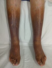
Venous Stasis
Wikipedia
A practical guide to hip surgery: from pre-op to recovery . Sunrise River Press.HFE, ESR2, BLOC1S2, BCL2, VEGFA, TGFB1, FAS, MYC, GADD45B, PGF, S100A8, SDC4, TGFBR2, MMP13, TP53, NRP1, GLRX3, SLC40A1, LGR4, MPO, MCC, MMP12, CSTA, ATF3, BAX, ZFP36L1, CASP3, CD44, COX8A, ENG, FASLG, ESR1, F13A1, F13B, FOXC2, HIF1A, SMAD7, MIR34A
-
Infectious Necrotic Hepatitis
Wikipedia
. ^ Mahin, Lucien, veterinary practitionner, personal communication. ^ Jensen & Brinton, op cit . ^ Stampfi, Henry (March 2014).

-
Party And Play
Wikipedia
'Social media and drug markets', The internet and drug markets (European Monitoring Centre for Drugs and Drug Addiction: Insights 21), Publications Office of the European Union, Luxembourg. v t e Lesbian , gay , bisexual , and transgender ( LGBT ) slang List Ace Bareback Banjee Bear Beard Beat Bi-curious Boi Top, bottom and versatile Bottom surgery Breeder Bugchasing Bulldagger Butch Castro clone Chicken Chickenhawk Chub Chubby chaser Cottaging Cruising Daddy Down-low Drag Dyke En femme En homme Fag (Faggot) Fag hag Fag stag Faux queen F2M Femme Flagging (hanky code) Friend of Dorothy Fruit Fruit fly Gay-for-pay Gaydar Gaymer Genderfuck Gold star lesbian Glory hole Heteroflexibility Lesbian until graduation Lipstick lesbian M2F Non-op Packing Party and play Passing Poppers Post-op Pre-op Queen RLE Shemale Soft butch Scissoring SRS Stone butch Stealth Swish T Tea-room TERF Top surgery Trache shave Trade Tranny Transfan Transition Tribbing Troll Twink U-Haul lesbian Womyn-born womyn Related Polari LGBT linguistics Terminology of homosexuality Category v t e Methamphetamine Enantiomers Dextromethamphetamine Levomethamphetamine Neuropharmacology Biomolecular targets TAAR1 (agonist) σ1R (agonist) σ2R (agonist) α 2A adrenoceptor (agonist) α 2B adrenoceptor (agonist) α 2C adrenoceptor (agonist) MAO ( competitive inhibitor ) Inhibited transporters DAT NET SERT VMAT1 VMAT2 EAAT1 EAAT2 SLC22A3 SLC22A5 Health Amphetamine dependence Meth mouth Prenatal methamphetamine exposure History and culture Amphetamine Crystal Darkness Crystal Meth Anonymous Faces of Meth History and culture of amphetamines Montana Meth Project No More Sunsets Party and play Rolling meth lab Ya ba Law Legal status Combat Methamphetamine Epidemic Act of 2005 Comprehensive Methamphetamine Control Act of 1996 Illinois Methamphetamine Precursor Control Act Ethnicity and nationality United States Native Americans Australia v t e Sexual slang General Anilingus Banjee Bareback Baseball metaphors for sex Blue balls Bottom Camel toe Chickenhead Circle jerk Cock tease Cornhole Cougar Cunt Deep-throating Dick Dirty Sanchez Dogging Donkey punch Douche Felching Fuck Girlfriend experience Glory hole Hogging Hot Karl Italian profanity Latin profanity Mama-san Mammary intercourse Mat Mile high club Motherfucker Nookie Party and play Pearl necklace Pegging Pirate Pussy Quickie Red wings Rusty trombone Serosorting Shemale Slut Snowballing Soggy biscuit Switch Teabagging Tits Top Top, bottom and versatile Turkey slap Twat Voulez-vous coucher avec moi?

-
Skin Infection
Wikipedia
. ^ In the WHO classification, it is noted that the infection classification "Excludes:... infective dermatitis...". See the WHO classification, op. cit. ^ Skin inflammation due to skin infection is called "infective dermatitis". See the WHO classifications, op. cit. ^ Global Burden of Disease Study 2013 Collaborators (22 August 2015).SLC9A6, TLR2, FLG, TNF, IL17A, PAGR1, IL1B, IL4, FAM3B, DOCK8, IL13, GAST, GALNS, VWF, TYR, TST, MAPK1, CFLAR, STAT6, STAT3, RFC1, RARRES2, AIMP2, ADAM10, GRAP2, CXCL14, PLG, AHSA1, CABIN1, RNF19A, POLDIP2, SETD2, IRAK4, TLR7, CD177, TNS3, NBEAL1, AGBL2, RNASE7, PPARG, MPO, PCP4, HP, CD36, LTB4R, CRK, CRP, MAPK14, CTLA4, EDNRB, EGFR, ELN, F5, FN1, GAPDH, GRN, HIF1A, HSPD1, MYO9B, IDE, IFNA1, IFNA13, IFNG, IL1A, IL2, IL6, IL9, IL15, LCT, LGALS3, MAP2, ADSS2, RNR1, COPD
-
Spastic Paraplegia 39, Autosomal Recessive
OMIM
The affected phenotype in each family conformed both to organophosphorous (OP) compound-induced delayed neuropathy (OPIDN) and to Troyer syndrome (275900), an autosomal recessive hereditary spastic paraplegia associated with distal muscle wasting.
-
Heterotaxy, Visceral, 6, Autosomal
OMIM
Narasimhan et al. (2015) reported a patient (OP-1069-II1), born of consanguineous parents, with situs inversus totalis.
-
Excess Skin
Wikipedia
People magazine covered the pre- and post- op experience. Erica told People the decision to get surgery wasn’t easy, but that she had a “new body” and was “floored” at the postoperative results. [8] [9] TLC Skin Tight TV Show [ edit ] TLC Skin Tight was a TV show with each episode following “two people who have lost massive amounts of weight and are about to undergo a full body transformation through skin removal surgery.” [10] “It’s not unusual, says the show, for there to be “up to 50 pounds” of sagging skin following massive weight loss.

-
Childhood Cataract
Wikipedia
Further reading [ edit ] Childhood cataracts at NHS Choices Cataracts in Children, Congenital and Acquired at EyeWiki v t e Diseases of the human eye Adnexa Eyelid Inflammation Stye Chalazion Blepharitis Entropion Ectropion Lagophthalmos Blepharochalasis Ptosis Blepharophimosis Xanthelasma Ankyloblepharon Eyelash Trichiasis Madarosis Lacrimal apparatus Dacryoadenitis Epiphora Dacryocystitis Xerophthalmia Orbit Exophthalmos Enophthalmos Orbital cellulitis Orbital lymphoma Periorbital cellulitis Conjunctiva Conjunctivitis allergic Pterygium Pseudopterygium Pinguecula Subconjunctival hemorrhage Globe Fibrous tunic Sclera Scleritis Episcleritis Cornea Keratitis herpetic acanthamoebic fungal Exposure Photokeratitis Corneal ulcer Thygeson's superficial punctate keratopathy Corneal dystrophy Fuchs' Meesmann Corneal ectasia Keratoconus Pellucid marginal degeneration Keratoglobus Terrien's marginal degeneration Post-LASIK ectasia Keratoconjunctivitis sicca Corneal opacity Corneal neovascularization Kayser–Fleischer ring Haab's striae Arcus senilis Band keratopathy Vascular tunic Iris Ciliary body Uveitis Intermediate uveitis Hyphema Rubeosis iridis Persistent pupillary membrane Iridodialysis Synechia Choroid Choroideremia Choroiditis Chorioretinitis Lens Cataract Congenital cataract Childhood cataract Aphakia Ectopia lentis Retina Retinitis Chorioretinitis Cytomegalovirus retinitis Retinal detachment Retinoschisis Ocular ischemic syndrome / Central retinal vein occlusion Central retinal artery occlusion Branch retinal artery occlusion Retinopathy diabetic hypertensive Purtscher's of prematurity Bietti's crystalline dystrophy Coats' disease Sickle cell Macular degeneration Retinitis pigmentosa Retinal haemorrhage Central serous retinopathy Macular edema Epiretinal membrane (Macular pucker) Vitelliform macular dystrophy Leber's congenital amaurosis Birdshot chorioretinopathy Other Glaucoma / Ocular hypertension / Primary juvenile glaucoma Floater Leber's hereditary optic neuropathy Red eye Globe rupture Keratomycosis Phthisis bulbi Persistent fetal vasculature / Persistent hyperplastic primary vitreous Persistent tunica vasculosa lentis Familial exudative vitreoretinopathy Pathways Optic nerve Optic disc Optic neuritis optic papillitis Papilledema Foster Kennedy syndrome Optic atrophy Optic disc drusen Optic neuropathy Ischemic anterior (AION) posterior (PION) Kjer's Leber's hereditary Toxic and nutritional Strabismus Extraocular muscles Binocular vision Accommodation Paralytic strabismus Ophthalmoparesis Chronic progressive external ophthalmoplegia Kearns–Sayre syndrome palsies Oculomotor (III) Fourth-nerve (IV) Sixth-nerve (VI) Other strabismus Esotropia / Exotropia Hypertropia Heterophoria Esophoria Exophoria Cyclotropia Brown's syndrome Duane syndrome Other binocular Conjugate gaze palsy Convergence insufficiency Internuclear ophthalmoplegia One and a half syndrome Refraction Refractive error Hyperopia Myopia Astigmatism Anisometropia / Aniseikonia Presbyopia Vision disorders Blindness Amblyopia Leber's congenital amaurosis Diplopia Scotoma Color blindness Achromatopsia Dichromacy Monochromacy Nyctalopia Oguchi disease Blindness / Vision loss / Visual impairment Anopsia Hemianopsia binasal bitemporal homonymous Quadrantanopia subjective Asthenopia Hemeralopia Photophobia Scintillating scotoma Pupil Anisocoria Argyll Robertson pupil Marcus Gunn pupil Adie syndrome Miosis Mydriasis Cycloplegia Parinaud's syndrome Other Nystagmus Childhood blindness Infections Trachoma Onchocerciasis v t e Optical illusions ( list ) Illusions Afterimage Ambiguous image Ames room Barberpole Bezold Café wall Checker shadow Chubb Cornsweet Delboeuf Ebbinghaus Ehrenstein Flash lag Fraser spiral Gravity hill Grid Hering Impossible trident Jastrow Lilac chaser Mach bands McCollough Müller-Lyer Necker cube Orbison Penrose stairs Penrose triangle Peripheral drift Poggendorff Ponzo Rubin vase Sander Schroeder stairs Shepard tables Spinning Dancer Ternus Vertical–horizontal White's Wundt Zöllner Popular culture Op art Trompe-l'œil Spectropia (1864 book) Ascending and Descending (1960 drawing) Waterfall (1961 drawing) The dress (2015 photograph) Related Accidental viewpoint Auditory illusions Tactile illusions Temporal illusion This article about the eye is a stub .
-
Head For Heights
Wikipedia
In: DAV Panorama 1/2008, ISSN 1437-5923 Pepi Stückl/Georg Sojer: Bergsteigen: Lehrbuch für alle Spielarten des Bergsteigens , Bruckmann, Munich, 1996, ISBN 3-7654-2859-0 v t e Climbing Types Aid Bouldering Clean Competition Crack Deep-water solo Direttissima Face Free Free solo Grass Ice Indoor Lead Rock Mixed Mountaineering Slab Speed Sport Top rope Trad Tree Lists Alpine clubs Climbers Deaths on eight-thousanders Equipment Everest deaths First ascents Knots Mount Hood incidents Terminology Terminology Abseiling Alpenstock Anchor Approach shoe Ascender Bachar ladder Belay device Belaying Bolt Bouldering mat Cam Carabiner Crampons Dry-tooling Dynamic rope Exposure Fifi hook Grades Grade (bouldering) Harness Head for heights Mountaineering boot Hex Ice axe Ice screw Ice tool Nut Picket Pitch Piton Protection Quickdraw Self-locking device Shoes Sling Snow fluke Snow protection Snowshoe Spotting Sure-footedness Tricam Webbing Media Climbing Rock & Ice Mountain film Companies Black Diamond CAMP Cascade Designs Deuter Early Winters Eastern Mountain Sports Five Ten Frostline Kits GERRY Mountain Sports Grivel Holubar Mountaineering JanSport Kelty La Sportiva Lowe Alpine Mammut Marmot Mountain Works Millet Mountain Safety Research Mountain Equipment Co-op Sierra Designs The North Face Therm-a-Rest Outdoor Research Petzl Rab REI Wild Country Organizations Alpine Club Alpine Club of Canada American Alpine Club Appalachian Mountain Club Austrian Alpine Club Austrian Tourist Club Club Alpin Français Club Alpino Italiano Den Norske Turistforening Federación Española de Deportes de Montaña y Escalada Fédération française de la montagne et de l'escalade German Alpine Club International Federation of Sport Climbing International Mountaineering and Climbing Federation South African National Climbing Federation South Tyrol Alpine Club Swedish Tourist Association Swiss Alpine Club USA Climbing Portal Category Commons WikiProject

-
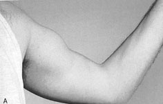
Biceps Tendon Rupture
Wikipedia
More severe injuries require surgery and post-op physical therapy to regain strength and functionality in the muscle.
-
Webbed Toes
Wikipedia
Treatment [ edit ] Partial simple syndactyly before surgery Partial simple syndactyly, 4 months post-op Webbed toes can be separated through surgery. ... Archived from the original on 2008-03-28 . Retrieved 2008-03-22 . [ unreliable source?CCNQ, APC, AUTS2, WDR60, NSUN2, DLL4, RIPK4, RAB23, OTUD6B, DYNC2LI1, DACT1, TBX22, UBE2T, PHGDH, IFT172, TCTN3, SIN3A, RFWD3, NIPBL, GRIP1, IQSEC2, RAI1, TBR1, DEAF1, MAD2L2, SF3B4, CD96, FIG4, BMS1, ATP6V1B2, FANCL, SETD5, NOG, FANCI, BHLHA9, KIF7, FREM2, CEP120, ARL6IP6, B3GLCT, TWIST2, NEK9, TICRR, WDR34, SLX4, BRIP1, FRAS1, TTC21B, PALB2, DYNC2H1, CPLANE1, PORCN, NXN, LMBR1, SMOC1, WDR19, FANCM, IFT80, ARHGAP31, SALL4, HDAC8, IFT140, HDAC4, SMC3, FANCC, TP63, FLNA, FLII, FLI1, FGFR2, FGFR1, GPC4, FBLN1, FANCG, FANCF, FANCB, FANCE, FANCD2, FANCA, GLI3, EVC, ERCC4, MEGF8, EFNB1, DYRK1A, CRMP1, CRKL, CREBBP, COL7A1, BRCA2, BRCA1, BMP4, BCR, GPC3, GJA1, HOXD13, RAD21, CUL4B, SMC1A, XRCC2, WNT10B, WNT7A, TWIST1, TFAP2B, TBX15, SMO, SHH, SC5D, SALL1, RAD51C, RAD51, NECTIN1, ROR2, IRF6, KCNJ2, LIG4, LRP4, MEF2C, KMT2A, PIK3CA, MAPK1, NEDD4L, PRKN, IDSP1, AKTIP, CBLL2, CUP2Q35, MUL1, IDS2

-
Atelectasis
Wikipedia
It is a common misconception and pure speculation that atelectasis causes fever. A study of 100 post-op patients followed with serial chest X-rays and temperature measurements showed that the incidence of fever decreased as the incidence of atelectasis increased. [4] A recent review article summarizing the available published evidence on the association between atelectasis and post-op fever concluded that there is no clinical evidence supporting this speculation. [5] Causes [ edit ] The most common cause is post-surgical atelectasis, characterized by splinting, i.e. restricted breathing after abdominal surgery. ... Retrieved 2018-02-02 . Updated: Nov 28, 2017 ^ Air Vice-Marshal John Ernsting (2008).ARVCF, CCDC114, DNAAF5, CCDC40, ARMC4, DNAAF2, CFAP298, DNAI2, TTC25, DNAL1, RSPH3, CFAP300, CCDC65, RSPH1, DRC1, LRRC56, COMT, CCDC151, DNAAF1, PIH1D3, DNAAF4, JMJD1C, RSPH9, GAS2L2, CCDC39, MCIDAS, RSPH4A, DNAAF3, NHLRC2, DNAJB13, HYDIN, RIPK4, ZMYND10, NME8, DNAH5, GAS8, GP1BB, LAMA2, OCRL, PAX3, PLEC, RPGR, RREB1, SPAG1, STAT3, TBX1, TSC1, TSC2, HIRA, UFD1, CXCR4, OFD1, DNAH11, SEC24C, CCNO, FBLN5, LRRC6, DNAH1, DNAI1, STK36, EFEMP2, CCDC103