• FindZebra
  • About
  • Contact
  • Help
  • Login
Deep Research Advanced
  • Fibrous Dysplasia Of Bone Wikipedia
    Fibrous Dysplasia/McCune-Albright Syndrome . Seattle (WA): University of Washington, Seattle. ... Journal of Bone and Mineral Research . 22 (9): 1468–1474. doi : 10.1359/jbmr.070511 . ISSN 0884-0431 . PMID 17501668 . ^ Cutler, Carolee M.; Lee, Janice S.; Butman, John A.; FitzGibbon, Edmond J.; Kelly, Marilyn H.; Brillante, Beth A.; Feuillan, Penelope; Robey, Pamela G.; DuFresne, Craig R. (2006-11-01). ... Journal of Bone and Mineral Research . 19 (4): 571–577. doi : 10.1359/JBMR.0301262 . ISSN 0884-0431 . PMID 15005844 . S2CID 37760051 . ^ Weinstein, L. ... Journal of Bone and Mineral Research . 19 (4): 571–577. doi : 10.1359/JBMR.0301262 . ISSN 0884-0431 . PMID 15005844 . S2CID 37760051 .
    GNAS, FGF23, COASY, FOLH1, GH1, IL6, TNFSF11, CREB1, MDM2, POSTN, MFAP1, CDK4, RUNX2, APRT, AR, HDAC8, DLEU7, S100A1, S100B, SH3BP2, SSTR4, LEPQTL1, ANBC, B3GAT1, LPAR2, ADAMTS2, ACKR3, PTH, CXCR6, SMUG1, PTGS2, ADRA1A, PRKAR1A, GHR, BGLAP, BMP2, BRS3, CAMP, COL1A1, CTNNB1, CFD, EDNRA, GPR42, CFP, IGF1, IGFBP3, SMAD6, MAS1, ADRA2B, COX2, NF1, FURIN, MTCO2P12
    • Fibrous Dysplasia GARD
      Fibrous dysplasia is a skeletal disorder that is characterized by the replacement of normal bone with fibrous bone tissue. It may involve one bone ( monostotic ) or multiple bones ( polyostotic ). Fibrous dysplasia can affect any bone in the body. The most common sites are the bones in the skull and face, the long bones in the arms and legs, the pelvis, and the ribs. Though many people with this disorder do not have any symptoms, others may have bone pain, abnormally shaped bones (deformities), or an increased risk of fractures (broken bones). The problems a person experiences depend on which bones are affected, and may arise from compression and displacement of adjacent structures to the lesions.
    • Fibrous Dysplasia Of Bone Orphanet
      A rare, benign, primary bone dysplasia characterized by progressive replacement of normal bone and marrow with fibrous connective tissue in either one (monostotic) or multiple (polyostotic) bones. Clinical manifestations depend on the anatomic location of the replacement and may include bone pain, deformities, pathological fractures, and cranial nerve deficits. Epidemiology The prevalence is unknown and is difficult to estimate due to the frequent asymptomatic lesions. Clinical description FD can involve the craniofacial, axial, and/or appendicular skeleton separately or simultaneously, and ranges from isolated asymptomatic monostotic lesions to severely incapacitating polyostotic lesions leading to pain, fracture, deformity or loss of vision and hearing. The monostotic form represents approximately 70% of cases, may present with pain or a pathologic fracture, and is usually diagnosed between 10 to 30 years of age.
  • Pacman Dysplasia Wikipedia
    Pediatr Radiol . 33 (4): 256–60. doi : 10.1007/s00247-002-0859-4 . PMID 12709756 . External links [ edit ] Classification D ICD - 10 : Q77.8 OMIM : 167220 MeSH : C538095 SNOMED CT : 722127006 External resources Orphanet : 1952 Online Mendelian Inheritance in Man (OMIM): 167220 This article about a congenital malformation is a stub .
    • Pacman Dysplasia OMIM
      Clinical Features Shohat et al. (1993) described a preterm (28 weeks) female fetus with a 'new' lethal skeletal dysplasia characterized by distinctive epiphyseal stippling, periosteal cloaking, and unusual microscopic morphology. Radiologically there was marked stippling of the coccygeal and sacral vertebral region as well as of the epiphyses. Long bones showed wide periosteal cloaking and bowing. Sagittal clefting was present in the upper spine. In contrast to the normal morphology of the epiphyses and growth plates, the marrow was filled with loose fibrous tissue containing numerous large multinucleated osteoclasts which were associated with Howship lacunae on the endosteal surface. The morphologic features were consistent with elevated bone resorption.
    • Pacman Dysplasia Orphanet
      A rare disorder characterized by epiphyseal stippling and osteoclastic overactivity. It has been described in less than 10 patients but may be underdiagnosed. It is characterized radiographically by severe stippling of the lower spine and long bones, and periosteal cloaking. Patients also have short metacarpals. The syndrome may be inherited as an autosomal recessive trait. This disorder should be included in the differential diagnosis of mucolipidosis type II.
  • Spillover Infection Wikipedia
    Biological Invasions . 17 (7): 2043–53. doi : 10.1007/s10530-015-0859-6 . ISSN 1387-3547 . ^ Durrer, Stephan; Schmid-Hempel, Paul (1994-12-22). ... Centers for Disease Control and Prevention , Infectious Disease Society of America (IDSA) v t e Concepts in infectious disease Transmission Basic concepts Asymptomatic carrier Host Incubation period Index case Infectious period Latent period Natural reservoir Subclinical infection Super-spreader Modes Human-to-human transmission Horizontal Vertical Cross-species transmission Spillover infection Vector Zoonosis Reverse zoonosis Routes Airborne disease Blood-borne disease Foodborne illness Waterborne disease Hospital-acquired infection Fomite Fecal-oral route Sexual Modelling Attack rate Basic reproduction number Compartmental models in epidemiology Critical community size Herd immunity Infection rate Serial interval Transmission risks and rates Medication Antimicrobial Antibiotic Antiviral drug Antimicrobial resistance Immunotherapy Phage therapy Vaccination Emerging infections Disease X Emergent virus Other Discovery of disease-causing pathogens Eradication of infectious diseases Pandemic
  • Tarantism Wikipedia
    Dancing for Health. Rowman Altamira. ISBN 0-7591-0859-5 , ISBN 978-0-7591-0859-2 . Rouget, Gilbert (1985) Music and Trance : a Theory of the Relations between Music and Possession .
  • Desmosis Wikipedia
    . : “Pathology of Chronic Constipation in Pediatric and Adult Coloproctology“, Karger 2005 ^ Meier-Ruge WA. (1998). "Desmosis of the colon: a working hypothesis of primary chronic constipation". Eur J Pediatr Surg.8; 299-303 ^ Meier-Ruge WA, Bruder E. (2007). "The morphological characteristics of aplastic and atrophic desmosis of the intestine". Pathologe 28: 149-54 ^ Meier-Ruge WA, Bruder E. (2005). "Atrophic desmosis as secondary connective tissue atrophy in muscularis propria".
  • Ovine Pulmonary Adenocarcinoma Wikipedia
    OPA has been eradicated in Iceland. [4] No breed or sex of sheep appears to be predisposed to OPA. [5] Most affected sheep show signs at 2 to 4 years of age. [4] OPA is not a notifiable disease , and therefore it is difficult to assess its prevalence . [4] History [ edit ] OPA was first described in the UK in 1888, [4] and described in detail in South Africa in 1891. [6] The disease was initially known as jaagsiekte [jɑːχˈsiktə] , a word derived from Afrikaans , meaning "chasing sickness", so called because animals are in respiratory distress as if they are out of breath from being chased. [6] It has also been known as sheep pulmonary adenomatosis and ovine pulmonary carcinoma. [7] Research [ edit ] OPA has been used as an animal model for human lung cancer, because OPA is histologically similar to human adenocarcinoma in situ of the lung . [8] Society and culture [ edit ] Dolly the sheep , the first mammal successfully cloned from an adult somatic cell, was euthanized after it was confirmed that she had OPA. [9] See also [ edit ] Enzootic nasal adenocarcinoma Jaagsiekte sheep retrovirus Enzootic nasal tumor virus References [ edit ] ^ "2.7.9 Ovine pulmonary adenocarcinoma (adenomatosis)" (PDF) . ... ISBN 9783642628979 . ^ Youssef, G; Wallace, WA; Dagleish, MP; Cousens, C; Griffiths, DJ (2015). "Ovine pulmonary adenocarcinoma: a large animal model for human lung cancer" . ILAR Journal . 56 (1): 99–115. doi : 10.1093/ilar/ilv014 .
  • Adrenal Crisis Wikipedia
    European Journal of Endocrinology . 162 (3): 597–602. doi : 10.1530/EJE-09-0884 . PMID 19955259 . External links [ edit ] Acute adrenal crisis on PubmedHealth Adrenal Crisis on Patient.info Classification D ICD - 10 : E27.2 External resources MedlinePlus : 000357 eMedicine : article/116716 article/765753 v t e Adrenal gland disorder Hyperfunction Aldosterone Hyperaldosteronism Primary aldosteronism Conn syndrome Bartter syndrome Glucocorticoid remediable aldosteronism AME Liddle's syndrome 17α CAH Pseudohypoaldosteronism Cortisol Cushing's syndrome Pseudo-Cushing's syndrome Steroid-induced osteoporosis Sex hormones 21α CAH 11β CAH Hypofunction Aldosterone Hypoaldosteronism 21α CAH 11β CAH Cortisol CAH Lipoid 3β 11β 17α 21α Sex hormones 17α CAH Inborn errors of steroid metabolism Adrenal insufficiency Adrenal crisis Adrenalitis Xanthogranulomatous Addison's disease Waterhouse–Friderichsen syndrome v t e Shock Distributive Septic shock Neurogenic shock Anaphylactic shock Toxic shock syndrome Obstructive Abdominal compartment syndrome Low volume Hemorrhage Hypovolemia Osmotic shock Other Spinal shock Cryptic shock Vasodilatory shock
    STAR, NR0B1, NR3C1, AAAS
    • Acute Adrenal Insufficiency Orphanet
      A primary adrenal insufficiency caused by a sudden defective production of adrenal steroids (cortisol and aldosterone). It represents an emergency, thus the rapid recognition and prompt therapy are critical for survival even before the diagnosis is made. Epidemiology Acute adrenal insufficiency (AAI) exact prevalence is unknown. Clinical description The disease may occur at any age. The onset is often sudden. The initial presentation may be non specific and may be limited to abdominal pain, nausea, vomiting, weight loss, tachycardia, and fever.
  • Galactose-1-Phosphate Uridylyltransferase Deficiency Wikipedia
    Long-term manifestations of the disease including ovarian failure in females, ataxia , and growth delays are not fully understood. [2] Routine monitoring of patients with GALT deficiency includes determining metabolite levels (galactose 1-phosphate in red blood cells and galactitol in urine) to measure the effectiveness of and adherence to dietary therapy, ophthalmologic examination for the detection of cataracts and assessment of speech, with the possibility of speech therapy if developmental verbal dyspraxia is evident. [2] Animal models [ edit ] Gal-1-P is assumed as to be a toxic agent, since the inhibition of the Galactokinase prevents toxicity in disease's models, [5] [6] although this is controversial for Drosophila models. [7] Phosphate depletion as a consequence of Gal-1-P is also proposed as a mechanism of toxicity in yeast models. [8] References [ edit ] ^ Online Mendelian Inheritance in Man (OMIM): Galactosemia - 230400 ^ a b c d e f g Elsas LJ (1993). ... In Pagon RA, Bird TD, Dolan CR, et al., eds. (1993). GeneReviews [Internet] . Seattle WA: University of Washington, Seattle. ^ a b Salway JG (2013). ... "The unfolded protein response has a protective role in yeast models of classic galactosemia" . Disease Models & Mechanisms . 7 (1): 55–61. doi : 10.1242/dmm.012641 . ... "Acute and long-term outcomes in a Drosophila melanogaster model of classic galactosemia occur independently of galactose-1-phosphate accumulation" . Disease Models & Mechanisms . 9 (11): 1375–1382. doi : 10.1242/dmm.022988 .
    GALT, GALE, GALK1, GAL, LGALS1, AMH, GALM, SPARC, S100A1, S100A8, S100B, BEST1, UGP2, UGT8, SEPTIN4, SDF4, LGALS7B, BRD2, OTC, PAH, CASP3, MAP1B, LGALS7, LEP, IL11, IGF1, GP2, G6PD, DMD, CYP51A1, CETN1, RN7SL263P
    • Epimerase Deficiency Galactosemia GeneReviews
      Summary Clinical characteristics. Epimerase deficiency galactosemia (GALE deficiency galactosemia) is a continuum comprising three forms: Generalized. Enzyme activity is profoundly decreased in all tissues tested. Peripheral. Enzyme activity is deficient in red blood cells (RBC) and circulating white blood cells, but normal or near normal in all other tissues. Intermediate. Enzyme activity is deficient in red blood cells and circulating white blood cells and less than 50% of normal levels in other cells tested. Infants with generalized epimerase deficiency galactosemia develop clinical findings on a regular milk diet (which contains lactose, a disaccharide of galactose and glucose); manifestations include hypotonia, poor feeding, vomiting, weight loss, jaundice, hepatomegaly, liver dysfunction, aminoaciduria, and cataracts.
    • Classic Galactosemia Orphanet
      A life-threatening metabolic disease with onset in the neonatal period. Infants usually develop feeding difficulties, lethargy, and severe liver disease. Epidemiology Global prevalence is unknown but estimated annual incidence has been reported to be between 1/40,000 and 1/60,000 in Western countries. The disorder appears to be more common in the Caucasian population than in other ethnic groups but figures in other populations may be underestimated. Males and females are equally affected. Clinical description When ingesting breast milk or lactose-containing formula, infants develop feeding problems, failure to thrive, and signs of liver damage (jaundice, bleeding tendency, hypoglycemia).
    • Classic Galactosemia And Clinical Variant Galactosemia GeneReviews
      While in vitro studies of GALT enzyme-deficient human fibroblasts demonstrated proof of concept, it is yet to be performed in an animal model. The GALT knockout mice generated by Leslie et al [1992] do not express the human phenotype of galactosemia and except for polyuria due to hypergalactosemia and hypergalactosuria are largely without disease. As mice have lost ARHI ( DIRAS3 ) during evolution [Lai et al 2008, Rubio-Gozalbo et al 2010, Tang et al 2010], a GALT enzyme-deficient mouse model that expresses an ARHI ( DIRAS3 ) signal is needed to test the hypothesis that ARHI gene expression plays a role in phenotypic expression of disease and determine whether inhibition of galactose-1-phosphate production limits or abrogates the "human phenotype."
  • Blue Toe Syndrome Wikipedia
    PMID 19103358 . ^ Blackshear JL, Oldenburg WA, Cohen MD (Dec 1994). "Making the diagnosis when the patient has 'blue toes ' ". ... PMID 22477301 . ^ Blackshear JL, Oldenburg WA, Cohen MD (Dec 1994). "Making the diagnosis when the patient has 'blue toes ' ". ... PMID 7982584 . ^ Blackshear JL, Oldenburg WA, Cohen MD (Dec 1994). "Making the diagnosis when the patient has 'blue toes ' ". ... PMID 7982584 . ^ Blackshear JL, Oldenburg WA, Cohen MD (Dec 1994). "Making the diagnosis when the patient has 'blue toes ' ".
  • Pinealoblastoma Wikipedia
    American Journal of Neuroradiology . 16 (1): 157–65. PMID 7900586 . ^ de Jong MC, Kors WA, de Graaf P, Castelijns JA, Moll AC, Kivelä T (December 2015). ... PMID 10561222 . ^ de Jong MC, Kors WA, de Graaf P, Castelijns JA, Kivelä T, Moll AC (September 2014).
    DICER1, ASMT, MYC, PDE4DIP, NIBAN1, CRB3, NEUROD4, PCDHGA3, DROSHA, PHOX2B, CRX, TPH1, SMARCB1, RB1, MYCN, CD99, INSM1, DICER1-AS1
    • Pineoblastoma Orphanet
      Pineoblastoma is a rare, malignant type of supratentorial primitive neuroectodermal tumor (sPNET), found mainly in children (less than 10% of cases are reported in adults), and located in the pineal region of the brain but that can metastasize along the neuroaxis. As it is the most aggressive of the pineal parenchymal tumors, it is usually associated with a poor prognosis.
    • Pineoblastoma GARD
      Pineoblastoma is a type of cancerous ( malignant ) tumor that grows in a part of the brain known as the pineal gland . It occurs mainly in children. Symptoms of pineoblastoma include a buildup of fluid around the brain (hydrocephalus), headaches, nausea, and difficulty with eye movement. Without treatment, pineoblastomas can cause weakness and difficulty controlling movement. The long term outcome depends on the age at diagnosis, the size of the tumor, and if the tumor has spread outside the brain ( metastasized ). The cause of pineoblastoma is unknown, but specific inherited genetic variants in two genes, RB1 and DICER1 can increase the risk for a pineoblastoma.
  • Prostatic Hyperplasia, Benign OMIM
    Although the small sample size prevented rigorous exclusion of nongenetic models, direct comparison of mendelian and nongenetic models showed that mendelian transmission provided the best overall explanation of the observed familial aggregation.
    KLK3, SRD5A2, FGF7, PRL, FGFR2, CYP19A1, TBX3, BCL2, NPEPPS, DLEU1, PTGS2, PROS1, PLAG1, UNC13A, CYP17A1, IL6, PSAT1, AR, BET1L, BCL11A, ODF3, ESR1, FGF2, IGF1, WDR11, CLPTM1L, GSTP1, ELOVL6, GATA5, GCLC, HNF1B, TGFB1, SYN3, PDE5A, VDR, STARD4-AS1, C5orf66, TP53, PCA3, PCNA, VEGFA, FOLH1, ARIH1, IGFBP3, ESR2, CXCL8, ERBB2, GSTM1, PTEN, ADRA1A, EGFR, TMPRSS2, ACE, HPGDS, CYP3A4, IL4, IGF2, COX2, TBC1D9, ABCB1, MMP9, CASP3, SRD5A1, IFNG, GHRH, NGF, MSMB, MTCO2P12, TRPV6, AKT1, RMC1, MIR21, DNMT1, GDF15, IL17A, CCN1, ADIPOQ, PSCA, GSTT1, IL2, GNRH1, PIK3CA, KDR, DKK3, THRB, STAT3, FLVCR1, FGFR1, CCL2, PPARG, CCL5, CD44, CD82, ELAC2, HSD17B7, MED15, EGF, IL18, MAPK7, IL10, PROM1, TGFB2, ACTB, CD34, MMP2, CIP2A, GPRC5A, PIK3CB, RARRES1, PIM1, NOS3, NOS2, MIR141, AMACR, HIF1A, MAPK14, CTNNB1, CYP1A1, PIK3CG, NCOA1, PIK3CD, IGFBP5, GLI2, NR1I2, IGFBP7, ZNF410, TRPV1, GPER1, KIDINS220, SOX11, GRP, HPN, KLF4, IL17RB, SPP1, TWIST1, IGF1R, GSR, LEP, SOX2, TGM4, MYC, ODC1, CBX4, RARB, RASSF1, PGF, PLK1, MST1, PON1, CXCR6, PRG2, MAPK1, TGFB3, POSTN, RNASEL, MSR1, LONP1, TIMP2, SHBG, TLR4, TGFA, ITGA4, TIMP3, KIT, KRAS, MMP14, LPL, AKAP12, S100A9, MLH1, MAGI2, FGF17, NTSR1, PCAP, GGCT, LMLN, NQO1, TSPO, MIR375, PWAR1, SLCO6A1, CXCR5, EDNRB, AZIN2, PPP1R14A, CCR2, ERG, ETS1, EZH2, F2R, HOTAIR, SPATA19, CYP11A1, CYP3A5, GSTK1, MIR145, CCR7, COMT, CGA, KLF6, CDKN2A, GADL1, ALKBH3, CDH1, CD81, CRP, CCNB1, VCAN, CSTA, APOB, CHRM3, ADCYAP1, ADRA1D, GATA6, ADCYAP1R1, SLC52A2, TMPRSS13, ALDH1A1, H4C6, KAT5, IFI44, MIR223, MIR25, H4C4, MIR221, CXCL13, TXNRD2, AHSA1, VAT1, PCAT1, FST, YAP1, TXNIP, MIR301A, STAG1, SPRY2, H4C11, H4C3, TSPAN1, RCE1, DLEC1, MFN2, RABGAP1L, H4C12, MIR205, MIR206, MIR15A, MIR143, FGF23, LILRA3, KLK11, IMMT, MIA, TAM, PRDX3, H3P10, HPSE, HBS1L, CTCF, MIR320A, MIR184, KHDRBS1, LINC02605, MIR187, H4C9, MIR191, AXIN1, MIR20A, H4C1, LOC110366354, NPRL2, SETDB1, KLK4, MIR34B, MIR615, SLC33A1, LPAR2, POTEF, SOCS3, SQSTM1, SPOP, GGT2, NCK1-DT, GGTLC4P, RECK, SCHLAP1, PGR-AS1, CDKN2B-AS1, PIK3R3, RIPK1, ARLNC1, EIF3H, DYNLL1, MIR708, FCGR1CP, PSMG1, MIR2909, LINC01672, CBR3-AS1, RNASEK-C17orf49, GGTLC3, DCLK1, AP3B1, GGTLC5P, H4C8, MIR34C, H4C2, CXCL14, MIR17HG, MIR373, TBPL1, H4C5, ADAMTS1, PAGE4, MAP4K4, H4C15, AIM2, GRAP2, H4C13, COX5A, MIR139, POU5F1P3, ITGBL1, PCSEAT, SLIT2, H4C14, RASSF10, POU5F1P4, AURKB, MIR193B, ENDOD1, MIR130B, ISYNA1, HAVCR2, IMP3, RMDN3, CEP55, ANO1, SLC25A21, TGIF2LX, GADD45GIP1, MTDH, TUG1, LEMD1, CGB5, CGB8, TRPM7, MARCHF5, UGT1A1, UGT1A4, UGT1A6, UGT1A7, UGT1A8, UGT1A10, TXNRD3, GNRHR2, KRT20, TLR9, PTOV1, UHRF2, ARL11, GDE1, KLK15, ACCS, ACSS2, ACE2, ATG9A, IL25, SMURF2, GOLPH3, WDR77, PCGEM1, PANK2, FUZ, CD276, ZFP91, OR51E2, AFAP1, LGR6, IL21, TDRD1, CXCL16, ARHGAP24, CCDC8, MIB1, QRFPR, NDRG2, NDRG3, CD177, AKR1B10, ACKR3, PRAC1, PMEPA1, SLC2A9, TMEM45B, H4-16, MIR126, ZBTB7A, ANKS4B, STEAP2, KLK5, BAMBI, H19, IL17RA, PLA2G15, NUP62, SSBP2, EPHA6, HAAO, HEY2, HEY1, NANOS2, ANGPTL2, SAP30L-AS1, SULF1, MIRLET7B, SEPTIN6, KDM4C, SWAP70, CYP3A43, MIRLET7C, DKK1, ELL2, SLC2A6, MIR106A, CDC37, PIM2, CTAG1A, RNF19A, GLCE, IL21R, FGD4, C17orf49, MZB1, OR51E1, DCTN4, ANGPTL4, ADIPOR1, PRLH, FOXP3, IRX4, PAQR3, KLB, IL22, SMARCAL1, POLDIP2, NRBP1, CREBRF, CD274, PYCARD, RANBP3L, GIT1, HTRA2, PDCD4, IL17B, VWCE, STEAP1, SNORD48, RFX6, A2M, SEMA3F, AIMP2, FLI1, FGF10, FGFR3, FGFR4, FGL1, FOXF1, FOXF2, FOXM1, FLT1, GATA3, FLT4, FN1, MTOR, FTH1, FTL, FYN, GABPA, FGF9, FCGR1B, FCGR1A, PTK2B, DUSP1, DUSP2, DUT, E2F1, LPAR1, EDN1, EDNRA, EFNA2, EGR1, ELN, MARK2, ENO2, EPHB2, EPOR, F2RL1, GALNT3, GDNF, ZNF165, HSPA1A, HOXD3, HP, HPS1, HES1, HSD17B1, HSD17B3, HSD17B2, HSPA1B, GGT1, HTC2, ID1, CFI, IFNA1, IFNA13, IGFBP2, IL1RN, HOXC6, HOXA7, HMMR, HMGA1, GH1, GLI1, GNRHR, SFN, GPC1, GPR42, GSTM3, GTF2H1, GTF2H2, GUCA2B, HDAC1, HHEX, HIC1, HLA-A, HLA-G, DRD2, DRD1, DPP4, ALDH7A1, ARG1, RHOA, STS, ATF3, ATM, RERE, ATP12A, BAX, CFD, CCND1, BMP2, BMP5, BMP6, BPI, BRCA1, BRS3, ARF6, APC, ANXA7, ANXA2, ABCA1, ABL2, ACR, ACP3, ADM, ADRA1B, ADRA2B, AGTR1, AHR, AKT2, ALOX12, ALOX5, ALOX15, ALOX15B, ANGPT2, CA9, CALCA, CANX, CNR1, COX8A, CLDN3, CPOX, CRAT, CRK, CRYZ, CSF2, CST3, CTAG1B, CTNND2, CTSB, CYP1B1, CYP2B6, DAPK1, DAXX, MAP3K8, CCR5, CASP9, CMA1, CAT, CAV1, CAV2, CAV3, CD28, CD63, CDC6, CDK4, CDKN1B, CDO1, CDX2, CGB3, CHRM2, CISH, CLU, IL6R, IL7, CXCR1, SFRP4, S100A11, SATB1, CCL3, CCL19, NAT2, SFRP1, SFRP2, SGK1, KLK7, SIM2, SLC7A1, SLC18A1, SLC22A3, SNAI1, SOD1, SOX9, S100A8, S100A6, S100A2, RPS10, PTK7, PTN, PXN, PYCR1, RARRES2, REN, RENBP, RNF2, RNY1, RNY3, RNY4, RNY5, ROCK1, RORA, RPL10, SPINK1, SPINT1, SRC, TNF, TP73, TPM1, TRAF6, TRPS1, TXN, UGT2B15, UGT2B17, USF2, VCL, VEGFB, VIM, VIPR1, WIPF1, YY1, ZFX, TOP2A, TLN1, SRF, TIMP1, SRY, SST, SSTR1, SSTR2, SSTR4, STAT5A, STC1, SULT1E1, TMBIM6, TERF2, TFF1, TFF3, TFRC, THBS1, THBS2, PTHLH, PPARD, IL10RA, MAS1, LPA, LTBR, LYZ, TM4SF1, SMAD2, SMAD4, SMAD9, MAZ, PPARA, MCAM, MEIS2, MAP3K3, MAP3K5, MET, KITLG, MGMT, LOX, LDHA, LASP1, LAMP2, IL10RB, IL13, IL15, ILK, IDO1, INHA, INSL3, INSR, ITGA6, JAK2, JUN, KCNK2, KCNMA1, KLK2, KRT15, MKI67, MMP3, MMP7, PAK1, PAX2, PAX5, ENPP1, ENPP2, SERPINF1, PGC, PGR, SERPINB5, PLAU, PLCL1, PLD1, FXYD3, PLXNA1, PMS1, POU5F1, PAM, P2RX1, MPO, OXTR, MRC1, MRE11, MSH2, MTHFR, MXI1, HNRNPM, NAIP, NELL2, NFE2L2, NFIB, NFKB1, NGFR, NME1, CCN3, OSM, H3P33
    • Benign Prostatic Hyperplasia (Bph) Mayo Clinic
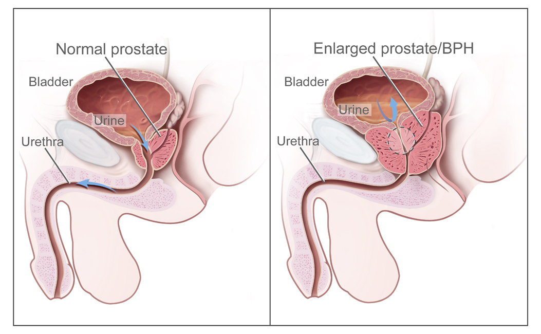
      Overview Benign prostatic hyperplasia (BPH) is a health issue that becomes more common with age. It's also called an enlarged prostate. The prostate is a small gland that helps make semen. It's found just below the bladder. And it often gets bigger as you get older. An enlarged prostate can cause symptoms that may bother you, such as blocking the flow of urine out of the bladder. It also can cause bladder, urinary tract or kidney problems. Many treatments can help BPH .
    • Benign Prostatic Hyperplasia Wikipedia
      Benign prostatic hyperplasia Other names Benign enlargement of the prostate (BEP, BPE), adenofibromyomatous hyperplasia, benign prostatic hypertrophy, [1] benign prostatic obstruction [1] Diagram of a normal prostate (left) and benign prostatic hyperplasia (right) Specialty Urology Symptoms Frequent urination, trouble starting to urinate, weak stream, inability to urinate , loss of bladder control [1] Complications Urinary tract infections , bladder stones , kidney failure [2] Usual onset Age over 40 [1] Causes Unclear [1] Risk factors Family history, obesity , type 2 diabetes , not enough exercise, erectile dysfunction [1] Diagnostic method Based on symptoms and examination after ruling out other possible causes [2] Differential diagnosis Heart failure , diabetes , prostate cancer [2] Treatment Lifestyle changes, medications, a number of procedures, surgery [1] [2] Medication Alpha blockers such as terazosin , 5α-reductase inhibitors such as finasteride [1] Frequency 105 million affected globally (2015) [3] Benign prostatic hyperplasia ( BPH ), also called prostate enlargement , is a noncancerous increase in size of the prostate gland . [1] Symptoms may include frequent urination, trouble starting to urinate, weak stream, inability to urinate , or loss of bladder control . [1] Complications can include urinary tract infections , bladder stones , and chronic kidney problems . [2] The cause is unclear. [1] Risk factors include a family history, obesity , type 2 diabetes , not enough exercise, and erectile dysfunction . [1] Medications like pseudoephedrine , anticholinergics , and calcium channel blockers may worsen symptoms. [2] The underlying mechanism involves the prostate pressing on the urethra thereby making it difficult to pass urine out of the bladder . [1] Diagnosis is typically based on symptoms and examination after ruling out other possible causes. [2] Treatment options include lifestyle changes, medications, a number of procedures, and surgery. [1] [2] In those with mild symptoms weight loss, exercise, and decreasing caffeine intake is recommended. [2] [4] In those with more significant symptoms, medications may include alpha blockers such as terazosin or 5α-reductase inhibitors such as finasteride . [1] Surgical removal of part of the prostate may be carried out in those who do not improve with other measures. [2] Phytotherapies that have been studied, such as saw palmetto , have not been shown to help. [2] About 105 million men are affected globally. [3] BPH typically begins after the age of 40. [1] Half of males age 50 and over are affected. [2] After the age of 80 about 90% of males are affected. [1] Although prostate specific antigen levels may be elevated in males with BPH, the condition does not increase the risk of prostate cancer . [5] Contents 1 Signs and symptoms 2 Causes 2.1 Hormones 2.2 Diet 2.3 Degeneration 3 Pathophysiology 4 Diagnosis 4.1 Differential diagnosis 4.1.1 Medical conditions 4.1.2 Medications 5 Management 5.1 Lifestyle 5.1.1 Voiding position 5.2 Medications 5.2.1 Alpha blockers 5.2.2 5α-Reductase inhibitors 5.2.3 Phosphodiesterase-5 inhibitors 5.2.4 Others 5.3 Self-catheterization 5.4 Surgery 5.5 Endovascular 5.6 Transurethral microwave thermotherapy 5.7 Alternative medicine 6 Epidemiology 7 References 8 External links Signs and symptoms [ edit ] BPH is the most common cause of lower urinary tract symptoms (LUTS), which are divided into storage, voiding , and symptoms which occur after urination. [6] Storage symptoms include the need to urinate frequently, waking at night to urinate , urgency (compelling need to void that cannot be deferred), involuntary urination , including involuntary urination at night, or urge incontinence (urine leak following a strong sudden need to urinate). [7] Voiding symptoms include urinary hesitancy (a delay between trying to urinate and the flow actually beginning), intermittency (not continuous), [8] involuntary interruption of voiding, weak urinary stream, straining to void, a sensation of incomplete emptying, and uncontrollable leaking after the end of urination. [9] [10] [11] These symptoms may be accompanied by bladder pain or pain while urinating, called dysuria . [12] Bladder outlet obstruction (BOO) can be caused by BPH. [13] Symptoms are abdominal pain, a continuous feeling of a full bladder, frequent urination, acute urinary retention (inability to urinate), pain during urination (dysuria), problems starting urination (urinary hesitancy), slow urine flow, starting and stopping (urinary intermittency), and nocturia. BPH can be a progressive disease, especially if left untreated. Incomplete voiding results in residual urine or urinary stasis, which can lead to an increased risk of urinary tract infection . [14] Causes [ edit ] Hormones [ edit ] Most experts consider androgens ( testosterone and related hormones ) to play a permissive role in the development of BPH. This means that androgens must be present for BPH to occur, but do not necessarily directly cause the condition. This is supported by evidence suggesting that castrated boys do not develop BPH when they age. In an unusual study of 26 eunuchs from the palace of the Qing dynasty still living in Beijing in 1960, the prostate could not be felt in 81% of the studied eunuchs. [15] The average time since castration was 54 years (range, 41–65 years).
  • Galactose Epimerase Deficiency Wikipedia
    This is largely due to a lack of functional animal models of classic galactosemia. The recent development of a Drosophila melanogaster GALE mutant exhibiting galactosemic symptoms may yield a promising future animal model. [3] Genetics [ edit ] Galactose epimerase deficiency has an autosomal recessive pattern of inheritance . ... High galactose-1-phosphate levels have been shown to interfere with phosphoglucomutase , [7] glycogen phosphorylase , [8] UDP-glycopyrophosphorylase, [9] activity in bacterial models and in vitro , yet in vivo mechanisms toxicity have yet to be confirmed. [3] Regardless, median galactose-1-phosphate levels act as the most accurate predictors of the severity of symptoms associated with Type III galactosemia. [10] Blockage of the Leloir pathway by GALE deficiency or dysfunction activates alternate pathways of glucose metabolism and leads to galactitol and galactonate formation. ... FEBS J . 272 (23): 6170–7. doi : 10.1111/j.1742-4658.2005.05017.x . PMID 16302980 . ^ de Jongh WA, Bro C, Ostergaard S, Regenberg B, Olsson L, Nielsen J (October 2008).
    GALE, GALK1, GALT
    • Galactose Epimerase Deficiency Orphanet
      A very rare, moderate to severe form of galactosemia characterized by moderate to severe signs of impaired galactose metabolism. Epidemiology Overall prevalence is not known but the disorder is thought to be very rare. Annual incidence is not known. Clinical description The disorder represents a continuum from peripheral to generalized states with corresponding disease severity. When ingesting breast milk or lactose-containing formula, patients may develop hypotonia, poor feeding, vomiting, weight loss, jaundice, hepatomegaly, splenomegaly, liver disorders, aminoaciduria, impaired growth, cataracts and cognitive deficiency. In severe cases, the disease can be life-threatening. Etiology Galactose epimerase deficiency is caused by mutations in the GALE gene (1p36) encoding the UDP-galactose 4-epimerase enzyme.
  • Cystinosis Wikipedia
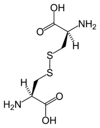
    Increased intracellular cystine profoundly disturbs cellular oxidative metabolism and glutathione status, [5] leading to altered mitochondrial energy metabolism, autophagy , and apoptosis . [6] Cystinosis is usually treated with cysteamine , which is prescribed to decrease intralysosomal cystine accumulation. [7] However, the discovery of new pathogenic mechanisms and the development of an animal model of the disease may open possibilities for the development of new treatment modalities to improve long-term prognosis. [3] Contents 1 Symptoms 1.1 Crystal morphology and identification 2 Genetics 3 Diagnosis 3.1 Types 4 Treatment 5 Cystinotic 6 See also 7 References 8 External links Symptoms [ edit ] There (are) three distinct types of cystinosis each with slightly different symptoms: nephropathic cystinosis, intermediate cystinosis, and non-nephropathic or ocular cystinosis. ... PMID 12110740 . ^ a b Nesterova G, Gahl WA. Cystinosis: the evolution of a treatable disease. Pediatr Nephrol 2012;28:51–9. ^ Gahl WA, Thoene JG, Schneider JA. Cystinosis.
    CTNS, LGALS3, TRPV1, SLC66A1, TFEB, APRT, SCN7A, CCL2, RAB7A, RAB11A, YBX3, PTH, NBAS, NLRP2, RILP, UNC13D, RAB27A, PRKAB1, CASP1, PRKAA2, PRKAA1, NAGLU, MFAP1, LRP2, LDLR, ITGAE, IL18, IL10, IL1B, IFNG, CTSD, RAB7B
    • Cystinosis GeneReviews
      Summary Clinical characteristics. Cystinosis comprises three allelic phenotypes: Nephropathic cystinosis in untreated children is characterized by renal Fanconi syndrome, poor growth, hypophosphatemic/calcipenic rickets, impaired glomerular function resulting in complete glomerular failure, and accumulation of cystine in almost all cells, leading to cellular dysfunction with tissue and organ impairment. The typical untreated child has short stature, rickets, and photophobia. Failure to thrive is generally noticed after approximately age six months; signs of renal tubular Fanconi syndrome (polyuria, polydipsia, dehydration, and acidosis) appear as early as age six months; corneal crystals can be present before age one year and are always present after age 16 months. Prior to the use of renal transplantation and cystine-depleting therapy, the life span in nephropathic cystinosis was no longer than ten years. With these interventions, affected individuals can survive at least into the mid-forties or fifties with satisfactory quality of life.
  • Disinhibition Wikipedia
    PBS also acknowledges the needs of support staff and includes strategies to manage crises when they arise. The following model is a brief guide to staff to remind them of key things to think about when planning support for a person with disabilities. ... "Tip Sheet – Positive Behaviour Support Model" (PDF) . Disability WA . Retrieved 2009-01-30 .
    TARDBP, CHMP2B, TREM2, TYROBP, VCP, PSEN2, PSEN1, SQSTM1, ABCA7, TOMM40, VPS13A, MAPT, SPAST, TBK1, SORL1, TMEM106B, CHCHD10, FUS, FTL, FMR1, APP, GRN, HGSNAT, C9orf72, SST, LRRK2, PVALB, TGFB1, VIP, CD200R1, HTR3B, EBPL, ALB, REN, SLC6A4, ROBO2, AR, BDNF, CCN2, ELK3, ETFA, GALNS, NR3C1, GRM2, HCRT, HTR2A, LRP1, MUC1, OPRM1, OXA1L, PDYN, PRKCG, PRL, ALDH2, ROBO1, STIN2-VNTR
  • Thrombophlebitis Wikipedia
    Journal of General Internal Medicine . 22 (1): 107–114. doi : 10.1007/s11606-006-0016-0 . ISSN 0884-8734 . PMC 1824715 . PMID 17351849 . ^ "Superficial Thrombophlebitis: Background, Pathophysiology, Etiology" . eMedicine .
    AKT1, PROS1, PTEN
    • Thrombophlebitis Mayo Clinic
      Overview Thrombophlebitis (throm-boe-fluh-BY-tis) is an inflammatory process that causes a blood clot to form and block one or more veins, usually in the legs. The affected vein might be near the surface of the skin (superficial thrombophlebitis) or deep within a muscle (deep vein thrombosis, or DVT). Blood clot in leg vein A blood clot in a leg vein may cause pain, warmth and tenderness in the affected area. Causes of thrombophlebitis include trauma, surgery or prolonged inactivity. deep vein thrombosis (DVT) increases the risk of serious health problems. It's usually treated with blood-thinning medications. Superficial thrombophlebitis is sometimes treated with blood-thinning medications, too.
  • Silent Sinus Syndrome Wikipedia
    AJR Am J Roentgenol . 178 (2): 503–6. doi : 10.2214/ajr.178.2.1780503 . PMID 11804926 . Full text Numa WA, Desai U, Gold DR, Heher KL, Annino DJ (2005).
    • Silent Sinus Syndrome Orphanet
      Silent sinus syndrome is characterised by adult-onset progressive enophthalmos due to collapse of some or all of the maxillary sinus walls. Epidemiology Its prevalence is unknown but around 100 cases have been reported in the literature so far. Clinical description The progressive enophthalmos may occasionally be associated with cheek pain, diplopia and blurred vision. Patients sometimes report a history of remote episodes of sinusitis. The syndrome may be idiopathic or occur following a bony orbital decompression resulting from Graves' ophthalmopathy or orbital floor fracture. Etiology The underlying mechanism involves obstruction of the maxillary antrum aeration followed by generation of negative antral pressure.
  • Arterial Tortuosity Syndrome Wikipedia
    .; Ledbetter, Nikki; Mefford, Heather C. (eds.). GeneReviews . Seattle (WA): University of Washington, Seattle. ... "The pathobiology of vascular malformations: insights from human and model organism genetics" . The Journal of Pathology . 241 (2): 281–293. doi : 10.1002/path.4844 .
    SLC2A10, MUS81, TGFBR1, TGFBR2, SMAD3, TGFB2, NT5E, COL4A4, SLC2A11, COL4A3, COL4A5, NXT1, ZNF197, MRPL28, SUB1, LAMTOR1, EFEMP2, ATRAID, MAFK, TPPP2, H3P12, EEF1E1, APOE, UBE2I, TNXB, CDKN2B, SLC2A1, PON1, P4HB, MYH9, MTHFR, FN1, ELN, COL4A1, CFL1, CDKN2C, H3P9
    • Arterial Tortuosity Syndrome GeneReviews
      Summary Clinical characteristics. Arterial tortuosity syndrome (ATS) is characterized by widespread elongation and tortuosity of the aorta and mid-sized arteries as well as focal stenosis of segments of the pulmonary arteries and/or aorta combined with findings of a generalized connective tissue disorder, which may include soft or doughy hyperextensible skin, joint hypermobility, inguinal hernia, and diaphragmatic hernia. Skeletal findings include pectus excavatum or carinatum, arachnodactyly, scoliosis, knee/elbow contractures, and camptodactyly. The cardiovascular system is the major source of morbidity and mortality with increased risk at any age for aneurysm formation and dissection both at the aortic root and throughout the arterial tree, and for ischemic vascular events involving cerebrovascular circulation (resulting in non-hemorrhagic stroke) and the abdominal arteries (resulting in infarctions of abdominal organs). Diagnosis/testing. The diagnosis of ATS is established in a proband with generalized arterial tortuosity and biallelic (homozygous or compound heterozygous) pathogenic variants in SLC2A10 identified on molecular genetic testing. Management. Treatment of manifestations: Individuals with ATS benefit from a coordinated approach of multidisciplinary specialists in a medical center familiar with ATS.
    • Arterial Tortuosity Syndrome MedlinePlus
      Arterial tortuosity syndrome is a disorder that affects connective tissue. Connective tissue provides strength and flexibility to structures throughout the body, including blood vessels, skin, joints, and the gastrointestinal tract. As its name suggests, arterial tortuosity syndrome is characterized by blood vessel abnormalities, particularly abnormal twists and turns (tortuosity) of the blood vessels that carry blood from the heart to the rest of the body (the arteries ). Tortuosity arises from abnormal elongation of the arteries; since the end points of the arteries are fixed, the extra length twists and curves. Other blood vessel abnormalities that may occur in this disorder include constriction (stenosis) and abnormal bulging (aneurysm) of vessels, as well as small clusters of enlarged blood vessels just under the skin (telangiectasia).
    • Arterial Tortuosity Syndrome GARD
      Arterial tortuosity syndrome is a disorder that affects connective tissue (a tissue that provides strength and flexibility to structures throughout the body). It is characterized by blood vessel abnormalities, particularly abnormal twists and turns (tortuosity) of the blood vessels that carry blood from the heart to the rest of the body (the arteries). Other blood vessel abnormalities that may occur in this disorder include constriction (stenosis) and abnormal bulging (aneurysm) of vessels, as well as small clusters of enlarged blood vessels just under the skin (telangiectasia). Other features include: Joints that are either loose and very flexible (hypermobile) or that have deformities limiting movement (contractures) Soft and stretchable skin Long, slender fingers and toes (arachnodactyly) Curvature of the spine (scoliosis) Sunken chest (pectus excavatum) or protruding chest (pectus carinatum) Protrusion of organs through gaps in muscles (hernias) Elongation of the intestines or pouches called diverticula in the intestinal walls People with arterial tortuosity syndrome often look older than their age and have distinctive facial features. The cornea, which is the clear front covering of the eye, may be cone-shaped and abnormally thin (keratoconus).
    • Arterial Tortuosity Syndrome OMIM
      A number sign (#) is used with this entry because of evidence that arterial tortuosity syndrome (ATORS) is caused by homozygous or compound heterozygous mutation in the gene encoding glucose transporter GLUT10 (SLC2A10; 606145) on chromosome 20q13. Description Arterial tortuosity syndrome is a rare connective tissue disorder characterized by generalized tortuosity, elongation, stenosis, and aneurysms of the major arteries. Skin and joint abnormalities, including hyperextensibility or hyperlaxity of the skin, joint laxity or contractures, and inguinal hernias, may also be observed. Other abnormalities include micrognathia, elongated face, high palate, beaked nose, sliding hernia, and ventricular hypertrophy (summary by Coucke et al., 2006). Clinical Features From Ankara, Turkey, Ertugrel (1967) described a 10-year-old girl with generalized tortuosity and elongation of all major arteries including the aorta.
    • Arterial Tortuosity Syndrome Orphanet
      A rare autosomal recessive connective tissue disorder characterized by tortuosity and elongation of the large and medium-sized arteries and a propensity towards aneurysm formation, vascular dissection, and stenosis of the pulmonary arteries. Epidemiology Approximately 100 patients have been described in the literature so far. The male to female ratio is 1:1. Clinical description The clinical manifestations are variable, depending on the arteries affected. Onset usually occurs in infancy or early childhood. The cardiovascular anomalies may lead to right ventricular hypertension, acute respiratory symptoms, ventricular hypertrophy and cardiac failure. Patients are prone to aneurysm formation, dissection and ischemic events.
  • Zettai Ryōiki Wikipedia
    Area of bare skin in the gap between overknee socks and a miniskirt or shorts An example of zettai ryōiki on a model at the 2012 Bologna Motor Show Zettai ryōiki ( Japanese : 絶対領域 , lit. ... ASIN 4758012121 . ^ a b "絶対領域(ゼッタイリョウイキ)とは - コトバンク" [Zettai ryōiki (zettairyouiki) to wa - kotobanku]. Kotobank.jp (in Japanese).
  • Protein Losing Enteropathy Wikipedia
    Journal of General Internal Medicine . 20 (10): C5–C7. doi : 10.1111/j.1525-1497.2005.0202.x . ISSN 0884-8734 . PMC 1490237 . PMID 16191147 .
    CD55, PLVAP, DGAT1, MPI, PIK3C2A, PTEN, COG8, ALG8, ADAMTS3, CCBE1, BMPR1A, SERPINA1, PTPN11, MEFV, IDO1, FGFR3, ALB
  • Trilateral Retinoblastoma Wikipedia
    PMID 10561222 . ^ De Jong MC, Kors WA, De Graaf P, Castelijns JA, Kivelä T, Moll AC (September 2014).
    RB1, MYCN, STK11
  • 1
  • 2
  • 3
  • 4
  • 5
  • ...
  • 142
  • 143
  • →

FindZebra

contact@findzebra.com