Load FindZebra Summary
Disclaimer:
FindZebra Search conducts a search using our specialized medical search engine.
FindZebra Summary uses the text completions API
(subject to OpenAI’s API data usage policies)
to summarize and reason about the search results.
The search is conducted in publicly available information on the Internet that we present “as is”.
You should be aware that FindZebra is not supplying any of the content in the search results.
FindZebra Summary is loading...
-
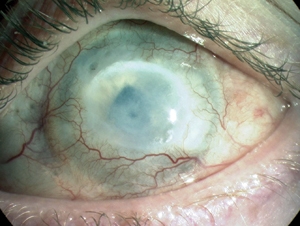
Chemical Eye Injury
Wikipedia
Specialty Ophthalmology Chemical eye injury are due to either an acidic or alkali substance getting in the eye. [1] Alkalis are typically worse than acidic burns. [2] Mild burns will produce conjunctivitis while more severe burns may cause the cornea to turn white. [2] Litmus paper is an easy way to rule out the diagnosis by verifying that the pH is within the normal range of 7.0—7.2. [1] Large volumes of irrigation is the treatment of choice and should continue until the pH is 6–8. [2] Local anesthetic eye drops can be used to decrease the pain. [2] Epidemiology [ edit ] In the United States , chemical eye injuries most commonly occur among working-age adults. [3] A 2016 analysis of emergency department visits from 2010-2013 reported over 36,000 visits annually for chemical burns to the eye, with a median age at presentation of 32 years. [4] By individual year of age, 1- and 2-year-old children have the highest incidence of these injuries, with rates approximately 50% higher than the highest-risk adult group (25 years), and 13 times higher than the rate among 7-year-olds. [4] Further research identified laundry detergent pods as a major source of injury among small children. [5] References [ edit ] ^ a b Zentani A, Burslem J (December 2009). "Towards evidence based emergency medicine: best BETs from the Manchester Royal Infirmary. BET 4: use of litmus paper in chemical eye injury". ... S.; Sheffield, I. D.; Frattaroli, S (2 February 2017). "Detergent Pod–Related Eye Injuries Among Preschool-Aged Children".
-
Leukemia, Acute Myeloid
OMIM
Fong et al. (2015) used primary mouse hematopoietic stem and progenitor cells immortalized with the fusion protein MLL-AF9 (see 159555) to generate several single-cell clones that demonstrated resistance, in vitro and in vivo, to the prototypical bromodomain and extra terminal protein (BET) inhibitor I-BET. Resistance to I-BET conferred cross-resistance to chemically distinct BET inhibitors such as JQ1, as well as resistance to genetic knockdown of BET proteins. ... Fong et al. (2015) demonstrated that resistance to BET inhibitors, in human and mouse leukemia cells, is in part a consequence of increased Wnt/beta-catenin (see 116806) signaling, and negative regulation of this pathway results in restoration of sensitivity to I-BET in vitro and in vivo. ... Rathert et al. (2015) performed a chromatin-focused RNAi screen in a sensitive MLL-AF9;Nras(G12D)-driven AML mouse model to identify factors involved in primary and acquired BET resistance in leukemia. The screen showed that suppression of the Polycomb repressive complex-2 (PRC2; see 606245), contrary to effects in other contexts, promotes BET inhibitor resistance in AML. ... Northern blot analysis showed that the 2 microRNAs targeted genes involved in apoptosis, the cell cycle, and cell proliferation. ... Mutations in NRAS (164790) and NPM1 (164040) had been previously identified in patients with AML, but 2 other mutations had not been identified.
-
Nut Midline Carcinoma
Wikipedia
Specialty Oncology NUT carcinoma (formerly NUT midline carcinoma), is a rare genetically defined, very aggressive squamous cell epithelial cancer that usually arises in the midline of the body and is characterized by a chromosomal rearrangement in the nuclear protein in testis gene . [2] In approximately 75% of cases, the coding sequence of NUTM1 on chromosome 15q14 is fused to BRD4 or BRD3 , which creates a chimeric gene that encodes the BRD-NUT fusion protein. The remaining cases, the fusion of NUTM1 is to an unknown partner gene, usually called NUT -variant. Contents 1 Signs and symptoms 2 Diagnosis 2.1 Differential diagnosis 3 Treatment 4 Prognosis 5 See also 6 References 7 External links Signs and symptoms [ edit ] In the United States, about 20-30 cases are reported each year. ... One of the most helpful and characteristic findings is the focal abrupt squamous differentiation, where stratification and gradual differentiation are absent, resembling a Hassall corpuscle of the thymus. [6] The defining feature of NMCs is rearrangement of the NUT gene. [4] Most common is a translocation involving the BRD4 gene and NUT gene (t(15;19)(q13;p13.1)). [5] [7] Treatment [ edit ] BET inhibitors are used for treatment [ citation needed ] Prognosis [ edit ] NUT midline carcinoma is very resistant to standard chemotherapy treatments. ... Specific molecular targeted therapies ( BET inhibitors and histone deacetylase inhibitors (HDACi)) may help to yield growth arrest of the neoplastic cells. [6] Overall, there is a mean survival of 6–9 months. [8] [9] See also [ edit ] BET inhibitor Mediastinum References [ edit ] ^ RESERVED, INSERM US14-- ALL RIGHTS.BRD4, NUTM1, MYC, AFP, CDK9, SMARCA4, DNER, NSD3, HDAC9, CIC, SF3B1, ZNF532, CD274, SYP, MIR145, MIR99A, MIR3140, TP63, SMARCB1, SOX2, CCNT1, ROS1, BRD2, PIK3CG, PIK3CD, PIK3CB, PIK3CA, EZH2, EP300, EGFR, CDKN2A, CDK4, H3P10

-
Problem Gambling
Wikipedia
Gambling Addiction: The Problem, the Pain, and the Path to Recovery . Vine Books. ISBN 978-0-8307-3425-2 . [ page needed ] ^ Petry, Nancy (September 2006). ... Journal of Gambling Studies . 31 (3): 1–20. doi : 10.1007/s10899-014-9449-2 . hdl : 10133/3418 . PMID 24627139 . ^ American Psychiatric Association (May 18, 2013). ... American Journal of Psychiatry . 163 (2): 297–302. doi : 10.1176/appi.ajp.163.2.297 . ... "Review of Self-exclusion from Gambling Venues as an Intervention for Problem Gambling" . Journal of Gambling Studies . 30 (2): 229–251. doi : 10.1007/s10899-013-9362-0 . ... "The Prevalence of Pathological Gambling in Canada". Journal of Gambling Studies . 12 (2): 129–142. doi : 10.1007/BF01539170 .DRD3, PLTP, DRD2, DRD4, SCLY, KRT7, TAL1, SLC6A3, BDNF, CHPT1, FZD10, CIT, TRIM31, BAG3, VLDLR, MAOB, OPRM1, COMT, MAOA, LEP, HTR2A, HTR1B, DRD1, DBH, CREB1, DHDDS

-
Costochondritis
Wikipedia
The Medical Clinics of North America . 94 (2): 259–273. doi : 10.1016/j.mcna.2010.01.007 . ... "Atypical chest pain in athletes". Curr Sports Med Rep . 8 (2): 52–8. doi : 10.1249/JSR.0b013e31819c7d01 . ... Journal of Manual and Manipulative Therapy . 18 (2): 64–68. doi : 10.1179/106698110X12640740712653 . ... "Towards evidence based emergency medicine: best BETs from the Manchester Royal Infirmary. BET 3: The use of corticosteroids in the management of costochondritis".
-
Covering Sickness
Wikipedia
The disease is caused by Trypanosoma equiperdum , which belongs to an important genus of parasitic protozoa , and is the only member of the genus that is spread through sexual intercourse. [2] The occurrence of dourine is notifiable in the European Union under legislation from the OIE. [3] There currently is no vaccine and although clinical signs can be treated, [2] there is no cure. [4] Contents 1 Parasite 2 Treatment 3 References 4 External links Parasite [ edit ] Trypanosoma equiperdum is one of three known strains from the Trypanosoma family; along with Trypanosoma evansi and Trypanosoma brucei . [2] Trypanosoma equiperdum has been discovered to be most closely linked to Trypanosoma evansi , so much so that even observation under microscope is not sufficient to differentiate between the two as their structure is very similar. [5] Dourine is a unique disease in the sense that it has no known vectors or fomites existing in the natural world, other than members of the equine family, including donkeys , mules , and horses. [2] In a laboratory setting, Trypanosoma equiperdum has been manipulated to adapt to and proliferate in other species, such as dogs, rabbits, mice and rats, but this has never been observed to occur naturally and without scientific manipulation. [4] Although this limits spread of the disease because it is restricted to the equine population alone, the organism has developed complex mechanisms over time to better equip itself for prolonged survival in the equine species. ... There are four main drugs on the market that are used to treat the clinical signs of dourine: Suramin, Diminazen, Cymerlarsan, and Quinapyramin. [2] [7] However, none of the listed drugs are a cure and even the individual animals that are treated will experience relapses. [4] Although this disease is not fatal in all cases and spontaneous recovery can occur, the death rate is relatively high and listed at a mortality rate of over fifty percent. [3] This lack of a cure or vaccine is a definite problem in the equine industry, especially in developing countries where equines are highly valuable for both agriculture and transportation. [5] Dourine is considered an endemic problem in developing countries, where over sixty percent of equines in the world are located. [4] The protocol for this disease, as stated by OIE, currently stands at slaughter of seropositive animals. [3] This is not an economically feasible option for many people who depend on horses for their livelihood. ... "Establishment of a panel of reference Trypanosoma evansi and Trypanosoma equiperdum strains for drug screening". Veterinary Parasitology . 148 (2): 114–121. doi : 10.1016/j.vetpar.2007.05.020 . PMID 17624671 . ^ a b c Codes of Practice 2017 (PDF) . Horserace Betting Levyboard. pp. 59–65 . Retrieved 19 May 2017 . ^ a b c d Hagos, A; Goddeeris, BM; Yilkal, K; Alemu, T; Fikru, R; Yacob, HT; Feseha, G; Claes, F (4 August 2010). ... "Trypanosoma evansi and T. equiperdum: distribution, biology, treatment and phylogenetic relationship (a review)". Veterinary Parasitology . 79 (2): 95–107. doi : 10.1016/S0304-4017(98)00146-0 .

-
Puppy Pregnancy Syndrome
Wikipedia
The syndrome is thought to be localized to villages in several states of India , including West Bengal , Assam , Bihar , Jharkhand , Orissa , and Chhattisgarh , and has been reported by tens of thousands of individuals. [1] It is far more prevalent in areas with little access to education. [1] People suffering from this condition believe that shortly after being bitten by a dog, puppies are conceived within their abdomen. [1] This is said to be especially likely if the dog is sexually excited at the time of the attack. [2] Victims are said to bark like dogs and have reported being able to see the puppies inside them when looking at water or hear them growling in their abdomen. [1] [2] [3] It is believed that the victims will eventually die – especially men, who will give birth to their puppies through the penis . [2] [3] Witch doctors offer oral cures, which they claim will dissolve the puppies, allowing them to pass through the digestive system and be excreted "without the knowledge of the patient". [1] [2] Doctors in India have tried to educate the public about the dangers of believing in this condition. [3] Most sufferers are referred to psychiatric services, but in rare instances patients fail to take anti- rabies medication in time, thinking that they are pregnant with a puppy and thus the witch doctor's medicine will cure them. Since rabies is so deadly, this is a very dangerous idea. [1] [2] This is further compounded by witch doctors stating that their medicine will fail if sufferers seek scientific treatment. [1] Some psychiatrists believe that the syndrome meets the criteria for a culture-bound disorder . [2] See also [ edit ] Clinical lycanthropy Superstition in India References [ edit ] ^ a b c d e f g Rahman, Shaikh Azizur (December 31, 2012). ... PMID 12793514 . v t e Superstition Main topics Amulet Evil eye Luck Omen Talismans Myth and ritual Lists List of superstitions List of lucky symbols List of bad luck signs Sailors' superstitions Theatrical superstitions Africa Buda Gris-gris Sampy Sleeping child Americas Ascalapha odorata Carranca Cooties Curupira Djucu Fortune cookie Groundhog Day I'noGo tied Oscar love curse Susto White lighter myth Witch window Asia Superstition in India Superstition in Pakistan Japanese superstitions Bhoot (ghost) Chhaupadi Churel Ghosts in Bengali culture Jackal's horn Kuai Kuai culture Muhurta Navaratna Nazar battu Pichal Peri Puppy pregnancy syndrome Akabeko Kanai Anzen Maneki-neko Okiagari-koboshi Omamori Fan death Agimat Arbularyo Barang Kulam Lihi Pagtatawas Pasma Usog Kuman Thong Palad khik Takrut Nang Kwak White elephant Curse of 39 Jin Chan Numbers in Chinese culture Superstitions of Malaysian Chinese Europe August curse Barbary macaques in Gibraltar Bayern-luck Blarney Stone Cimaruta Cornicello The Goodman's Croft Himmelsbrief Icelandic magical staves In bocca al lupo Kitchen witch Klabautermann Mooncalf Nazar Need-fire Painted pebbles Powder of sympathy Rabbit rabbit rabbit Ravens of the Tower of London Russian traditions and superstitions Spilling water for luck The Scottish Play Troll cross Tycho Brahe days Witch post Wolfssegen General 11:11 4 ( Four-leaf clover , Tetraphobia ) 7 ( Seventh son of a seventh son ) 8 9 13 ( Friday the 13th , The Thirteen Club , Thirteenth floor , Triskaidekaphobia ) 108 111 666 ( Number of the Beast ) Ace of spades Auspicious wedding dates Baseball superstition Beginner's luck Black cat Bread and butter Break a leg Chain letter Cramp-ring Curse Davy Jones' Locker Dead man's hand End-of-the-day betting effect Fear of frogs Fear of ghosts First-foot Flying Dutchman Four Eleven Forty Four Gambler's conceit Good luck charm Human sacrifice Jinx Knocking on wood Law of contagion Literomancy Lock of hair Maternal impression Miasma theory Nelson Numismatic charm Penny Rabbit's foot Rainmaking Ship sponsor Shoes on a table Sign of the horns Something old Spilling salt Statue rubbing Three on a match Threshold Toi toi toi 27 Club Wishing well Witch ball Witching hour Related Apotropaic magic Astrology and science Coincidence Debunker Divination Folk religion Fortune-telling Magic and religion Magical thinking Numerology Perceptions of religious imagery in natural phenomena Post hoc ergo propter hoc Traditional medicine Urban legend Jew Muslim
-
Tricyclic Antidepressant Overdose
Wikipedia
Tricyclic anti-depressant overdose Other names TCA poisoning, TCA overdose, TCA toxicity Chemical structure of the tricyclic antidepressant amitriptyline Specialty Emergency medicine Symptoms Elevated body temperature , large pupils, irregular heart beat , seizures [1] Usual onset Within 6 hours [2] Causes Accidental or purposeful [2] [3] Treatment Supportive , sodium bicarbonate , lipid emulsion [2] Frequency Relatively common [1] [4] Deaths 270 per year (UK) [1] Tricyclic antidepressant overdose is poisoning caused by excessive medication of the tricyclic antidepressant (TCA) type. Symptoms may include elevated body temperature , blurred vision, dilated pupils , sleepiness, confusion, seizures , rapid heart rate , and cardiac arrest . [1] If symptoms have not occurred within six hours of exposure they are unlikely to occur. [2] TCA overdose may occur by accident or purposefully in an attempt to cause death . [2] The toxic dose depends on the specific TCA. [2] Most are non-toxic at less than 5 mg/kg except for desipramine , nortriptyline , and trimipramine , which are generally non-toxic at less than 2.5 mg/kg. [5] [2] In small children one or two pills can be fatal. [6] An electrocardiogram (ECG) should be included in the assessment when there is concern of an overdose. [2] In overdose activated charcoal is often recommended. [1] People should not be forced to vomit . [2] In those who have a wide QRS complex ( > 100 ms ) sodium bicarbonate is recommended. [2] If seizures occur benzodiazepines should be given. [2] In those with low blood pressure intravenous fluids and norepinephrine may be used. [1] The use of intravenous lipid emulsion may also be tried. [3] In the early 2000s TCAs were one of the most common cause of poisoning. [1] In the United States in 2004 there was more than 12,000 cases. [2] In the United Kingdom they resulted in about 270 deaths a year. [1] An overdose from TCAs was first reported in 1959. [1] Contents 1 Signs and symptoms 2 Cause 3 Pathophysiology 4 Diagnosis 5 Treatment 5.1 Decontamination 5.2 Medication 5.3 Dialysis 6 Epidemiology 7 References 8 External links Signs and symptoms [ edit ] The peripheral autonomic nervous system , central nervous system and the heart are the main systems that are affected following overdose. [1] Initial or mild symptoms typically develop within 2 hours and include tachycardia , drowsiness , a dry mouth, nausea and vomiting , urinary retention, confusion, agitation, and headache . [7] More severe complications include hypotension , cardiac rhythm disturbances, hallucinations , and seizures . ... S2CID 44532041 . ^ Bartram, Tom (1 March 2008). "Best BETs from the Manchester Royal Infirmary. Bet 3. Toxic levels of tricyclic drugs in accidental overdose". ... Tricyclic antidepressant exposure in toddlers". J Emerg Med . 28 (2): 169–74. doi : 10.1016/j.jemermed.2004.08.018 .
-
Numerophobia
Wikipedia
Find sources: "Numerophobia" – news · newspapers · books · scholar · JSTOR ( April 2016 ) ( Learn how and when to remove this template message ) Numerophobia Other names Math anxiety Specialty Psychiatry Numerophobia , arithmophobia or mathematics anxiety is an anxiety disorder , [1] [2] [ page needed ] where the condition is fear of dealing with numbers or mathematics. ... ISBN 978-1-4381-2098-0 . v t e Superstition Main topics Amulet Evil eye Luck Omen Talismans Myth and ritual Lists List of superstitions List of lucky symbols List of bad luck signs Sailors' superstitions Theatrical superstitions Africa Buda Gris-gris Sampy Sleeping child Americas Ascalapha odorata Carranca Cooties Curupira Djucu Fortune cookie Groundhog Day I'noGo tied Oscar love curse Susto White lighter myth Witch window Asia Superstition in India Superstition in Pakistan Japanese superstitions Bhoot (ghost) Chhaupadi Churel Ghosts in Bengali culture Jackal's horn Kuai Kuai culture Muhurta Navaratna Nazar battu Pichal Peri Puppy pregnancy syndrome Akabeko Kanai Anzen Maneki-neko Okiagari-koboshi Omamori Fan death Agimat Arbularyo Barang Kulam Lihi Pagtatawas Pasma Usog Kuman Thong Palad khik Takrut Nang Kwak White elephant Curse of 39 Jin Chan Numbers in Chinese culture Superstitions of Malaysian Chinese Europe August curse Barbary macaques in Gibraltar Bayern-luck Blarney Stone Cimaruta Cornicello The Goodman's Croft Himmelsbrief Icelandic magical staves In bocca al lupo Kitchen witch Klabautermann Mooncalf Nazar Need-fire Painted pebbles Powder of sympathy Rabbit rabbit rabbit Ravens of the Tower of London Russian traditions and superstitions Spilling water for luck The Scottish Play Troll cross Tycho Brahe days Witch post Wolfssegen General 11:11 4 ( Four-leaf clover , Tetraphobia ) 7 ( Seventh son of a seventh son ) 8 9 13 ( Friday the 13th , The Thirteen Club , Thirteenth floor , Triskaidekaphobia ) 108 111 666 ( Number of the Beast ) Ace of spades Auspicious wedding dates Baseball superstition Beginner's luck Black cat Bread and butter Break a leg Chain letter Cramp-ring Curse Davy Jones' Locker Dead man's hand End-of-the-day betting effect Fear of frogs Fear of ghosts First-foot Flying Dutchman Four Eleven Forty Four Gambler's conceit Good luck charm Human sacrifice Jinx Knocking on wood Law of contagion Literomancy Lock of hair Maternal impression Miasma theory Nelson Numismatic charm Penny Rabbit's foot Rainmaking Ship sponsor Shoes on a table Sign of the horns Something old Spilling salt Statue rubbing Three on a match Threshold Toi toi toi 27 Club Wishing well Witch ball Witching hour Related Apotropaic magic Astrology and science Coincidence Debunker Divination Folk religion Fortune-telling Magic and religion Magical thinking Numerology Perceptions of religious imagery in natural phenomena Post hoc ergo propter hoc Traditional medicine Urban legend Jew Muslim This article about a mental disorder is a stub .
-
Fear Of Frogs
Wikipedia
Psychiatric speciality literature uses the simple term "fear of frogs" rather than any specialized term. [1] The term batrachophobia has also been recorded in a 1953 psychiatric dictionary. [2] Contents 1 Popular beliefs 2 As a phobia 3 See also 4 References Popular beliefs [ edit ] According to some, the sight of a frog may be a bad omen . ... Retrieved February 4, 2019 . v t e Superstition Main topics Amulet Evil eye Luck Omen Talismans Myth and ritual Lists List of superstitions List of lucky symbols List of bad luck signs Sailors' superstitions Theatrical superstitions Africa Buda Gris-gris Sampy Sleeping child Americas Ascalapha odorata Carranca Cooties Curupira Djucu Fortune cookie Groundhog Day I'noGo tied Oscar love curse Susto White lighter myth Witch window Asia Superstition in India Superstition in Pakistan Japanese superstitions Bhoot (ghost) Chhaupadi Churel Ghosts in Bengali culture Jackal's horn Kuai Kuai culture Muhurta Navaratna Nazar battu Pichal Peri Puppy pregnancy syndrome Akabeko Kanai Anzen Maneki-neko Okiagari-koboshi Omamori Fan death Agimat Arbularyo Barang Kulam Lihi Pagtatawas Pasma Usog Kuman Thong Palad khik Takrut Nang Kwak White elephant Curse of 39 Jin Chan Numbers in Chinese culture Superstitions of Malaysian Chinese Europe August curse Barbary macaques in Gibraltar Bayern-luck Blarney Stone Cimaruta Cornicello The Goodman's Croft Himmelsbrief Icelandic magical staves In bocca al lupo Kitchen witch Klabautermann Mooncalf Nazar Need-fire Painted pebbles Powder of sympathy Rabbit rabbit rabbit Ravens of the Tower of London Russian traditions and superstitions Spilling water for luck The Scottish Play Troll cross Tycho Brahe days Witch post Wolfssegen General 11:11 4 ( Four-leaf clover , Tetraphobia ) 7 ( Seventh son of a seventh son ) 8 9 13 ( Friday the 13th , The Thirteen Club , Thirteenth floor , Triskaidekaphobia ) 108 111 666 ( Number of the Beast ) Ace of spades Auspicious wedding dates Baseball superstition Beginner's luck Black cat Bread and butter Break a leg Chain letter Cramp-ring Curse Davy Jones' Locker Dead man's hand End-of-the-day betting effect Fear of frogs Fear of ghosts First-foot Flying Dutchman Four Eleven Forty Four Gambler's conceit Good luck charm Human sacrifice Jinx Knocking on wood Law of contagion Literomancy Lock of hair Maternal impression Miasma theory Nelson Numismatic charm Penny Rabbit's foot Rainmaking Ship sponsor Shoes on a table Sign of the horns Something old Spilling salt Statue rubbing Three on a match Threshold Toi toi toi 27 Club Wishing well Witch ball Witching hour Related Apotropaic magic Astrology and science Coincidence Debunker Divination Folk religion Fortune-telling Magic and religion Magical thinking Numerology Perceptions of religious imagery in natural phenomena Post hoc ergo propter hoc Traditional medicine Urban legend Jew Muslim
-
Susto
Wikipedia
It is described by Razzouk et al. as a condition of being frightened and "chronic somatic suffering stemming from emotional trauma or from witnessing traumatic experiences lived by others". [1] Susto is classified as a culture-bound syndrome , [2] a symptom that occurs and is recognized within an ethnic group. [3] Contents 1 Symptoms 2 Treatment 3 Classification 4 See also 5 References 6 Further reading Symptoms [ edit ] Among the indigenous peoples of Latin America , in which this illness is most common, susto may be conceptualized as a case of spirit attack. [4] Symptoms of susto are thought to include nervousness, anorexia , insomnia, listlessness, fever, depression, and diarrhea. [1] Treatment [ edit ] The treatment used among the indigenous people was all natural. ... Retrieved 6 March 2013 . v t e Superstition Main topics Amulet Evil eye Luck Omen Talismans Myth and ritual Lists List of superstitions List of lucky symbols List of bad luck signs Sailors' superstitions Theatrical superstitions Africa Buda Gris-gris Sampy Sleeping child Americas Ascalapha odorata Carranca Cooties Curupira Djucu Fortune cookie Groundhog Day I'noGo tied Oscar love curse Susto White lighter myth Witch window Asia Superstition in India Superstition in Pakistan Japanese superstitions Bhoot (ghost) Chhaupadi Churel Ghosts in Bengali culture Jackal's horn Kuai Kuai culture Muhurta Navaratna Nazar battu Pichal Peri Puppy pregnancy syndrome Akabeko Kanai Anzen Maneki-neko Okiagari-koboshi Omamori Fan death Agimat Arbularyo Barang Kulam Lihi Pagtatawas Pasma Usog Kuman Thong Palad khik Takrut Nang Kwak White elephant Curse of 39 Jin Chan Numbers in Chinese culture Superstitions of Malaysian Chinese Europe August curse Barbary macaques in Gibraltar Bayern-luck Blarney Stone Cimaruta Cornicello The Goodman's Croft Himmelsbrief Icelandic magical staves In bocca al lupo Kitchen witch Klabautermann Mooncalf Nazar Need-fire Painted pebbles Powder of sympathy Rabbit rabbit rabbit Ravens of the Tower of London Russian traditions and superstitions Spilling water for luck The Scottish Play Troll cross Tycho Brahe days Witch post Wolfssegen General 11:11 4 ( Four-leaf clover , Tetraphobia ) 7 ( Seventh son of a seventh son ) 8 9 13 ( Friday the 13th , The Thirteen Club , Thirteenth floor , Triskaidekaphobia ) 108 111 666 ( Number of the Beast ) Ace of spades Auspicious wedding dates Baseball superstition Beginner's luck Black cat Bread and butter Break a leg Chain letter Cramp-ring Curse Davy Jones' Locker Dead man's hand End-of-the-day betting effect Fear of frogs Fear of ghosts First-foot Flying Dutchman Four Eleven Forty Four Gambler's conceit Good luck charm Human sacrifice Jinx Knocking on wood Law of contagion Literomancy Lock of hair Maternal impression Miasma theory Nelson Numismatic charm Penny Rabbit's foot Rainmaking Ship sponsor Shoes on a table Sign of the horns Something old Spilling salt Statue rubbing Three on a match Threshold Toi toi toi 27 Club Wishing well Witch ball Witching hour Related Apotropaic magic Astrology and science Coincidence Debunker Divination Folk religion Fortune-telling Magic and religion Magical thinking Numerology Perceptions of religious imagery in natural phenomena Post hoc ergo propter hoc Traditional medicine Urban legend Jew Muslim
-
Morning Sickness
Wikipedia
Journal of Population Therapeutics and Clinical Pharmacology . 20 (2): e163–170. ISSN 1710-6222 . PMID 23863545 . ^ a b "Pregnancy" . ... "Morning sickness: a mechanism for protecting mother and embryo". Quarterly Review of Biology . 75 (2): 113–148. doi : 10.1086/393377 . ... Current Opinion in Obstetrics and Gynecology . 23 (2): 87–93. doi : 10.1097/GCO.0b013e328342d208 . ... "Towards evidence-based emergency medicine: Best BETs from the Manchester Royal Infirmary. BET 2: Steroid therapy in the treatment of intractable hyperemesis gravidarum".
-
Mooncalf
Wikipedia
Wells ' 1901 novel The First Men in the Moon , large creatures domesticated by the Selenites are referred to as "mooncalves." [2] Mooncalf is used as a derogatory term indicating someone is a dullard, fool or otherwise not particularly bright or sharp. ... Chapter X, p. 86, et infra . v t e Superstition Main topics Amulet Evil eye Luck Omen Talismans Myth and ritual Lists List of superstitions List of lucky symbols List of bad luck signs Sailors' superstitions Theatrical superstitions Africa Buda Gris-gris Sampy Sleeping child Americas Ascalapha odorata Carranca Cooties Curupira Djucu Fortune cookie Groundhog Day I'noGo tied Oscar love curse Susto White lighter myth Witch window Asia Superstition in India Superstition in Pakistan Japanese superstitions Bhoot (ghost) Chhaupadi Churel Ghosts in Bengali culture Jackal's horn Kuai Kuai culture Muhurta Navaratna Nazar battu Pichal Peri Puppy pregnancy syndrome Akabeko Kanai Anzen Maneki-neko Okiagari-koboshi Omamori Fan death Agimat Arbularyo Barang Kulam Lihi Pagtatawas Pasma Usog Kuman Thong Palad khik Takrut Nang Kwak White elephant Curse of 39 Jin Chan Numbers in Chinese culture Superstitions of Malaysian Chinese Europe August curse Barbary macaques in Gibraltar Bayern-luck Blarney Stone Cimaruta Cornicello The Goodman's Croft Himmelsbrief Icelandic magical staves In bocca al lupo Kitchen witch Klabautermann Mooncalf Nazar Need-fire Painted pebbles Powder of sympathy Rabbit rabbit rabbit Ravens of the Tower of London Russian traditions and superstitions Spilling water for luck The Scottish Play Troll cross Tycho Brahe days Witch post Wolfssegen General 11:11 4 ( Four-leaf clover , Tetraphobia ) 7 ( Seventh son of a seventh son ) 8 9 13 ( Friday the 13th , The Thirteen Club , Thirteenth floor , Triskaidekaphobia ) 108 111 666 ( Number of the Beast ) Ace of spades Auspicious wedding dates Baseball superstition Beginner's luck Black cat Bread and butter Break a leg Chain letter Cramp-ring Curse Davy Jones' Locker Dead man's hand End-of-the-day betting effect Fear of frogs Fear of ghosts First-foot Flying Dutchman Four Eleven Forty Four Gambler's conceit Good luck charm Human sacrifice Jinx Knocking on wood Law of contagion Literomancy Lock of hair Maternal impression Miasma theory Nelson Numismatic charm Penny Rabbit's foot Rainmaking Ship sponsor Shoes on a table Sign of the horns Something old Spilling salt Statue rubbing Three on a match Threshold Toi toi toi 27 Club Wishing well Witch ball Witching hour Related Apotropaic magic Astrology and science Coincidence Debunker Divination Folk religion Fortune-telling Magic and religion Magical thinking Numerology Perceptions of religious imagery in natural phenomena Post hoc ergo propter hoc Traditional medicine Urban legend Jew Muslim
-
Croup
Wikipedia
"Croup – assessment and management". Aust Fam Physician . 39 (5): 280–2. PMID 20485713 . ^ a b c d e f g h i j k l m n o p Johnson D (2009). ... North Am . 46 (6): 1167–78. doi : 10.1016/S0031-3955(05)70180-2 . PMID 10629679 . ^ Port C (April 2009). "Towards evidence based emergency medicine: best BETs from the Manchester Royal Infirmary. BET 4. Dose of dexamethasone in croup". Emerg Med J . 26 (4): 291–2. doi : 10.1136/emj.2009.072090 . PMID 19307398 .

-
Aplastic Anemia
Wikipedia
Blood cells are produced in the bone marrow by stem cells that reside there. [2] Aplastic anaemia causes a deficiency of all blood cell types : red blood cells , white blood cells , and platelets . [3] [4] It is more frequent in people in their teens and twenties but is also common among the elderly. ... Hematopoietic stem cell transplantation is also used, especially for patients under 30 years of age with a related matched marrow donor. [3] [4] The disease is also known as the cause of death of Eleanor Roosevelt and Marie Curie . Contents 1 Signs and symptoms 2 Causes 3 Diagnosis 4 Treatment 4.1 Follow-up 5 Prognosis 6 Etymology 7 Epidemiology 8 Notable cases 9 See also 10 References 11 External links Signs and symptoms [ edit ] Anemia may lead to feeling tired , pale skin and a fast heart rate . [5] Low platelets are associated with an increased risk of bleeding , bruising and petechiae . ... Marie Curie , famous for her pioneering work in the field of radioactivity , died of aplastic anemia after working unprotected with radioactive materials for a long period of time; the damaging effects of ionizing radiation were not then known. [7] Aplastic anemia is present in up to 2% of patients with acute viral hepatitis . [8] One known cause is an autoimmune disorder in which white blood cells attack the bone marrow. [1] Acquired aplastic anemia is a T-cell mediated autoimmune disease, in which regulatory T cells are decreased in patients, and T-bet, a transcription factor and key regulator of Th1 development and function, is upregulated in affected T-cells. As a result of active transcription of the IFN-gamma gene by T-bet, IFN-gamma levels are increased, which reduces colony formation of hematopoietic progenitor cells in vitro by inducing apoptosis of CD34+ cells of bone marrow. [9] Short-lived aplastic anemia can also be a result of parvovirus infection. [10] In humans, the P antigen (also known as globoside), one of the many cellular receptors that contribute to a person's blood type, is the cellular receptor for parvovirus B19 virus that causes erythema infectiosum (fifth disease) in children. ... "Clinical management of aplastic anemia" . Expert Review of Hematology . 4 (2): 221–230. doi : 10.1586/ehm.11.11 .IFNG, TERT, NBN, SBDS, PRF1, CSF3, CSF2, KIT, PIGA, RASA3, TERC, SRP72, TINF2, HLA-DRB1, TNF, DNAJC21, EFL1, RPL5, HOXA11, NHP2, ACD, NOP10, ELANE, DIPK1A, TCIRG1, SRP54, GFI1, PALB2, THPO, GATA2, CD34, RBM45, TP53, FOXP3, CD59, IL6, CDR3, STAT3, RPS19, IL10, GSTT1, HLA-A, CD55, GSTM1, TBX21, CD247, TGFB1, HLA-B, PDCD1, CXCL8, GEM, SH2D1A, GATA3, ID4, MPL, HPGDS, GPI, HLA-DQA1, MSN, KIR3DL1, DKC1, NRAS, FCGR3B, FAS, FASLG, ITGA2B, MIR204, IL27, STAT4, CXCL10, ASXL1, CXCR4, IL17A, CD48, HAVCR2, TNFSF10, CEACAM8, CSF3R, PPARG, CTLA4, MYDGF, NLRP2, NQO1, VEGFA, IL17D, IL23A, FCGR3A, YWHAE, WT1, ALDH2, VDR, UROD, TRAF1, THBD, TGFBI, TFR2, TERF2, TERF1, TRB, TAL1, SPARC, SNCA, TFRC, BRD4, LOH19CR1, DGKZ, QRSL1, HAMP, IL21, BCORL1, NHEJ1, SLCO6A1, PPARGC1B, IL23R, HBFQTL4, APLF, FOLH1B, GSTK1, MIR126, MIR34A, CCR2, RPL17-C18orf32, KLRC4-KLRK1, TET2, SCLY, WT1-AS, CAP1, CD164, SPHK1, HACD1, MSC, SPATA2, GDF11, ECI2, SORBS1, SLC25A37, KLRK1, SF3B1, SELPLG, SH2B1, PTPN22, CD274, ANAPC2, SMARCA1, NFE2L2, SELP, F9, CSF1R, CTAA1, CYP1A1, CYP2B6, CYP2D6, CYP2E1, EGF, ETV6, EZH2, FANCA, GSTP1, FANCD2, FANCG, FGF1, FGF2, FLT1, FOLH1, GABPA, GATA1, CBLIF, CCR6, TPP1, CDKN2D, CDKN2A, AR, SERPINC1, B2M, BCL2, BGLAP, BMP4, BMP6, BRCA2, ZFP36L1, BTK, CA1, RUNX1, CBLB, CD3G, CD19, CD27, TNFRSF8, CD40LG, CD70, CXCR3, GYPA, CCL20, PMS1, SMAD4, MDM4, NCAM1, NFATC2, APC, NOS2, PAFAH1B1, SLC26A4, SERPINB6, PRTN3, GYPB, PTGDS, RAG2, REG1A, RMRP, RPL17, S100A8, S100A9, SCT, CCL2, LNPEP, LGALS9, LEPR, LEP, GYPE, HFE, HLA-DQB1, HOXB4, HP, HES1, ICAM1, IGH, IL1B, IL2, IL3, IL4, IL11, IL15, IL18, ITGB3, KIR3DL2, KRT7, KTN1, H3P13
-
Hyperemesis Gravidarum
Wikipedia
Res. Clin. Endocrinol. Metab . 18 (2): 249–65. doi : 10.1016/j.beem.2004.03.010 . ... Current Opinion in Obstetrics and Gynecology . 23 (2): 87–93. doi : 10.1097/GCO.0b013e328342d208 . ... Current Opinion in Obstetrics and Gynecology . 23 (2): 87–93. doi : 10.1097/GCO.0b013e328342d208 . ... "Towards evidence-based emergency medicine: Best BETs from the Manchester Royal Infirmary. BET 2: Steroid therapy in the treatment of intractable hyperemesis gravidarum".
-
Fear Of Ghosts
Wikipedia
The fear of ghosts is sometimes referred to as phasmophobia [1] and erroneously spectrophobia , the latter being an established term for fear of mirrors and one's own body. Contents 1 Typical character 2 Fears of ghosts among various cultures 2.1 Wari' 2.2 Papuans 2.3 Japanese 3 Literature and arts 4 See also 5 References Typical character [ edit ] The fear of ghosts is widespread even in post-industrial societies. Philosopher Peter van Inwagen wrote: [2] "...I am perfectly aware that the fear of ghosts is contrary to science, reason and religion. ... Conklin ( 2001) ISBN 0-292-71236-7 , p. 161, "Ghost Fears and Dissociation" ^ "The Belief in Immortality and the Worship of the Dead", by James George Frazer (1913), [ p. 305] in Google Books v t e Ghosts and ghostlore List of ghosts Manifestations Ancestral spirits Haunted locations Haunted highways Haunted house Haunted trains Haunted ships Hungry ghost Phantom vehicle Poltergeist Residual haunting Vengeful ghost By continent and culture African South Africa Asian Burmese Chinese locations Ghost Festival Tibetan Filipino locations Indian locations Bengali Japanese Onryō Korean Malay Thai locations Vietnamese Europe France Slavic religion Romania United Kingdom Scotland North America Canada Caribbean Navajo Ghost sickness Mexican locations Day of the Dead United States District of Columbia South America Colombia Oceania Maori Polynesian History Mesopotamian Ancient Egyptian culture Classical Antiquity Ghosts in English-speaking cultures Ghosts in Spanish-speaking cultures Parapsychology Apparitional experience Electronic voice phenomenon kaidan Ghost hunting Séance Mediumship Spirit photography Popular culture Films about ghosts India Stories about ghosts Halloween Samhain Paranormal television Court cases Booty v Barnaby Related Fear of ghosts Spectrophilia Spiritualism Spiritism The Ghost Club Category v t e Superstition Main topics Amulet Evil eye Luck Omen Talismans Myth and ritual Lists List of superstitions List of lucky symbols List of bad luck signs Sailors' superstitions Theatrical superstitions Africa Buda Gris-gris Sampy Sleeping child Americas Ascalapha odorata Carranca Cooties Curupira Djucu Fortune cookie Groundhog Day I'noGo tied Oscar love curse Susto White lighter myth Witch window Asia Superstition in India Superstition in Pakistan Japanese superstitions Bhoot (ghost) Chhaupadi Churel Ghosts in Bengali culture Jackal's horn Kuai Kuai culture Muhurta Navaratna Nazar battu Pichal Peri Puppy pregnancy syndrome Akabeko Kanai Anzen Maneki-neko Okiagari-koboshi Omamori Fan death Agimat Arbularyo Barang Kulam Lihi Pagtatawas Pasma Usog Kuman Thong Palad khik Takrut Nang Kwak White elephant Curse of 39 Jin Chan Numbers in Chinese culture Superstitions of Malaysian Chinese Europe August curse Barbary macaques in Gibraltar Bayern-luck Blarney Stone Cimaruta Cornicello The Goodman's Croft Himmelsbrief Icelandic magical staves In bocca al lupo Kitchen witch Klabautermann Mooncalf Nazar Need-fire Painted pebbles Powder of sympathy Rabbit rabbit rabbit Ravens of the Tower of London Russian traditions and superstitions Spilling water for luck The Scottish Play Troll cross Tycho Brahe days Witch post Wolfssegen General 11:11 4 ( Four-leaf clover , Tetraphobia ) 7 ( Seventh son of a seventh son ) 8 9 13 ( Friday the 13th , The Thirteen Club , Thirteenth floor , Triskaidekaphobia ) 108 111 666 ( Number of the Beast ) Ace of spades Auspicious wedding dates Baseball superstition Beginner's luck Black cat Bread and butter Break a leg Chain letter Cramp-ring Curse Davy Jones' Locker Dead man's hand End-of-the-day betting effect Fear of frogs Fear of ghosts First-foot Flying Dutchman Four Eleven Forty Four Gambler's conceit Good luck charm Human sacrifice Jinx Knocking on wood Law of contagion Literomancy Lock of hair Maternal impression Miasma theory Nelson Numismatic charm Penny Rabbit's foot Rainmaking Ship sponsor Shoes on a table Sign of the horns Something old Spilling salt Statue rubbing Three on a match Threshold Toi toi toi 27 Club Wishing well Witch ball Witching hour Related Apotropaic magic Astrology and science Coincidence Debunker Divination Folk religion Fortune-telling Magic and religion Magical thinking Numerology Perceptions of religious imagery in natural phenomena Post hoc ergo propter hoc Traditional medicine Urban legend Jew Muslim
-
Alveolar Rhabdomyosarcoma
Wikipedia
Alveolar rhabdomyosarcoma Specialty Oncology Alveolar rhabdomyosarcoma ( ARMS ) is a sub-type of the rhabdomyosarcoma soft tissue cancer family whose lineage is from mesenchymal cells and are related to skeletal muscle cells. [1] ARMS tumors resemble the alveoli tissue that can be found in the lungs. [1] Tumor location varies from patient to patient, but is commonly found in the head and neck region, male and female urogenital tracts, the torso, and extremities. [2] Two fusion proteins can be associated with ARMS, but are not necessary, PAX3 -FKHR (now known as FOXO1 ). [3] [4] and PAX7 -FKHR. [5] [6] In children and adolescents ARMS accounts for about 1 percent of all malignancies, has an incidence rate of 1 per million, and most cases occur sporadically with no genetic predisposition. [1] PAX3-FOXO1 is now known to drive cancer-promoting gene expression programs through creation of distant genetic elements called super enhancers . [7] Contents 1 Genetics 2 Pathology 2.1 Embryonic origin and epidemiology 2.2 Clinical 3 Prognosis 4 See also 5 References 6 External links Genetics [ edit ] There is no genetic predisposition for developing ARMS, but there are a few genetic recombination events that occurs to cause the fusion protein to be synthesized. In order to have the PAX3-FOXO1 fusion there needs to be a recombination event that translocates part of chromosome 13 to chromosome 2, and for PAX7-FOXO1 fusion there must be a translocation of part of chromosome 13 to chromosome 1. [1] The 2;13 translocation reciprocal is often balanced and not amplified, while the 1;13 translocation reciprocal is sometimes viewed as balanced and sometimes not, so it is often amplified. [1] The PAX7-FOXO1 fusion is often amplified in tumors (about 70 percent of all PAX7-FOXO1 fusion positive tumors) and the PAX3-FOXO1 fusion is rarely amplified (only in 5 percent of all PAX3-FOXO1 fusion positive tumors). [1] About 60 percent of all ARMS cases are positive for PAX3-FOXO1 fusion gene, 20 percent are positive for PAX7-FOXO1 fusion gene, and the remaining 20 percent are fusion negative ARMS cases. [1] Both fusion genes are composed of either the PAX3 or PAX7 DNA binding domains and the FOXO1 transactivation domain . [1] This fusion causes a dysregulation of transcription and acts as an oncogene promoting cancer formation. ... The four year survival rate without remission for local ARMS tumors is 65 percent, while the four year survival rate with metastatic ARMS is only 15 percent. [1] Patients who have metastatic ARMS positive with PAX3-FOXO1 fusion often have a poorer outcome than patients positive with PAX7-FOXO1 fusion, with a four-year survival rate of 8 percent and 75 percent respectively. [1] Other variables affect the four year survival rate, such as primary tumor site, size of primary tumor, amount of local invasion, number of distal lymph nodes spread to, and whether metastasis has occurred. [1] Prognosis for patients who have primary tumor sites within the bones often have higher survival rates and respond well to treatment options. [2] While patients who have primary tumor sites within the nasopharynx region with metastases to the breast have very poor outcomes. [8] Patients who are fusion protein negative with low risk clinical features should be treated with reduced therapy, while patients who are fusion protein positive with low risk clinical features should be treated as an intermediate risk and have more intensive therapy regimens. [9] See also [ edit ] FOXO genes Oncology Pax genes References [ edit ] ^ a b c d e f g h i j k l m n o p q r s t u v Barr, FG (2009-01-01). ... "Global gene expression profiling of PAX-FKHR fusion-positive alveolar and PAX-FKHR fusion-negative embryonal rhabdomyosarcomas". J. Pathol . 212 (2): 143–51. doi : 10.1002/path.2170 . ... "PAX3-FOXO1 Establishes Myogenic Super Enhancers and Confers BET Bromodomain Vulnerability" . Cancer Discovery . 7 (8): 884–899. doi : 10.1158/2159-8290.CD-16-1297 .PAX3, FOXO1, PAX7, WWTR1, TP73, DICER1, KEAP1, RHD, MYCN, KIDINS220, MYOG, TP53, MET, EWSR1, FGFR4, MYOD1, NCOA2, MDM2, PDGFRA, IGF1R, SNAI1, CCND1, FGFR1, NR0B1, RASSF4, ALK, RAC1, TFAP2B, CDK4, CAV1, OLIG2, HDAC5, TAZ, ARHGAP25, TEAD1, CCNE2, IRS4, DLK1, TGFBR2, VEGFA, NCOA1, TFPI2, AP2B1, SPRY1, CCDC88A, XAGE1A, XAGE1B, MIR486-1, MIR324, MIR17HG, MIR221, MIR200C, ARHGEF25, ALKBH6, LRFN3, HES6, MYCNOS, MEG3, HEATR3, FAM193B, CD274, PANX1, SMUG1, HEY1, SUV39H1, IL24, YAP1, TBC1D9, PLK1, SMARCB1, ERBB2, IGF2, NR4A1, HGF, GLI1, FRZB, FLI1, FOXF2, FOXF1, ERBB3, EGFR, SLC2A4, E2F1, CPT1A, CNR1, CHD4, CDKN2A, CDKN1C, CD34, APEX1, AKT1, IGFBP2, IGFBP3, IGHG4, ILK, RB1, MAPK8, MAPK3, MAPK1, PRKCI, PIK3CA, ABCB1, PAX5, NOS2, NOS1, NCAM1, MYF6, MYF5, MYC, MMP2, MGMT, KDR, KCNN2, INSM1, DUX4
-
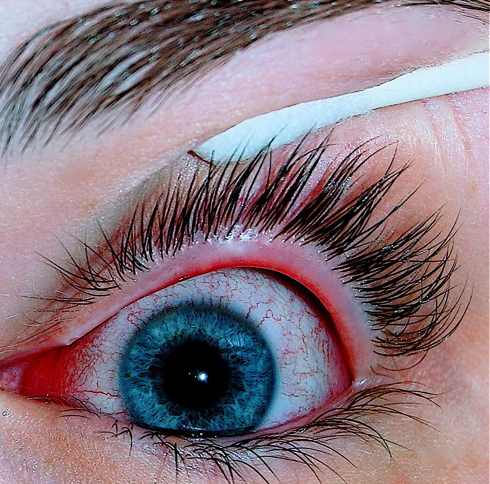
Conjunctivitis
Wikipedia
"Towards evidence based emergency medicine: best BETs from the Manchester Royal Infirmary. BET 4: use of litmus paper in chemical eye injury". ... The Egyptian Journal of Hospital Medicine . 71 (2): 2484–2489. doi : 10.12816/0045645 . ... "Relapsing polychondritis" . Joint, Bone, Spine . 81 (2): 118–24. doi : 10.1016/j.jbspin.2014.01.001 . ... Centers for Disease Control and Prevention . 2 October 2017 . Retrieved 18 October 2017 . ^ a b Isenberg SJ (2003).MMP2, PTAFR, PSMB9, LRRC8A, FERMT1, SAMD9, MMP1, MBTPS2, PIK3R1, PLG, POLH, PSMB4, PSMB8, RAG1, RAG2, ICOS, BLNK, TCF3, TNFRSF13B, GJB6, TNFRSF1A, UROS, WAS, WIPF1, XPA, XPC, TP63, STX16, SLC39A4, NLRC4, ERCC6, BTD, BTK, CD19, CD79A, CD79B, LRBA, COL7A1, CR2, DDB2, DKC1, DNASE1L3, USB1, ERCC2, ERF, FGFR2, FOXC2, GNAS, HLA-B, TNFRSF13C, IGHM, IGLL1, NLRP3, IKZF1, MBL3P, PLD3, IMMP1L, IGF2BP1, AD11, HM13, ALB, AOC3, IMPA1, CD14, GYPA, IFNGR1, IL4, CXCL8, IL10, IL12B, IL13, LGALS1, TYR, MBL2, PCOS1, ALK, CCL17, SOD3, STAT6, TEK, TNF, ERVK-19
-
Diffuse Large B-Cell Lymphoma
Wikipedia
DH/THL). RO6870810, another BET inhibitor, in combination with Venetoclax , an inhibitor of the Bcl-2 protein, is likewise in a phase I clinical trial [43] for the treatment of DH/THL. [41] Subtypes of diffuse large B-cell lymphoma [ edit ] DLBCL subtypes have been sorted into groups based on their distinctive morphology or immunophenotype , distinctive clinical issues, and distinctive virus-driven etiology. ... Best Practice & Research. Clinical Haematology . 30 (1–2): 131–138. doi : 10.1016/j.beha.2016.08.029 . ... Best Practice & Research. Clinical Haematology . 30 (1–2): 50–55. doi : 10.1016/j.beha.2016.11.001 . ... Best Practice & Research. Clinical Haematology . 31 (2): 135–146. doi : 10.1016/j.beha.2018.04.001 . ... "CAR-T Cell Therapy in Diffuse Large B Cell Lymphoma: Hype and Hope" . HemaSphere . 3 (2): e185. doi : 10.1097/HS9.0000000000000185 .EZH2, MYD88, CARD11, PIK3CD, CD79B, CD79A, PRDM1, CD274, PIK3CG, MYC, CDKN2A, PIK3CA, ALK, PIK3CB, ABCB1, BCL2, MALT1, CREBBP, EP300, JAK2, FAS, FOXO1, GLI1, B2M, KMT2D, ABCG2, CDK2, RPS6KB1, GNA13, STAT6, CDK1, IRF8, FBXO11, IKZF1, NOTCH2, SKP2, CLTC, PDPK1, PRDM11, RANGAP1, NECTIN2, SOD2, SGK1, SOD3, NOM1, PRDX3, SPEN, CAT, TRAF5, TP53, TXNIP, BCAS2, SEC31A, CCDC86, PRDX4, IMMP2L, SMURF2, HLA-B, CD86, SMUG1, PWWP3A, TNFRSF8, ARAP3, MIR34A, GBA, NCOA5, BCL10, AICDA, DDIT3, CDC42BPB, FOXP1, RTEL1, IGH, MIR155, NPR2, NOS2, TNF, KRT20, TNPO2, STAT3, MIR21, LMO2, NOS1, AKT1, SYK, CCND1, KALRN, CXCR4, MCL1, MME, IGHV3-69-1, PVT1, MMEL1, MGMT, IL10, IRF4, BTK, DLEU1, ATM, ABCB6, BCL6, CD19, MS4A1, FCGR3A, LOC102723407, H3P10, PAX5, PRKAR1A, PTEN, IL6, LOC102724971, SPG7, CXADR, ARR3, FOXP3, CASR, PDLIM7, TRIM13, NR1I3, BIRC3, NFKB1, CXADRP1, IL2, CCND2, CD40, SOAT1, TCL1A, MEF2B, REL, MET, GSTP1, MCTS1, PDE4B, PDCD1, SLC16A1, CDKN1B, PDCD1LG2, PIM1, IGHV3OR16-7, HDAC9, IL4, HDAC6, SDC1, DNER, TBC1D9, MCAT, MDM2, ALB, CDKN2B, CDR3, TNFAIP3, TCL1B, HTC2, CMA1, MTHFR, CCND3, ZC3H12D, DAPK1, WEE1, VEGFA, TP63, CTNNB1, MIR17HG, GCSAM, CD44, CD38, PLK1, GSTT1, PTPN6, GSTM1, FHIT, MAPK3, ERCC2, MAPK1, MTOR, CDK4, BCL3, SOCS1, SMAD1, CASP3, LTA, XBP1, CD22, TNFSF13B, LAIR1, KMT2B, TET2, BCL2A1, BACH2, IFNG, CD163, S1PR2, LIG4, IL21, MIR15A, HSP90AA1, NFKB2, PARP9, CDK9, KRAS, UHMK1, MTA3, POU2F2, IL21R, PDE4A, MLH1, JAK1, ACKR3, MKI67, HIF1A, KMT2A, MIR155HG, MTX1, MUC1, NEK2, NRAS, HLA-DRB1, KLHL6, HLA-C, NCAM1, LGALS1, CDKN1C, CXCL12, SERPINA9, BCR, SLC22A2, ARID1A, LOC390714, CUX1, SOX11, CIB1, SPIB, SPN, SPP1, MRPL28, CR2, CASP1, TPX2, CCK, TRBV20OR9-2, CHEK2, FOXM1, MIR224, SUB1, CD37, TXN, SIRT1, CD58, H3P9, PTPN1, EPHB2, NXT1, FCGR2A, PTPRC, LOC105379528, SART3, RHOH, CES2, PARP1, CCL2, CCL5, APC, MSH3, WRN, MIB1, LPP, EZR, VIM, MTAP, SLC16A4, UVRAG, DCTN6, SLC16A3, XPO1, MBTPS1, XRCC1, PRMT5, MAL, EBI3, CFLAR, EFS, UCHL1, MAD2L1, LYN, EXOC2, MDM4, SOCS3, ZNRD2, HIP1R, ZAP70, MTA1, DIABLO, PRKCB, TYMS, HPGDS, SLC19A1, PMEL, BRD4, CRBN, SCT, IBTK, RPE65, SND1, RFC1, PLCG2, MXI1, IRAK4, PTPRO, TMED7, POLD1, POU2AF1, PTPN2, IGK, IL22, PSMD9, PCNA, SLPI, ABL2, SMO, TRAF6, TRAF3, NR2C2, TP73, CKAP4, NFATC1, NFE2L2, PSIP1, NME1, ZEB1, MAP3K7, KDM6B, TLR9, STAT1, ACSBG1, NOTCH1, ROR1, OAS3, SPI1, SLC7A4, SMARCA4, LGALS3, GRN, FKBP4, CCNE1, CASP9, FLT1, MIR497, GABPA, GAPDH, C1QA, ANTXR2, GNA12, GOT2, TNFRSF13C, GPX4, BTG1, HSPA4, GTF2H4, GZMB, H2AX, BRCA1, MTDH, HK2, BPI, HLA-DQB1, HMGB1, HAVCR2, TMED7-TICAM2, BDNF, NR4A1, FCGR3B, FASN, ETS1, CD28, CRP, CSF2, MIR146A, MIRLET7B, MALAT1, MIR17, MIR195, TICAM2, DNMT1, DNMT3A, MIR214, E2F1, MIR223, EIF4B, CDKN1A, EIF4E, MIR23A, EIF4EBP1, CDH1, MIR31, CD70, ERCC5, BTLA, DTX3L, ESR2, CD36, ETFA, OPN1SW, MIR142, ASCC2, PPP1R2C, IL2RA, IL4R, CXCL8, IL10RA, ID3, IL17A, VTCN1, BIRC5, IRF1, ISG20, RAPH1, ITGAX, ITPR2, H3P23, H3P42, ADA, KDR, ACVRL1, GAS5, LAG3, LAMC2, LBR, ACTB, ATP7A, TNFRSF9, ATR, BCL2L2-PABPN1, BCL2L2, IFI27, HDAC4, IL24, WIF1, RASSF1, MIR221, CORO1A, GLMN, IL1RAPL1, MIR222, CCR2, PIM2, BCL2L10, METAP2, H3P47, TNIP1, COPS5, SH3BP5, MIR25, MIR27A, MIR27B, MELK, MIR28, MIR22, VSIG4, CD300A, SNHG16, NCR2, RCOR1, AIM2, PASK, MIR145, RAD50, LINC01672, MIR150, MIR16-1, SPART, KDM1A, MIR30A, MAP4K4, MIR1234, MIR197, MLXIP, BCL2L11, MIR200C, SYCE1L, MIR210, PHB2, HOTAIR, MIR29C, NAPSA, POTEF, MIR33A, MIR494, RACK1, RBCK1, NCOR2, AHSA1, CCT4, MIR504, CXCL13, SNHG14, MIR4638, POU5F1P3, TP53COR1, ANP32B, YAP1, BATF, POU5F1P4, PANDAR, MIR144, USP34, HULC, D2HGDH, CIB2, MAD2L2, LUNAR1, MIR146B, NCOR1, KEAP1, BAG3, HSPH1, MASP2, WARS2, MNS16A, MIR99A, MTCO2P12, MIR135B, LRP1-AS, NPM3, MTSS1, SMAD5-AS1, BCLAF1, MIR148B, CXCR6, MIR328, MIR431, PRDX6, MIR323B, MIR451A, MIR511, WTAP, LINC00273, NEAT1, MDN1, KDM2B, MAP3K20, BCL11A, IL17D, FCRLA, PRKAG3, RIPK4, GLIS2, MIR127, MIXL1, FSD1L, ARHGAP24, PARP14, LY6K, DOHH, ORAI1, IL23A, CHRDL1, UCHL5, NAPRT, C1RL, MZB1, CGB5, FOXP2, CGB8, PRRT2, LEF1, CARD16, RMDN1, ASCC1, RASGRP4, MSI2, FCRL4, DOCK8, TRPM4, EAF2, KLHL1, PCDH10, GRAP2, SLC5A7, NDRG2, NSD1, GMCL1, PCBP4, GORASP1, P2RY12, WNK1, MYDGF, LMO3, FSD1, PAG1, MAP1LC3B, MORC4, CCAR1, RNF34, NLRP2, SYBU, MEG3, LAPTM4B, FBXW7, RMDN3, TUG1, ELL3, MPIG6B, DEFB126, PGPEP1, IL31RA, F11R, SLCO6A1, SIRT3, CNRIP1, FOXK1, CNOT6L, RNF19A, SSBP2, IL27, AMACR, PATZ1, NUTM1, SLAMF7, LOC283177, PRAME, LYPD5, FIRRE, ILDR1, TES, EIF4E3, DICER1, TSPAN33, ZACN, GSTK1, SLC27A1, CRTC1, TMEM189, TMEM189-UBE2V1, NEDD4L, SIK2, MIRLET7G, MIR10A, MIR125A, POLDIP2, PHGDH, PPARGC1B, COMMD1, ADGRE2, CD109, PASD1, HT, VPREB3, BLNK, IL34, LGALS13, MYLIP, PYCARD, APCDD1, LYPD4, LGALS16, SETD2, IGHV3-23, CKAP2, IGHV3-52, RMDN2, IGHV4-34, EML4, SGSM3, NKX6-3, UBR1, CRTC2, AGO2, KLHDC8B, MORC1, MARCHF8, RASGEF1A, HAVCR1, CXCL16, NAT2, KL, GCLC, FLI1, FN1, FOS, FOSB, KDSR, XRCC6, GATA2, GATA3, GEM, GLI2, LIF, GPI, XCR1, MKNK2, GPX1, NR3C1, GSTA1, GUCA1B, HDAC2, HELLS, FBP1, FABP7, ETV6, ETV5, ATN1, DUSP4, DVL3, S1PR1, EFNA5, EFNB2, EGF, EGR3, EIF4A1, EIF4A2, ELAVL2, ELF3, ENO2, EPHA4, EPHX1, EPO, ERG, ERN1, ESR1, HGF, HIC1, HLA-DQA1, IL9, IL13, IDO1, CXCL10, INPP5D, IRAK1, IRF2, ISL1, ITGAM, ITPR1, ITPR3, JAK3, JUN, JUNB, JUND, KIF2A, KIR3DL1, LAMP1, LCK, LEP, IL10RB, IL1B, HLA-G, IL1A, HLX, HMMR, HMOX1, HNMT, HNRNPK, TLX1, HSPA8, MRPL58, IDH1, IDH2, IFNA1, IFNA13, IFNB1, IGF1, IGHA1, IGHM, JCHAIN, IGL, IKBKB, DPP4, DOCK2, DNMT3B, BLK, BMI1, BMP6, BRAF, BRCA2, BUB1B, BYSL, TSPO, SERPING1, CACNA1C, CALD1, CALR, CASP8, RUNX3, CCNH, CCNT1, CD247, CD5, CD14, CD27, CXCR5, BIK, CD59, BGN, ABO, ADAM10, AHR, ALDH1A1, AMCN, AMPD1, ANXA5, APAF1, XIAP, APP, APRT, RHOA, ARNT, STS, ATF3, BAX, BCL2L1, BCL7A, NBEAP1, CD47, CD63, NQO1, CCR8, CNC2, CNP, COL3A1, CRK, CRY1, MAPK14, CSE1L, CSF1R, CSF3, CSNK2B, CTLA4, CX3CR1, CYBA, CYP1A1, CYP17A1, CYP24A1, DAP, DAXX, DCK, ABCC2, CCR7, CD68, CCR1, CD74, CD81, ADGRE5, CDC20, CDC25C, CDH11, CDH13, CDK3, CDK5, CDK6, CDS1, CEBPA, CGA, CGB3, CHEK1, FOXN3, CHGB, CHM, CLU, LEPR, LMNA, SLIT2, UBE2V1, TNFRSF1B, TOP1, TOP2A, TP53BP1, TP53BP2, TPM3, TPT1, UBA52, UBE2N, UCN, LMNB1, UGCG, UMOD, VAV1, WARS1, YY1, ZNF134, ZBTB17, PRDM2, BTG2, TMSB4X, TLR1, TLE1, NKX2-1, SPARC, SPRR1A, SRM, STAT5A, STAT5B, STIM1, SYN2, SYT1, TAP2, TAT, TCF4, TCF7, TRA, TRB, TRG, TFF3, TGFB1, TGFBR1, THOP1, BAG6, AIMP2, ARHGEF5, TNFRSF14, FADD, TNFRSF10B, HDAC3, CDK5R1, PER3, PER2, ARHGEF7, SPHK1, TIMELESS, MBD2, BTRC, P4HA2, NOL3, MAP3K14, AIP, PIAS2, USP14, CD83, RPL23, TNFRSF6B, TNFSF10, NR4A3, TNFSF13, MLLT10, HMGA2, CLLS2, USP11, USP9X, SMC1A, PICALM, CDC7, SPOP, NR0B2, CUL4B, OGT, CAMK1, MKNK1, RFXANK, NCOA1, EIF4G3, EED, RIPK1, SOX2, SNAP25, SLC8A1, NBN, NEDD8, NFATC2, NFE2L1, NGF, NODAL, NOTCH3, NPM1, NRF1, NT5E, NTRK1, P2RY1, SERPINB2, PCYT1A, PDK1, PECAM1, PHB, PLAGL1, PLG, PLOD2, NCL, MTRR, POLA1, MTR, LPL, LRMP, SH2D1A, MXD1, SMAD5, MAP4, MBP, MC1R, BORCS8-MEF2B, MFAP1, CIITA, CD99, CXCL9, FOXO4, ABCC1, MSH2, MSN, MST1R, COX2, PMAIP1, POU5F1, SIAH1, RDX, RELB, RGS1, RNASEL, BRD2, RPS6, RPS6KA1, RPS6KA2, RPS6KA3, RPS20, RREB1, S100A8, CCL3, CCL4, CCL13, CCL18, CCL22, SELL, SELPLG, SET, RELA, OPN1LW, PPA1, RBL1, PTPA, PPP2R5E, PRKAA2, PRKCA, MAPK11, MAP2K7, LGMN, PSMB5, PSMB9, PTH, PTGS2, PTMS, PTPN13, PTPRD, PTPRJ, RAD1, RAD23B, RAD51, RARA, H3P40


