• FindZebra
  • About
  • Contact
  • Help
  • Login
Advanced
  • Childhood Cataract Wikipedia
    Further reading [ edit ] Childhood cataracts at NHS Choices Cataracts in Children, Congenital and Acquired at EyeWiki v t e Diseases of the human eye Adnexa Eyelid Inflammation Stye Chalazion Blepharitis Entropion Ectropion Lagophthalmos Blepharochalasis Ptosis Blepharophimosis Xanthelasma Ankyloblepharon Eyelash Trichiasis Madarosis Lacrimal apparatus Dacryoadenitis Epiphora Dacryocystitis Xerophthalmia Orbit Exophthalmos Enophthalmos Orbital cellulitis Orbital lymphoma Periorbital cellulitis Conjunctiva Conjunctivitis allergic Pterygium Pseudopterygium Pinguecula Subconjunctival hemorrhage Globe Fibrous tunic Sclera Scleritis Episcleritis Cornea Keratitis herpetic acanthamoebic fungal Exposure Photokeratitis Corneal ulcer Thygeson's superficial punctate keratopathy Corneal dystrophy Fuchs' Meesmann Corneal ectasia Keratoconus Pellucid marginal degeneration Keratoglobus Terrien's marginal degeneration Post-LASIK ectasia Keratoconjunctivitis sicca Corneal opacity Corneal neovascularization Kayser–Fleischer ring Haab's striae Arcus senilis Band keratopathy Vascular tunic Iris Ciliary body Uveitis Intermediate uveitis Hyphema Rubeosis iridis Persistent pupillary membrane Iridodialysis Synechia Choroid Choroideremia Choroiditis Chorioretinitis Lens Cataract Congenital cataract Childhood cataract Aphakia Ectopia lentis Retina Retinitis Chorioretinitis Cytomegalovirus retinitis Retinal detachment Retinoschisis Ocular ischemic syndrome / Central retinal vein occlusion Central retinal artery occlusion Branch retinal artery occlusion Retinopathy diabetic hypertensive Purtscher's of prematurity Bietti's crystalline dystrophy Coats' disease Sickle cell Macular degeneration Retinitis pigmentosa Retinal haemorrhage Central serous retinopathy Macular edema Epiretinal membrane (Macular pucker) Vitelliform macular dystrophy Leber's congenital amaurosis Birdshot chorioretinopathy Other Glaucoma / Ocular hypertension / Primary juvenile glaucoma Floater Leber's hereditary optic neuropathy Red eye Globe rupture Keratomycosis Phthisis bulbi Persistent fetal vasculature / Persistent hyperplastic primary vitreous Persistent tunica vasculosa lentis Familial exudative vitreoretinopathy Pathways Optic nerve Optic disc Optic neuritis optic papillitis Papilledema Foster Kennedy syndrome Optic atrophy Optic disc drusen Optic neuropathy Ischemic anterior (AION) posterior (PION) Kjer's Leber's hereditary Toxic and nutritional Strabismus Extraocular muscles Binocular vision Accommodation Paralytic strabismus Ophthalmoparesis Chronic progressive external ophthalmoplegia Kearns–Sayre syndrome palsies Oculomotor (III) Fourth-nerve (IV) Sixth-nerve (VI) Other strabismus Esotropia / Exotropia Hypertropia Heterophoria Esophoria Exophoria Cyclotropia Brown's syndrome Duane syndrome Other binocular Conjugate gaze palsy Convergence insufficiency Internuclear ophthalmoplegia One and a half syndrome Refraction Refractive error Hyperopia Myopia Astigmatism Anisometropia / Aniseikonia Presbyopia Vision disorders Blindness Amblyopia Leber's congenital amaurosis Diplopia Scotoma Color blindness Achromatopsia Dichromacy Monochromacy Nyctalopia Oguchi disease Blindness / Vision loss / Visual impairment Anopsia Hemianopsia binasal bitemporal homonymous Quadrantanopia subjective Asthenopia Hemeralopia Photophobia Scintillating scotoma Pupil Anisocoria Argyll Robertson pupil Marcus Gunn pupil Adie syndrome Miosis Mydriasis Cycloplegia Parinaud's syndrome Other Nystagmus Childhood blindness Infections Trachoma Onchocerciasis v t e Optical illusions ( list ) Illusions Afterimage Ambiguous image Ames room Barberpole Bezold Café wall Checker shadow Chubb Cornsweet Delboeuf Ebbinghaus Ehrenstein Flash lag Fraser spiral Gravity hill Grid Hering Impossible trident Jastrow Lilac chaser Mach bands McCollough Müller-Lyer Necker cube Orbison Penrose stairs Penrose triangle Peripheral drift Poggendorff Ponzo Rubin vase Sander Schroeder stairs Shepard tables Spinning Dancer Ternus Vertical–horizontal White's Wundt Zöllner Popular culture Op art Trompe-l'œil Spectropia (1864 book) Ascending and Descending (1960 drawing) Waterfall (1961 drawing) The dress (2015 photograph) Related Accidental viewpoint Auditory illusions Tactile illusions Temporal illusion This article about the eye is a stub .
    TGM3, ADNP, LEMD2, CRYAB, CRYGD, SLC16A12, CRYGA, CRYGB, EPHA2, KCNJ13, PAX6, RECQL4, SIPA1L3, ABHD12, TRNT1, UNC45B
  • Androgen Insensitivity, Partial OMIM
    ., 2015). Clinical Features Reifenstein (1946) reported a family in which 9 of 10 male members over 2 generations exhibited abnormally high follicle-stimulating hormone secretion, hypospadias, sterility, gynecomastia, small testes, absent beard, height of 65 +/- inches, normal sized phallus, normal 17-ketosteroid excretion, late puberty, and normal libido. ... On the basis of this and another reported pedigree, they suggested that the affected members in the kindred reported by Gilbert-Dreyfus et al. (1957), Lubs et al. (1959) and Rosewater et al. (1965) had the same condition as that reported by Reifenstein (1946). Wilson et al. (1974) chose to refer to the condition as type 1 familial incomplete male pseudohermaphroditism (type 2 is autosomal recessive; 264600).
    AR, GART, PAICS, NR5A1, MECP2, AMH, CD38, GH1, RNF4, MAMLD1, NCOA2
    • Partial Androgen Insensitivity Syndrome Orphanet
      A disorder of sex development (DSD) distinct from complete AIS (CAIS) characterized by the presence of abnormal genital development in a 46,XY individual with normal testis development and partial responsiveness to age-appropriate levels of androgens. Epidemiology There are no available data on the prevalence of PAIS in the general population. Nevertheless, resistance to androgens is the most frequent cause of XY DSD. Clinical description Patients have a highly variable genital appearance. The prototypic form of presentation is severe hypospadias, micropenis, and bifid scrotum in which the testes may or may not be descended.
    • Partial Androgen Insensitivity Syndrome Wikipedia
      Partial androgen insensitivity syndrome Other names Partial androgen resistance syndrome; Reifenstein syndrome AIS results when the function of the androgen receptor (AR) is impaired. The AR protein (pictured) mediates the effects of androgens in the human body. Specialty Endocrinology Partial androgen insensitivity syndrome ( PAIS ) is a condition that results in the partial inability of the cell to respond to androgens . [1] [2] [3] It is an X linked recessive condition. The partial unresponsiveness of the cell to the presence of androgenic hormones impairs the masculinization of male genitalia in the developing fetus, as well as the development of male secondary sexual characteristics at puberty , but does not significantly impair female genital or sexual development. [3] [4] As such, the insensitivity to androgens is clinically significant only when it occurs in individuals with a Y chromosome (or more specifically, an SRY gene ). [1] Clinical features include ambiguous genitalia at birth and primary amenhorrhoea with clitoromegaly with inguinal masses. Mullerian structures are not present in the individual. PAIS is one of three types of androgen insensitivity syndrome , which is divided into three categories that are differentiated by the degree of genital masculinization : complete androgen insensitivity syndrome (CAIS) is indicated when the external genitalia is that of a normal female, mild androgen insensitivity syndrome (MAIS) is indicated when the external genitalia is that of a normal male, and partial androgen insensitivity syndrome (PAIS) is indicated when the external genitalia is partially, but not fully masculinized. [1] [2] [5] [6] [7] [8] [9] [10] [11] Androgen insensitivity syndrome is the largest single entity that leads to 46,XY undermasculinization . [12] There are differing opinions on whether treatment is necessary.
    • Partial Androgen Insensitivity Syndrome GARD
      Partial androgen insensitivity syndrome (PAIS) is a disorder of sex development that affects the growing reproductive and genital organs of a fetus. Androgen insensitivity refers to the inability of the body of an individual with a 46, XY karyotype (usually leading to normal male development) to properly respond to male sex hormones (androgens). In PAIS, the body partially responds to these hormones. Signs and symptoms of PAIS can vary greatly, causing a range of differences in genital appearance. Some individuals have severe hypospadias , an unusually small penis, and bifid scrotum . More severely affected individuals may have female external genitalia with an abnormally large clitoris, partial fusion of the labia and gynecomastia (excessive development of male breasts).
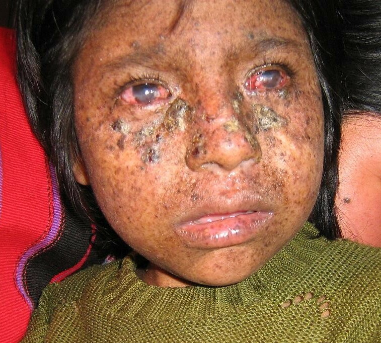
  • Xeroderma Pigmentosum, Autosomal Dominant, Mild OMIM
    Although the disorder in this kindred was milder than ordinary xeroderma pigmentosum ('George was in a tropical climate for many years, being invalided home on two occasions on account of the disease' and 'Douglas was invalided home from North Africa in 1943'), 'Douglas was considered to be a case of xeroderma pigmentosum by all the members present when he was shown at a meeting of the North British Dermatological Society in 1946.' Imray et al. (1986) described an Australian kindred with persons in 5 generations affected by a mild form of XP.
    XPA, XPC, POLH, ERCC5, ERCC2, ERCC1, DDB2, ERCC4, ERCC3, TERF2, GTF2H1, TP53, NR1H2, BIVM-ERCC5, GTF2H2, GTF2H3, GTF2H5, GTF2H4, POLQ, CAT, XRCC1, CSH2, XRCC6P5, HPRT1, LIG4, ERCC6, CSH1, HRAS, OGG1, HPGDS, PCNA, PTEN, XRCC3, ENDOV, CDKN2A, GSTM1, SIRT1, MC1R, LCE2B, HSPA9, RAD23A, NRAS, FUS, WDR76, ATR, BRIP1, CHEK1, DDB1, BCL2, DECR1, ERCC8, CCNH, ATM, TYMS, BTN2A2, PTF1A, IRF9, EXO1, HERC2, RAD54L, H3P38, PDIK1L, HFM1, UBE3B, USP7, IFI44, XRCC2, CATSPER1, TDG, SLX4, XYLT2, VPS11, RTEL1, CRLF3, DEFB103A, DEFB103B, MEPE, WDR77, LRRC59, RNU1-1, VEPH1, PRDX5, SMUG1, AAGAB, KAT7, ZC3H12D, NAT1, TALDO1, CGA, FEN1, EGFR, NQO1, TIMM8A, DDX11, GADD45A, CYP1B1, CSNK2A1, CRYZ, COMT, CETN2, SYCP1, CDK7, CDK4, DDR1, BRAF, BRCA1, BAX, ATF3, ASIP, FAS, APEX1, GOT1, GSTM2, GSTT1, HGF, SULT1A1, STAT3, ST13, SMO, RNU2-1, RNU1-4, RECQL, RAD23B, PTCH1, PTAFR, MAPK8, PRKDC, POMC, AKT1, ODC1, NPM1, NCAM1, MYC, MPO, LBR, KIT, H3P33
    • Xeroderma Pigmentosum GeneReviews
      Summary Clinical characteristics. Xeroderma pigmentosum (XP) is characterized by: Sun sensitivity (severe sunburn with blistering, persistent erythema on minimal sun exposure in ~60% of affected individuals), with marked freckle-like pigmentation of the face before age two years in most affected individuals; Sunlight-induced ocular involvement (photophobia, keratitis, atrophy of the skin of the lids); Greatly increased risk of sunlight-induced cutaneous neoplasms (basal cell carcinoma, squamous cell carcinoma, melanoma). Approximately 25% of affected individuals have neurologic manifestations (acquired microcephaly, diminished or absent deep tendon stretch reflexes, progressive sensorineural hearing loss, and progressive cognitive impairment). The most common causes of death are skin cancer, neurologic degeneration, and internal cancer. The median age at death in persons with XP with neurodegeneration (29 years) was found to be younger than that in persons with XP without neurodegeneration (37 years). Diagnosis/testing. The diagnosis of XP is made on the basis of clinical findings and family history and/or by the identification of biallelic pathogenic variants in DDB2 , ERCC1 , ERCC2 , ERCC3 , ERCC4 , ERCC5 , POLH , XPA , or XPC .
    • Xeroderma Pigmentosum, Complementation Group D OMIM
      A number sign (#) is used with this entry because xeroderma pigmentosum complementation group D (XPD) is caused by homozygous or compound heterozygous mutation in the excision repair gene ERCC2 (126340) on chromosome 19q13. Description Xeroderma pigmentosum is a rare autosomal recessive disorder characterized by acute photosensitivity and a predisposition to skin cancer on sun-exposed areas of the body. The primary defect in XP involves nucleotide excision repair (NER) (summary by Flejter et al., 1992). Clinical Features In a 31-year-old patient with XP classified as complementation group D by cell-fusion complementation methods, Ichihashi et al. (1988) described mild skin lesions and no apparent neurologic abnormalities despite the characteristic group D level of DNA repair deficiency. Johnson and Squires (1992) stated that more than 30 unrelated individuals with XPD were known and that less than half of them showed major abnormalities of the central nervous system, once considered to be the hallmark of XPD.
    • Xeroderma Pigmentosum, Complementation Group A OMIM
      A number sign (#) is used with this entry because xeroderma pigmentosum complementation group A (XPA) is caused by homozygous or compound heterozygous mutation in the XPA gene (611153) on chromosome 9q22. Description Xeroderma pigmentosum is a genetically heterogeneous autosomal recessive disorder characterized by increased sensitivity to sunlight with the development of carcinomas at an early age. Some patients develop neurologic symptoms or a more severe clinical phenotype known as de Sanctis-Cacchione syndrome (278800) (Satokata et al., 1992). See also XPB (610651), XPC (278720), XPD (278730), XPE (278740), XPF (278760), XPG (278780), and variant XP (XPV; 278750). Clinical Features Ruder (cited by Cockayne, 1933) observed the condition in 7 out of 13 sibs.
    • Xeroderma Pigmentosum Orphanet
      Xeroderma pigmentosum (XP) is a rare genodermatosis characterized by extreme sensitivity to ultraviolet (UV)-induced changes in the skin and eyes, and multiple skin cancers. It is subdivided into 8 complementation groups, according to the affected gene: classical XP (XPA to XPG) and XP variant (XPV) (see these terms). Epidemiology It has an estimated prevalence of 1/1,000,000 in the USA and Europe, with higher figures in other countries (e.g. Japan, North Africa and Pakistan), particularly in communities with a high degree of consanguinity. Clinical description The severity of the clinical manifestations and the age of onset are extremely variable and are in part dependent on exposure to sunlight and the complementation group.
    • Xeroderma Pigmentosum GARD
      Xeroderma pigmentosum (XP) causes the skin and eyes to be extra sensitive to exposure to ultraviolet radiation from the sun and other sources. Symptoms begin in early childhood. People with XP can develop bad sunburns, blistering, and freckling in response to sunlight. The eyes may develop light sensitivity, corneal clouding, and swelling. Some people with XP have nervous system involvement as well. People with XP are at very high risk of developing skin cancer and other types of cancers. XP is caused by variants in one of at least nine genes involved in repairing damaged DNA.
    • Xeroderma Pigmentosum, Complementation Group C OMIM
      A number sign (#) is used with this entry because xeroderma pigmentosum complementation group C (XPC) is caused by mutation in the XPC gene (613208) on chromosome 3p25. Description Xeroderma pigmentosum is a genetically heterogeneous condition characterized by increased sensitivity to ultraviolet (UV) irradiation and increased risk of skin cancer resulting from a defect in DNA repair. XPC is the most common form of XP in the white population, accounting for over a third of all cases in this group (review by Li et al., 1993). For a general discussion of xeroderma pigmentosum, see XPA (278700). Clinical Features Lynch et al. (1984) suggested that complementation group C patients may be particularly prone to malignant melanoma. Li et al. (1993) identified 2 patients with XPC confirmed by genetic analysis (613208.0003).
    • Xeroderma Pigmentosum, Complementation Group B OMIM
      A number sign (#) is used with this entry because the phenotype associated with xeroderma pigmentosum complementation group B (XPB) is caused by mutation in the DNA excision repair gene ERCC3 (133510) on chromosome 2q14. Description For a general discussion of xeroderma pigmentosum, see XPA (278700), and of Cockayne syndrome, see CSA (216400). Cleaver (1990) provided a review of the causes of xeroderma pigmentosum. Clinical Features Lehmann (1982) performed cell fusion studies on cultured cells from 11 patients with Cockayne syndrome. The 11 cell lines were assigned to 3 complementation groups: 2 to group A, 8 to group B, and 1 to group C.
    • Xeroderma Pigmentosum, Complementation Group F OMIM
      A number sign (#) is used with this entry because of evidence that the disorder is caused by homozygous or compound heterozygous mutation in the ERCC4 gene (133520) on chromosome 16p13. Description Xeroderma pigmentosum is an autosomal recessive disorder characterized by sun sensitivity and increased skin sensitivity to UV light, as well as an increased risk of skin cancer associated with a defect in nucleotide excision repair (NER). The XPF form of XP is usually relatively mild compared to other forms. Patients with XPF tend to have later onset of skin cancer. Some patients with XPF may develop neurologic impairment or growth defects, and are then classified as having Cockayne syndrome (summary by Kashiyama et al., 2013). For a general phenotypic description and a discussion of genetic heterogeneity of xeroderma pigmentosa, see XPA (278700), and of Cockayne syndrome, see CSA (216400).
    • Xeroderma Pigmentosum, Complementation Group E OMIM
      A number sign (#) is used with this entry because xeroderma pigmentosum complementation group E is caused by homozygous mutation in the DDB2 gene (600811) on chromosome 11p11. For background information on xeroderma pigmentosum, see 278700. Clinical Features Kondo et al. (1988) studied 3 Japanese patients, aged 50, 42, and 41 years, with mild XP symptoms and no neurologic abnormalities. Two had developed basal cell carcinomas at ages 46 and 41 years. Chu and Chang (1988) studied cells from 2 second-cousin patients with group E xeroderma pigmentosum who had a skin disease less severe than that seen in patients of several other complementation groups (de Weerd-Kastelein et al., 1974). Using an extension of the gel electrophoresis binding assay, Chu and Chang (1988) identified at least 1 nuclear factor that binds to DNA damaged by ultraviolet radiation or the antitumor drug cisplatin, but is notably absent in group E cells. The defect was not the result of a failure of protein transport to the nucleus since binding activity was absent in both the nuclear and cytoplasmic extracts.
    • Xeroderma Pigmentosum Wikipedia
      Xeroderma pigmentosum Other names DeSanctis-Cacchione syndrome [1] [2] XP1 / XP2 / XP3 / XP4 / XP5 / XP6 / XP7 [3] Xeroderma pigmentosum I/II/III/IV/V/VI/VII [3] Xeroderma pigmentosum complementation group A/B/C/D/E/F/G [3] xeroderma pigmentosum group A/B/C/D/E/F/G [3] An eight-year-old girl from Guatemala with xeroderma pigmentosum [4] Specialty Medical genetics Symptoms Severe sunburn after only a few seconds in the sun, freckling in sun-exposed areas, dry skin, changes in skin pigmentation [1] Complications Skin cancer , brain cancer , cataracts [1] Usual onset Becomes visible ~6 months of age [2] Duration Lifelong Causes Genetic disorder ( autosomal recessive ) [1] Diagnostic method Based on symptoms and confirmed by genetic testing [5] Differential diagnosis Trichothiodystrophy , Cockayne syndrome , cerebrooculofacioskeletal syndrome , erythropoietic protoporphyria [6] Prevention No cure available Treatment Completely avoiding sun or UV rays, retinoid creams , vitamin D [5] [6] Prognosis Life expectancy is shortened by about 30 years. [7] Frequency • 1 in 100,000 (worldwide) [3] • 1 in 370 (India) [8] • 1 in 22,000 (Japan) [3] • 1 in 250,000 (USA) [9] • 1 in 430,000 (Europe) • 1 in 1,000,000 (UK) [3] Xeroderma pigmentosum ( XP ) is a genetic disorder in which there is a decreased ability to repair DNA damage such as that caused by ultraviolet (UV) light. [1] Symptoms may include a severe sunburn after only a few minutes in the sun, freckling in sun exposed areas, dry skin and changes in skin pigmentation. [1] Nervous system problems, such as hearing loss , poor coordination, loss of intellectual function and seizures , may also occur. [1] Complications include a high risk of skin cancer , with about half having skin cancer by age 10 without preventive efforts, and cataracts . [1] There may be a higher risk of other cancers such as brain cancers . [1] XP is autosomal recessive , with at least nine specific mutations able to result in the condition. [1] [6] Normally, the damage to DNA which occurs in skin cells from exposure to UV light is repaired by nucleotide excision repair . [1] In people with xeroderma pigmentosum, this damage is not repaired. [1] As more abnormalities form in DNA, cells malfunction and eventually become cancerous or die. [1] Diagnosis is typically suspected based on symptoms and confirmed by genetic testing . [5] There is no cure for XP. [6] Treatment involves completely avoiding the sun . [6] This includes protective clothing, sunscreen and dark sunglasses when out in the sun. [6] Retinoid creams may help decrease the risk of skin cancer. [6] Vitamin D supplementation is generally required. [5] If skin cancer occurs, it is treated in the usual way. [6] The life expectancy of those with the condition is about 30 years less than normal. [7] The disease affects about 1 in 100,000 worldwide. [3] By region, it affects about 1 in 370 in India, [8] 1 in 20,000 in Japan, 1 in 250,000 people in the United States and 1 in 430,000 in Europe. [9] It occurs equally commonly in males and females. [10] Xeroderma pigmentosum was first described in the 1870s by Moritz Kaposi . [5] [10] In 1882, Kaposi coined the term xeroderma pigmentosum for the condition, referring to its characteristic dry, pigmented skin. [10] Individuals with the disease have been referred to as "children of the night" or "moon children". [11] [12] Contents 1 Signs and symptoms 2 Genetics 2.1 XP repair proteins 3 Diagnosis 3.1 Types 4 Treatment 5 Prognosis 6 History 7 Culture 8 Research directions 9 See also 10 References 11 External links Signs and symptoms [ edit ] Child with xeroderma pigmentosum in Nepal Signs and symptoms of xeroderma pigmentosum may include: [ citation needed ] Severe sunburn when exposed to only small amounts of sunlight. These often occur during a child's first exposure to sunlight. Development of many freckles at an early age Rough-surfaced growths ( solar keratoses ), and skin cancers Eyes that are painfully sensitive to the sun and may easily become irritated, bloodshot and clouded Blistering or freckling on minimum sun exposure Telangiectasia (spider veins) Limited growth of hair on chest and legs Scaly skin Xeroderma (dry skin) Irregular dark spots on the skin Corneal ulcerations Genetics [ edit ] Xeroderma pigmentosum has an autosomal recessive pattern of inheritance. One of the most frequent defects in xeroderma pigmentosum is an autosomal recessive genetic defect in which nucleotide excision repair (NER) enzymes are mutated, leading to a reduction in or elimination of NER. [13] If left unchecked, damage caused by ultraviolet light can cause mutations in individual cell's DNA. The causes of the neurological abnormalities are poorly understood and are not connected with exposure to ultraviolet light. The most current theories suggest that oxidative DNA damage is generated during normal metabolism in the central nervous system, and that some types of this damage must be repaired by NER. [14] Since DNA repair is under genetic control, it can mutate.
  • West Nile Fever Wikipedia
    Prevention [ edit ] Low-cost, ceiling hung mosquito netting for a bed Many of the guidelines for preventing occupational West Nile virus exposure are common to all mosquito-borne diseases . [57] Public health measures include taking steps to reduce mosquito populations. ... DEET formulations as high as 30% are recommended for children over two months of age. [58] The CDC also recommends the use of: IR3535, oil of lemon eucalyptus, para-menthane-diol, or 2-undecanone. [59] Protect infants less than two months of age by using a carrier draped with mosquito netting with an elastic edge for a tight fit. ... Repellents containing permethrin ( e.g. , Permanone) or other insect repellents may be applied to clothing, shoes, tents, mosquito nets, and other gear. (Permethrin is not suitable for use directly on skin.) ... They are now widespread in the United States, and in Florida they have been found in all 67 counties. [60] In an at-risk area, staying in air-conditioned or well- screened room, or sleeping under an insecticide-treated bed net is recommended. Bed nets should be tucked under mattresses, and can be sprayed with a repellent if not already treated with an insecticide. [57] Monitoring and control [ edit ] West Nile virus can be sampled from the environment by the pooling of trapped mosquitoes via ovitraps , carbon dioxide -baited light traps, and gravid traps, testing blood samples drawn from wild birds, dogs, and sentinel monkeys, and testing brains of dead birds found by various animal control agencies and the public. ... Retrieved 28 October 2017 . ^ Gompf, Sandra. "West Nile Virus" . Medicine Net . MedicineNet Inc . Retrieved 15 January 2019 . ^ "Symptoms, Diagnosis, & Treatment" .
    CCR5, ERVK-32, ROBO3, MAVS, DDX58, PLAAT4, IFIT2, ERVK-6, STAT1, SPP1, OAS1, IL1B, IFNB1, RNASEL, CASP8, HLA-DRB1, PELI1, SELENBP1, ARHGEF2, LRRFIP1, NAMPT, TRAIP, RIPK3, SEC14L2, CSF1R, LAMP3, ERVW-1, FOXP3, ZMYND10, DDX56, CCR7, VCP, CDKN2A, IFIH1, DHX58, ZBP1, HAVCR2, PIK3IP1, NLRP3, TNFRSF13C, TRIM6, RBM45, CCR2, ERVK-20, ERVK-18, VAMP8, TNFRSF1A, IFNA1, TNF, IFNA13, HLA-DQA1, IL1A, HLA-C, IL10, IL17A, IL18, IRF3, IRF5, KIR2DL2, KIR3DL1, KIR3DS1, LSAMP, CD180, SMAD4, MMP9, HLA-A, PIK3CA, PIK3CB, PIK3CD, PIK3CG, PZP, GLS, CASP1, SNCA, GEM, DDX3X, TAP1, TLR3, ATF4
  • Retiform Parapsoriasis Wikipedia
    Retiform parapsoriasis Specialty Dermatology Retiform parapsoriasis is a cutaneous condition, considered to be a type of large-plaque parapsoriasis . [1] It is characterized by widespread, ill-defined plaques on the skin, that have a net-like or zebra-striped pattern. [2] Skin atrophy , a wasting away of the cutaneous tissue , usually occurs within the area of these plaques. [1] See also [ edit ] Parapsoriasis Poikiloderma vasculare atrophicans List of cutaneous conditions References [ edit ] ^ a b Lambert WC, Everett MA (Oct 1981).
    • Poikiloderma Vasculare Atrophicans Wikipedia
      Poikiloderma vasculare atrophicans Other names Parapsoriasis variegata [1] or Parapsoriasis lichenoides [2] Typical skin changes and discoloration described as poikiloderma vasculare atrophicans Specialty Dermatology Poikiloderma vasculare atrophicans ( PVA ), is a cutaneous condition ( skin disease ) characterized by hypo- or hyperpigmentation (diminished or heightened skin pigmentation , respectively), telangiectasia and skin atrophy . [3] [4] [5] Other names for the condition include prereticulotic poikiloderma and atrophic parapsoriasis . [6] The condition was first described by pioneer American pediatrician Abraham Jacobi in 1906. [7] PVA causes areas of affected skin to appear speckled red and inflamed, yellowish and/or brown, gray or grayish-black, with scaling and a thinness that may be described as "cigarette paper". [3] On the surface of the skin, these areas may range in size from small patches, to plaques (larger, raised areas), to neoplasms (spreading, tumor-like growths on the skin). [3] [6] Mycosis fungoides, a type of skin lymphoma , may be a cause of PVA. The condition may also be caused by, associated with or accompany any of the following conditions or disorders: other skin lymphomas, dermatomyositis , lupus erythematosus , Rothmund–Thomson syndrome , Kindler syndrome , dyskeratosis congenita , and chronic radiodermatitis . [4] Rare causes include arsenic ingestion, and the condition can also be idiopathic . [1] [3] [5] PVA may be considered a rare variant of cutaneous T-cell lymphoma , a non-Hodgkin's form of lymphoma affecting the skin. [7] It may also be included among a number of similar conditions that are considered as precursors to mycosis fungoides . PVA is believed to be a syndrome closely associated with large-plaque parapsoriasis and its cohort retiform parapsoriasis ; including PVA, all three conditions fit within an updated view of the once ambiguous classification scheme known as parapsoriasis . [5] Contents 1 Presentation 2 Cause 3 Diagnosis 3.1 Classification 4 Management 5 See also 6 References 7 External links Presentation [ edit ] The layers of the epidermis (left). Melanocytes (rlght), located in the bottom epidermal layer, produce melanin. PVA can be characterized by speckled, combined hyper- and hypopigmentation in the plaques or patches of affected skin. [5] Hyperpigmentation is excess coloration, or darkening of the skin, [8] while hypopigmentation is a diminished or pallid coloring to the skin.
  • Epileptic Encephalopathy, Early Infantile, 45 OMIM
    In vitro functional studies in HEK293 cells showed that the mutation altered the kinetic properties of the channel, resulting in the net loss of GABAergic inhibition. In a boy with EIEE45, Lien et al. (2016) identified a de novo heterozygous missense mutation in the GABRB1 gene (T287I; 137190.0002).
    GABRB1
  • Head For Heights Wikipedia
    In: DAV Panorama 1/2008, ISSN 1437-5923 Pepi Stückl/Georg Sojer: Bergsteigen: Lehrbuch für alle Spielarten des Bergsteigens , Bruckmann, Munich, 1996, ISBN 3-7654-2859-0 v t e Climbing Types Aid Bouldering Clean Competition Crack Deep-water solo Direttissima Face Free Free solo Grass Ice Indoor Lead Rock Mixed Mountaineering Slab Speed Sport Top rope Trad Tree Lists Alpine clubs Climbers Deaths on eight-thousanders Equipment Everest deaths First ascents Knots Mount Hood incidents Terminology Terminology Abseiling Alpenstock Anchor Approach shoe Ascender Bachar ladder Belay device Belaying Bolt Bouldering mat Cam Carabiner Crampons Dry-tooling Dynamic rope Exposure Fifi hook Grades Grade (bouldering) Harness Head for heights Mountaineering boot Hex Ice axe Ice screw Ice tool Nut Picket Pitch Piton Protection Quickdraw Self-locking device Shoes Sling Snow fluke Snow protection Snowshoe Spotting Sure-footedness Tricam Webbing Media Climbing Rock & Ice Mountain film Companies Black Diamond CAMP Cascade Designs Deuter Early Winters Eastern Mountain Sports Five Ten Frostline Kits GERRY Mountain Sports Grivel Holubar Mountaineering JanSport Kelty La Sportiva Lowe Alpine Mammut Marmot Mountain Works Millet Mountain Safety Research Mountain Equipment Co-op Sierra Designs The North Face Therm-a-Rest Outdoor Research Petzl Rab REI Wild Country Organizations Alpine Club Alpine Club of Canada American Alpine Club Appalachian Mountain Club Austrian Alpine Club Austrian Tourist Club Club Alpin Français Club Alpino Italiano Den Norske Turistforening Federación Española de Deportes de Montaña y Escalada Fédération française de la montagne et de l'escalade German Alpine Club International Federation of Sport Climbing International Mountaineering and Climbing Federation South African National Climbing Federation South Tyrol Alpine Club Swedish Tourist Association Swiss Alpine Club USA Climbing Portal Category Commons WikiProject
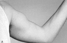
  • Biceps Tendon Rupture Wikipedia
    More severe injuries require surgery and post-op physical therapy to regain strength and functionality in the muscle.
  • Ideas And Delusions Of Reference Wikipedia
    Contents 1 Psychoanalytic views 2 Anti-psychiatry 3 Delusions of reference 4 Examples 5 Literary analogues 6 See also 7 References Psychoanalytic views [ edit ] Main article: Psychoanalytic theory In Sigmund Freud 's view, "Delusions of being watched present this power in a regressive form, thus revealing its genesis...voices, as well as the undefined multitude, are brought into the foreground again by the [ paranoid ] disease, and so the evolution of conscience is reproduced regressively." [7] As early as 1928, Freud's contemporary, Carl Jung , introduced the concept of synchronicity , a theory of "meaningful coincidences". [8] In 1946, Otto Fenichel concluded that "the projection of the superego is most clearly seen in ideas of reference and of being influenced....Delusions of this kind merely bring to the patient from the outside what his self-observing and self-critical conscience actually tells him." [9] Jacques Lacan similarly saw ideas of reference as linked to "the unbalancing of the relation to the capital Other and the radical anomaly that it involves, qualified, improperly, but not without some approximation to the truth, in old clinical medicine, as partial delusion" [10] —the "big other, that is, the other of language, the Names-of-the-Father , signifiers or words", [11] in short, the realm of the superego. ... ISBN 978-0-670-03292-1 . ^ Fenichel, Otto (1946). The Psychoanalytic Theory of Neurosis (London) pp. 430–1 ^ Jacques Lacan, Ecrits: A Selection (London 1996) p. 214 ^ Hill, Philip (1997).
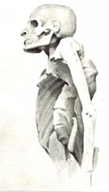
  • Muselmann Wikipedia
    Boder assisted in identifying the term musselman when in 1946 he conducted interviews with camp survivors in Europe. ... ISBN 978-0-19-533955-0 . ^ Alan Rosen (18 October 2010). The Wonder of Their Voices: The 1946 Holocaust Interviews of David Boder .
  • Pott Disease Wikipedia
    Imogen, in the novella "The Princess with the Golden Hair", part of Memoirs of Hecate County by Edmund Wilson (1946), has Pott disease. Jane Addams , social activist and Nobel Peace Prize winner, had Pott disease. ... Willem Ten Boom, brother of Corrie Ten Boom , died of tuberculosis of the spine in December 1946 [5] English writer Denton Welch (1915–1948) died of spinal tuberculosis after being involved in a motor accident (1935) that irreparably damaged his spine.
    TNF, CCL2, IL10, VDR, IL4, MAP3K7, THEMIS, SNHG15, SIL1, NCAPG2, SMUG1, PHB2, VCAM1, NR2C2, TLR2, BMP4, SPP1, CD14, P2RX7, MT1JP, MMP13, MMP9, MMP1, MBL2, IL6, IFNG, HSPE1, HLA-DQA1, MIR155
  • Murrain Wikipedia
    Retrieved 28 July 2008 . ^ Mullett, Charles F. (1946). "The Cattle Distemper in Mid-Eighteenth-Century England" .
  • Nipples, Supernumerary OMIM
    Rather extensive literature supporting dominant inheritance was reviewed by Gates (1946). Klinkerfuss (1924) found polymastia in 5 females in 4 generations.
    GPC3, ACTB, PORCN, PGAP2, RNF216, TMCO1, PIGV, HDAC8, KLHL7, ARHGAP31, COLEC11, COLEC10, CSPP1, PIGO, PIGY, PGAP3, CKAP2L, ASXL1, PNPLA6, ZEB2, TFAP2A, KIAA0586, DHODH, MEGF8, GPC4, KRAS, NONO, MASP1, TCF4, PIGW, TFAP2B, KAT6A, TRRAP, IKBKG, TP63, PIGL, HDAC4, BRCA2
    • Familial Supernumerary Nipples Orphanet
      Familial supernumerary nipples is a rare breast malformation characterized by the presence, in various members of a single family, of one or more nipple(s) and/or their related tissue, in addition to the normal bilateral chest nipples. The anomaly is usually situated along the embryonic milk line, from axillae to inguinal regions, but other locations are also possible. Association with dental abnormalities, Becker nevus, renal or underlying breast tissue malignancy and genitourinary malformations has been reported.
    • Supernumerary Nipple Wikipedia
      Supernumerary nipple Other names Third nipple , Triple nipple , Accessory nipple , [1] Polythelia Specialty Medical genetics , dermatology A supernumerary nipple is an additional nipple occurring in mammals , including humans . They are often mistaken for moles . Studies variously report the prevalence of supernumerary nipples as approximately 1 in 18 and 1 in 40. [2] [3] [1] The nipples appear along the two vertical " milk lines ", which start in the armpit on each side, run down through the typical nipples and end at the groin. They are classified into eight levels of completeness from a simple patch of hair to a milk-bearing breast in miniature. [4] [5] Contents 1 Types 2 Clinical significance 2.1 Clinical presentation 2.2 Associations 2.3 Treatment and prognosis 3 Society and culture 3.1 Television 4 See also 5 References 6 External links Types [ edit ] Type Glandular tissue Nipple Areola Fat tissue Hair patch 1 yes yes yes yes 2 yes yes 3 yes yes 4 yes 5 ("pseudomamma") yes yes yes 6 ("polythelia") yes 7 ("polythelia areolaris") yes 8 ("polythelia pilosa") yes [6] Polythelia refers to the presence of an additional nipple alone while polymastia denotes the much rarer presence of additional mammary glands . Although usually presenting on the milk line, pseudomamma can appear as far away as the foot. [7] A possible relationship with mitral valve prolapse has been proposed. [8] Clinical significance [ edit ] Clinical presentation [ edit ] May remain undetected. Occasionally, the supernumerary nipple is noticed when hormonal changes during adolescence, menstruation, or pregnancy cause increased pigmentation, fluctuating swelling, tenderness, or even lactation.
  • Maple Syrup Urine Disease GeneReviews
    Acute metabolic decompensation is corrected by treating the precipitating stress while delivering sufficient calories, insulin, free amino acids, isoleucine, and valine to achieve sustained net protein synthesis in tissues. Some centers use hemodialysis/hemofiltration to remove BCAAs from the extracellular compartment, but this intervention does not alone establish net protein accretion. ... Thus, leucine tolerance reflects a balance between unmeasured protein losses (e.g., sloughed skin, hair, and nails) and the net accretion of body protein, which in turn is linked to growth rate [Strauss et al 2010]. ... The risk for metabolic crisis in any ill person with MSUD depends on residual in vivo BCKD enzyme activity in relation to the net liberation of free leucine from protein catabolism. ... Following the neonatal period, acute metabolic intoxication (leucinosis) and neurologic deterioration can develop rapidly at any age as a result of net protein degradation precipitated by infection, surgery, injury, or psychological stress (see Figure 1). ... Plasma leucine levels rise predictably as a result of net protein catabolism provoked by a variety of physiologic stresses, including (more...)
    DBT, BCKDHB, BCKDHA, BCAT2, PPM1K, DLD, ARID4B, BDNF, CTSD, SERPINE1, TNS3, CACNA2D2, MKRN3, UMOD, SPN, NME1, PAH, NBN, MEA1, IL1B, GPR4, GLI2, F2, MECP2
    • Maple Syrup Urine Disease Wikipedia
      Metabolic disorder affecting branched-chain amino acids. It is one type of organic acidemia.[2] The condition gets its name from the distinctive sweet odor of affected infants' urine Maple syrup urine disease Other names Branched-chain ketoaciduria Isoleucine (pictured above), leucine , and valine are the branched-chain amino acids that build up in MSUD. Specialty Medical genetics Maple syrup urine disease ( MSUD ) is an autosomal recessive [1] metabolic disorder affecting branched-chain amino acids . It is one type of organic acidemia . [2] The condition gets its name from the distinctive sweet odor of affected infants' urine, particularly prior to diagnosis and during times of acute illness. [3] Contents 1 Classification 2 Signs and symptoms 2.1 Classic MSUD 2.2 Intermediate MSUD 2.3 Intermittent MSUD 2.4 Thiamine-response MSUD 2.5 Later onset 3 Causes 4 Pathophysiology 5 Diagnosis 6 Prevention 7 Treatment 7.1 Monitoring 7.2 Diet control 7.3 Liver transplantation 7.4 Pregnancy 8 Prognosis 9 Epidemiology 10 Research directions 10.1 Gene therapy 10.2 Phenylbutyrate therapy 11 See also 12 References 13 External links Classification [ edit ] Maple syrup urine disease can be classified by its pattern of signs and symptoms, or by its genetic cause. The most common and severe form of this disease is the classic type, which appears soon after birth, and as long as it remains untreated, gives rise to progressive and unremitting symptoms. Variant forms of the disorder may become apparent only later in infancy or childhood, with typically less severe symptoms that may only appear during times of fasting, stress or illness, but still involve mental and physical problems if left untreated.
    • Maple Syrup Urine Disease, Mild Variant OMIM
      A number sign (#) is used with this entry because of evidence that a mild variant of maple syrup urine disease (MSUDMV) is caused by homozygous mutation in the PPM1K gene (611065) on chromosome 4q22. One such family has been reported. Description The mild variant of MSUD is characterized by increased plasma levels of branched-chain amino acids (BCAA) apparent at birth. Treatment with a low-protein diet free of BCAA can result in normal psychomotor development and lack of metabolic episodes; however, plasma levels of BCAA may remain elevated (summary by Oyarzabal et al., 2013). For a general description and a discussion of genetic heterogeneity of maple syrup urine disease, see 248600. Clinical Features Oyarzabal et al. (2013) reported a 21-year-old woman with a mild variant of maple syrup urine disease.
    • Maple Syrup Urine Disease Orphanet
      A rare inherited disorder of branched-chain amino acid metabolism classically characterized by poor feeding, lethargy, vomiting and a maple syrup odor in the cerumen (and later in urine) noted soon after birth, followed by progressive encephalopathy and central respiratory failure if untreated. The four overlapping phenotypic subtypes are: classic, intermediate, intermittent and thiamine-responsive MSUD. Epidemiology The estimated prevalence is around 1/150,000 live births, from published and unpublished newborn screening data. Clinical description Classic MSUD presents in the first days of life with poor feeding and drowsiness followed by a worsening encephalopathy with lethargy, intermittent apnea, stereotypic movements ("fencing" and ''bicycling") and opisthotonus. Coma and central respiratory failure supervene 7 to 10 days after birth.
    • Maple Syrup Urine Disease GARD
      Maple syrup urine disease (MSUD) occurs when the body is unable to breakdown certain parts of proteins. This leads to the build-up of toxic substances that can cause organ and brain damage. There are several forms of MSUD. The most common is the classic or infantile form. Symptoms of the classic form of MSUD start in early infancy and include poor feeding, irritability, extra sleepiness, and muscle spasms. If untreated, respiratory failure (lack of oxygen getting to the blood) may occur.
  • Afterdepolarization Wikipedia
    They are due to elevated cytosolic calcium concentrations, classically seen with digoxin toxicity. [3] [4] The overload of the sarcoplasmic reticulum may cause spontaneous Ca 2+ release after repolarization, causing the released Ca 2+ to exit the cell through the 3Na + /Ca 2+ -exchanger. This results in a net depolarizing current. The classical feature is Bidirectional ventricular tachycardia .
  • Krukenberg's Spindle Wikipedia
    The sign was described in 1899 by Friedrich Ernst Krukenberg (1871-1946), who was a German pathologist specialising in ophthalmology . [1] Contents 1 Diagnosis 1.1 Differential diagnosis 1.1.1 Iritis 1.1.2 Vortex keratopathy 1.1.3 Corneal guttata 2 See also 3 References 4 External links Diagnosis [ edit ] Differential diagnosis [ edit ] Iritis [ edit ] Painful red eye with photophobia associated with inflammation Vortex keratopathy [ edit ] Corneal deposits also known as cornea verticillata , caused by netarsudil eye drops or chronic amiodarone use for cardiac arrhythmias . [2] Corneal guttata [ edit ] Non-transparent collagen deposits appearing following loss of corneal endothelial cells [3] See also [ edit ] Pigment dispersion syndrome References [ edit ] ^ Krukenberg F. (1899) Beiderseitige angeborene Melanose der Hornhaut.
  • Bugchasing Wikipedia
    , 10 April 2006 v t e Lesbian , gay , bisexual , and transgender ( LGBT ) slang List Ace Bareback Banjee Bear Beard Beat Bi-curious Boi Top, bottom and versatile Bottom surgery Breeder Bugchasing Bulldagger Butch Castro clone Chicken Chickenhawk Chub Chubby chaser Cottaging Cruising Daddy Down-low Drag Dyke En femme En homme Fag (Faggot) Fag hag Fag stag Faux queen F2M Femme Flagging (hanky code) Friend of Dorothy Fruit Fruit fly Gay-for-pay Gaydar Gaymer Genderfuck Gold star lesbian Glory hole Heteroflexibility Lesbian until graduation Lipstick lesbian M2F Non-op Packing Party and play Passing Poppers Post-op Pre-op Queen RLE Shemale Soft butch Scissoring SRS Stone butch Stealth Swish T Tea-room TERF Top surgery Trache shave Trade Tranny Transfan Transition Tribbing Troll Twink U-Haul lesbian Womyn-born womyn Related Polari LGBT linguistics Terminology of homosexuality Category v t e Sexual slang General Anilingus Banjee Bareback Baseball metaphors for sex Blue balls Bottom Camel toe Chickenhead Circle jerk Cock tease Cornhole Cougar Cunt Deep-throating Dick Dirty Sanchez Dogging Donkey punch Douche Felching Fuck Girlfriend experience Glory hole Hogging Hot Karl Italian profanity Latin profanity Mama-san Mammary intercourse Mat Mile high club Motherfucker Nookie Party and play Pearl necklace Pegging Pirate Pussy Quickie Red wings Rusty trombone Serosorting Shemale Slut Snowballing Soggy biscuit Switch Teabagging Tits Top Top, bottom and versatile Turkey slap Twat Voulez-vous coucher avec moi?
  • Nephrolithiasis, Calcium Oxalate OMIM
    In vitro flux studies indicated that mice lacking Slc26a6 have a defect in intestinal oxalate secretion resulting in enhanced net absorption of oxalate. Jiang et al. (2006) concluded that the anion exchanger, SLC26A6, has a major constitutive role in limiting net intestinal absorption of oxalate, thereby preventing hyperoxaluria and calcium oxalate urolithiasis.
    SLC26A1, F2, FN1, AMBP, AHSG, HOGA1, HAO1, AGXT, SLC36A2, PAQR6, ADCY10, SLC6A20, OPLAH, GRHPR, SLC6A19, CD44, ABCB1, MSH3, CD9, SLC22A12, CDH1, SLC26A6, CHD1, GGCX, IL1B, IL1RN, TBC1D9, MDM4, ABCG2, ABCC1, KL, VDR, SPP1
    • Kidney Stones MedlinePlus
      Kidney stones (also called renal stones or urinary stones) are small, hard deposits that form in one or both kidneys; the stones are made up of minerals or other compounds found in urine. Kidney stones vary in size, shape, and color. To be cleared from the body (or "passed"), the stones need to travel through ducts that carry urine from the kidneys to the bladder (ureters) and be excreted. Depending on their size, kidney stones generally take days to weeks to pass out of the body. Kidney stones can cause abdominal or back pain (known as renal colic). Renal colic usually begins sporadically but then becomes constant and can lead to nausea and vomiting.
  • Steroid Diabetes Wikipedia
    Mechanism [ edit ] Glucocorticoids oppose insulin action and stimulate gluconeogenesis , especially in the liver , resulting in a net increase in hepatic glucose output.
  • Naegeli-Franceschetti-Jadassohn Syndrome/dermatopathia Pigmentosa Reticularis MedlinePlus
    Among the most common signs of NFJS/DPR is a net-like pattern of dark brown or gray skin coloring, known as reticulate hyperpigmentation.
    KRT14, TARDBP, C9orf72
    • Dermatopathia Pigmentosa Reticularis OMIM
      A number sign (#) is used with this entry because of evidence that dermatopathia pigmentosa reticularis (DPR) is caused by heterozygous mutation in the keratin-14 gene (KRT14; 148066) on chromosome 17q21. One such family has been reported. A closely related disorder, Naegeli-Franceschetti-Jadassohn syndrome (NFJS; 161000), is also caused by heterozygous mutation in the KRT14 gene. Description Dermatopathia pigmentosa reticularis is a rare heritable disorder consisting of a triad of cutaneous findings including reticulate hyperpigmentation, noncicatricial alopecia, and onychodystrophy. Variable features include adermatoglyphia, hypohidrosis or hyperhidrosis, and palmoplantar hyperkeratosis (Heimer et al., 1992). Clinical Features Heimer et al. (1992) described a family with 9 cases of dermatopathia pigmentosa reticularis distributed through 6 sibships of 4 generations.
    • Dermatopathia Pigmentosa Reticularis Orphanet
      A rare, genetic, ectodermal dysplasia characterized by a widespread, early-onset, reticulate hyperpigmentation that persists throughout life, mild, diffuse non-cicatricial alopecia, and onychodystrophy. There are no dental anomalies. Patients may also present with adermatoglyphia, palmoplantar hyperkeratosis, acral dorsal blistering, and hypohidrosis or hyperhidrosis.
    • Dermatopathia Pigmentosa Reticularis Wikipedia
      Dermatopathia pigmentosa reticularis Other names Dermatopathic pigmentosa reticularis [1] : 511 Dermatopathia pigmentosa reticularis has an autosomal dominant pattern of inheritance Specialty Medical genetics Dermatopathia pigmentosa reticularis is a rare, autosomal dominant [2] congenital disorder that is a form of ectodermal dysplasia . Dermatopathia pigmentosa reticularis is composed of the triad of generalized reticulate hyperpigmentation , noncicatricial alopecia , and onychodystrophy . [3] : 856 Contents 1 Presentation 2 Cause 3 Treatment 4 See also 5 References 6 External links Presentation [ edit ] Symptoms include lack of sweat glands, thin hair, brittle nails, mottled skin, and lack of fingerprints. [4] DPR is very similar to the related Naegeli-Franceschetti-Jadassohn syndrome . Both cause an affected person to lack fingerprints, have a lace-like pattern of hyperpigmentation and hyperkeratosis of the palms of the hands and soles of the feet. DPR is distinguished from NFJS by the duration of hyperpigmentation and lack of dental abnormalities. [5] Cause [ edit ] DPR is caused by a mutation in the keratin 14 gene. [6] Treatment [ edit ] This section is empty. You can help by adding to it . ( April 2017 ) See also [ edit ] List of cutaneous conditions caused by mutations in keratins References [ edit ] ^ Freedberg, et al. (2003).
  • ←
  • 1
  • 2
  • 3
  • 4
  • 5
  • 6
  • 7
  • 8
  • ...
  • 21
  • 22
  • →

FindZebra

contact@findzebra.com