Load FindZebra Summary
Disclaimer:
FindZebra Search conducts a search using our specialized medical search engine.
FindZebra Summary uses the text completions API
(subject to OpenAI’s API data usage policies)
to summarize and reason about the search results.
The search is conducted in publicly available information on the Internet that we present “as is”.
You should be aware that FindZebra is not supplying any of the content in the search results.
FindZebra Summary is loading...
-
Rice Allergy
Wikipedia
Although some reactions might lead to severe health problems, doctors can diagnose rice allergy with many methods and help allergic people to avoid reactions. Contents 1 Symptoms 2 Diagnosis 3 Treatment 4 Prevalence 5 References Symptoms [ edit ] Some rice proteins are regarded as the causes of allergy in people. [1] [2] People allergic to rice might have sneezing , runny nose , itching , asthma , stomachache or eczema after they eat rice. [2] [3] [4] [5] Besides eating rice, people with a rice allergy can have reactions breathing rice steam from cooked rice. ... This is regarded as a new choice for rice allergic people. [2] [13] Reactions might lessen by staying away from rice long-term. [3] Prevalence [ edit ] Unlike other food allergies, rice allergy is relatively uncommon. [4] It has been reported worldwide but mostly in China , Japan or Korea . [13] [14] Because rice is a major food in Asia , people from Asia are exposed to higher allergy risk than people from other areas. [15] References [ edit ] ^ YH Jeon, et al, "Identification of major rice allergen and their clinical significance in children," Korean J Pediatric 54, no. 10 (2011): 414. ^ a b c Yuko Ogo, et al, "Generation of transgenic rice with reduced content of major and novel high molecular weight allergens," Rice 7, no. 1(2014): 19. ^ a b B. ... Ballmer-Weber, "Isolated allergy to rice," Allergy 57, no. 3(2002): 264. ^ a b Mitsuhiko Nambu, Noriaki Shintaku and Shigeru Ohta, "Rice Allergy," Pediatrics 117, no. 6 (June, 2006): 2331. ^ Richard Lockey, "Food Allergy," World Allergy Organization, accessed February 29, 2016, http://www.worldallergy.org/public/allergic_diseases_center/foodallergy/. ^ Raj Kumar, et al, "Rice (Oryza sativa) allergy in rhinitis and asthma patients: A clinico-immunological study," Immunobiology 212, no. 2 (2007): 142. ^ F. Orhan and B. E.Sekerel, "A case of isolated rice allergy," Allergy 58, no.5(2003): 457. ^ Alessandro Fiocchi, et al, "Anaphylaxis to rice by inhalation," Journal of Allergy and Clinical Immunology 111, no. 1 (2003): 193. ^ Food Allergy Research & Education, "Food Elimination Diet," Food Allergy Research & Education, accessed March 3, 2016, http://www.foodallergy.org/diagnosis-and-testing/food-elimination-diet. ^ Food Allergy Research & Education, "Blood Tests," Food Allergy Research & Education, accessed March 3, 2016, http://www.foodallergy.org/diagnosis-and-testing/blood-tests. ^ Food Allergy Research & Education ,"Skin Prick Tests," Food Allergy Research & Education, accessed March 3, 2016, https://www.foodallergy.org/diagnosis-and-testing/skin-tests. ^ American Academy of Allergy, Asthma & Immunology, "ALLERGY SHOTS (IMMUNOTHERAPY)," American Academy of Allergy, Asthma & Immunology, accessed February 16, 2016, https://www.aaaai.org/conditions-and-treatments/treatments/allergy-shots-(immunotherapy).aspx. ^ a b YH. Jeon, et al, "A Study for Allergenicity and Cross-reactivity of Rice," Journal of Allergy and Clinical Immunology 123, no. 2(February 2009): S190. ^ Caroline Hadley, "Food allergies on the rise? Determining the prevalence of food allergies, and how quickly it is increasing, is the first step in tackling the problem," EMBO Report 7, no. 11 (2006): 1082. ^ YH Jeon, et al, "Identification of major rice allergen and their clinical significance in children," Korean J Pediatric 54, no. 10 (2011): 419.
-
Neuroendocrine Tumor
Wikipedia
G Mitotic count (per 10 HPF ) Ki-67 index (%) GX Grade cannot be assessed G1 < 2 < 3% G2 2 to 20 3–20% G3 > 20 > 20% If mitotic count and Ki-67 are discordant, the figure which gives the highest grade is used. ... Not all cells are immediately killed; cell death can go on for up to two years. [ citation needed ] PRRT was initially used for low grade NETs. It is also very useful in more aggressive NETs such as Grade 2 and 3 NETs [83] [84] provided they demonstrate high uptake on SSTR imaging to suggest benefit. ... World Journal of Clinical Oncology . 2 (1): 28–43. doi : 10.5306/wjco.v2.i1.28 . ... World Journal of Gastrointestinal Surgery . 2 (5): 153–56. doi : 10.4240/wjgs.v2.i5.153 . ... Archived from the original on January 2, 2010. ^ Walsh, Bryan (January 1, 2010).MEN1, CDKN1B, SSTR2, DAXX, ATRX, BRAF, TYMS, PTHLH, SSTR3, SSTR1, BAP1, MTOR, SST, GAST, SLC6A2, INSM1, CTNNB1, RET, PIK3CA, DNMT3A, POMC, EPHB1, PIK3CG, PIK3CD, CHGA, ELK3, CHEK2, PIK3CB, GRN, CD274, SMUG1, AKT1, GNA12, TP53, SYP, VEGFA, CDKN2A, ASCL1, BCL2, ENO2, NCAM1, GCG, MYCN, EGFR, MGMT, KIT, RASSF1, VHL, SCLC1, SSTR5, FOLH1, NKX2-1, KRAS, CALCA, CCND1, TAC1, PTPRF, VIP, NTS, PAX5, RHBDF2, GRP, IGF1, SDHD, GOT1, MAP2K7, CCK, ERBB2, DLL3, PPY, CXCL12, TP63, SMAD4, MUC1, INS, GCGR, CKAP4, NEUROD1, ISL1, MYC, NGF, SATB2, GLP1R, HSP90AA1, H3P10, HRAS, CHGB, CALR, NTRK1, TEK, DLK1, CDK4, CDX2, TGFA, UCHL1, RPE65, PGR, PDGFRA, CARTPT, CRH, UVRAG, SLC5A5, CXCR4, IGF1R, OTP, IL6, PHLDA3, TTF1, PAX8, TACR1, STK11, TRIM21, PLA2G15, SCG2, SQLE, SLC18A2, TERT, HDAC9, SLC2A1, PROM1, BCL2L11, NTSR1, PAX6, NAMPT, NOCT, INA, PLCB3, CD200, MKI67, PDX1, MAPK1, NES, HPSE, PTEN, STMN1, ABO, RIPK1, RORC, RAF1, IL1B, TRPV1, GATA3, ANGPT2, FOXM1, PTK2B, SDHAF2, ACCS, BDNF, EPAS1, EGF, ACSS2, MIB1, DNMT1, CCN2, TRPM8, CLDN4, CPE, CD34, CD44, FLNA, CEACAM5, B3GAT1, GH1, GIP, GHSR, GIPR, ADCY2, ALB, H3P28, TPPP2, H4C5, GGH, MIR1290, TMEM209, ELOA3, H4C13, H4C14, GPR151, SRPX, LGR5, TNFSF11, PSMG1, DCBLD2, H4-16, NRP1, MRGPRX4, SOCS1, H4C2, MIR3137, MRGPRX3, TNFRSF25, H3P12, CYYR1, AZIN2, DNER, AK6, MLIP, LMLN, NRP2, GPR68, MIR1246, H4C8, MAFK, MIR150, MIR155, MBOAT4, H4C9, MIR21, POTEKP, VN1R17P, SNORD95, GPR166P, ARID1A, EID3, SLC7A5, MIR375, H4C15, FZD4, MIRLET7C, OXER1, H4C12, HMGA2, H4C3, ARX, ELOA3B, GPRC6A, H4C11, H4C6, C17orf97, POTEM, MRGPRX1, ARMH1, H4C1, GADL1, ACTBL2, H4C4, BRI3, SQSTM1, ISYNA1, GHRL, ACOT7, KLF12, KRT20, SLC27A4, TET2, BCOR, EBNA1BP2, RALBP1, PGRMC1, LAMTOR1, FBXW7, MEG3, MAML3, TMEM127, NTNG1, ATRAID, KHDRBS1, DCTN4, SNORD61, NUP62, SNORD48, NTSR2, LPAR3, MAPK8IP2, SRRM2, BRD4, TRAM1, SPINK4, XIST, PPWD1, RBMS3, SETD1B, ZHX2, TNFSF13B, USE1, MAK16, UBE2Z, ONECUT2, FHL5, GCM2, DCLK1, ZBED1, ARHGEF2, PALB2, ALG9, SNED1, TET1, PDCD1LG2, TMPRSS13, MTA1, RPAIN, H1-10, EEF1E1, LGR6, PRMT5, NEUROD4, YAP1, SCML2, LANCL1, PAK4, RABEPK, ZNF197, CTNNBL1, PNO1, INSL5, EPB41L5, HDAC5, AKT3, CD302, GBA3, DCAF1, ATAT1, SERPINA3, VCL, CGA, ESR1, ERBB4, EPHB2, E2F1, DUSP2, DSG3, DPT, DPP4, DMBT1, DDC, DAD1, VCAN, CREB1, CRABP1, KLF6, CLU, FOXN3, CEACAM7, CEACAM3, ESR2, ETFA, EZH2, GHRH, HSPA4, AGFG1, HMOX1, HMGA1, GTF2H1, GSN, GNAS, GNA15, GFRA1, F3, GDNF, FSHR, FLT4, FLII, FLI1, FOXO1, FHIT, FGFR4, CGB3, CFL1, UQCRFS1, CDKN2C, FAS, APRT, APLP1, XIAP, APC, SLC25A6, SLC25A4, ANGPT1, ALK, AKT2, AFP, PARP1, ADCYAP1R1, ADCYAP1, ACVRL1, ACTN4, ACTG2, ACTG1, ACR, AQP4, ARF1, ATM, CASP3, CDK6, CD40LG, CD36, CD33, CCNE1, CCKBR, SERPINA6, CAV1, CA9, ATOH1, VPS51, C5, BRS3, BRCA2, DST, BAX, AVP, ATP4A, HTC2, HTR2A, TNC, IAPP, SDC1, SCT, SORT1, RNASE3, RARB, PTPRZ1, PTPRM, PTBP1, PSMD7, PSG2, PRKAR1A, PPP4C, POU4F1, PNN, PKD2, PITX2, PCYT1A, SERPINA5, PAX4, SDCBP, SDHB, SDHC, ST2, UBE2I, TPM3, TPH1, TNF, TM7SF2, TERC, TAT, STAT3, SSTR4, SEMA3F, SSR2, SOX11, SOX4, SOX2, SLPI, SLC3A2, SLC1A5, SFRP1, PAK3, PAK1, TNFRSF11B, KIF11, MDK, MAOA, LCN2, RPSA, L1CAM, KRT19, KRT7, KRT5, IL12A, MET, IL9, CXCL8, IL2, IL1A, IGFBP1, IGF2, IFNA13, IFNA1, MDM2, MFAP1, ODC1, MUTYH, NTRK2, NT5E, NRAS, NOTCH3, NPY, NOTCH1, NFKB1, NEFM, MUC4, CD99, NUDT1, COX2, MTAP, MST1R, MST1, MSMB, MMP7, MLH1, PTPRC

-
Descending Perineum Syndrome
Wikipedia
Descending perineum syndrome (also known as levator plate sagging ) [1] refers to a condition where the perineum "balloons" several centimeters below the bony outlet of the pelvis during strain, although this descent may happen without straining. [2] The syndrome was first described in 1966 by Parks et al . [3] Contents 1 Signs and symptoms 2 Cause 3 Diagnosis 4 Treatment 5 Epidemiology 6 References Signs and symptoms [ edit ] Abnormal descent of the perineum may be asymptomatic, but otherwise the following may feature: perineodynia (perineal pain) [1] Colo-proctological symptoms, e.g. obstructed defecation , [4] dyschesia (constipation), [1] or degrees of fecal incontinence [1] gynaecological symptoms, e.g. cystocele (prolapse of the bladder into the vagina) and rectocele (prolapse of the rectum into the vagina) [1] lower urinary tract symptoms, e.g. dysuria (painful urination), dyspareunia (pain during sexual intercourse), urinary incontinence and urgency [1] Other researchers concluded that abnormal perineal descent did not correlate with constipation or perineal pain, and there are also conflicting reports of the correlation of fecal incontinence with this condition. [2] Cause [ edit ] One of the main causes is suggested to be excessive and repetitive straining during defecation. [2] Other causes include weakness of the pelvic floor muscles (secondary to age-related neuropathic degeneration or traumatic injury during pregnancy and labor. ... CS1 maint: extra text: authors list ( link ) ^ Baek, HN; Hwang, YH; Jung, YH (December 2010). "Clinical Significance of Perineal Descent in Pelvic Outlet Obstruction Diagnosed by using Defecography" .
-
Yao Syndrome
Wikipedia
Please introduce links to this page from related articles ; try the Find link tool for suggestions. ( January 2020 ) Yao syndrome (YAOS) (formerly called NOD2-associated autoinflammatory disease [1] ) is an autoinflammatory syndrome involving episodes of fever and abnormal inflammation affecting many parts of the body , particularly the skin , joints , and gastrointestinal system. [2] [3] [4] Contents 1 Signs and symptoms 2 Diagnostic criteria 3 Treatment 4 Inheritance 5 References 6 External links Signs and symptoms [ edit ] Arthralgia (Arthritis) Fever Diarrhea Abdominal Pain Keratoconjunctivitis Sicca (Dry eyes) Pleurisy Idiopathic Pericarditis Xerostomia (Dry mouth) Erythematous Plaques Dermatitis Diagnostic criteria [ edit ] Yao syndrome is diagnosed if 2 major criteria, at least one minor criterion, the molecular criterion, and exclusion criteria are fulfilled. [5] Clinical Criteria Comments Major 1 Periodic occurrence ≥ twice 2 Recurrent fever or dermatitis or both Minor 1 Polyarthralgia / inflammatory arthritis , or distal extremity swelling 2 Abdominal pain or diarrhea or both 3 Sicca -like symptoms 4 Pericarditis or pleuritis or both Molecular Criterion NOD2 IVS8+158 or R702W or both, or other rare variants Exclusion Criteria High titer antinuclear antibodies, inflammatory bowel disease , Blau syndrome , adult sarcoidosis , primary Sjögren syndrome and monogenic autoinflammatory diseases Treatment [ edit ] A study to determine the effectiveness of Novartis pharmaceutical drug Canakinumab was conducted. ... The American Journal of Medicine . 130 (3): 365.e13–365.e18. doi : 10.1016/j.amjmed.2016.09.028 .
-
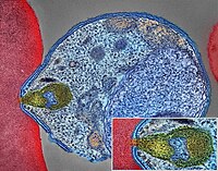
Malaria
Wikipedia
Malaria Malaria parasite connecting to a red blood cell Pronunciation / m ə ˈ l ɛər i ə / Specialty Infectious disease Symptoms Fever, vomiting, headache, yellow skin [1] Complications Seizures , coma [1] Usual onset 10–15 days post exposure [2] Causes Plasmodium spread by mosquitoes [1] Diagnostic method Examination of the blood, antigen detection tests [1] Prevention Mosquito nets , insect repellent , mosquito control , medications [1] Medication Antimalarial medication [2] Frequency 228 million (2018) [3] Deaths 405,000 in 2018 [3] Malaria is a mosquito-borne infectious disease that affects humans and other animals. [2] Malaria causes symptoms that typically include fever , tiredness , vomiting , and headaches . [1] In severe cases, it can cause yellow skin , seizures , coma , or death . [1] Symptoms usually begin ten to fifteen days after being bitten by an infected mosquito . [2] If not properly treated, people may have recurrences of the disease months later. [2] In those who have recently survived an infection , reinfection usually causes milder symptoms. [1] This partial resistance disappears over months to years if the person has no continuing exposure to malaria. [1] Malaria is caused by single-celled microorganisms of the Plasmodium group. [2] The disease is most commonly spread by an infected female Anopheles mosquito. [2] The mosquito bite introduces the parasites from the mosquito's saliva into a person's blood . [2] The parasites travel to the liver where they mature and reproduce . [1] Five species of Plasmodium can infect and be spread by humans. [1] Most deaths are caused by P. falciparum , whereas P. vivax , P. ovale , and P. malariae generally cause a milder form of malaria. [1] [2] The species P. knowlesi rarely causes disease in humans. [2] Malaria is typically diagnosed by the microscopic examination of blood using blood films , or with antigen-based rapid diagnostic tests . [1] Methods that use the polymerase chain reaction to detect the parasite's DNA have been developed, but are not widely used in areas where malaria is common due to their cost and complexity. [4] The risk of disease can be reduced by preventing mosquito bites through the use of mosquito nets and insect repellents or with mosquito-control measures such as spraying insecticides and draining standing water . [1] Several medications are available to prevent malaria in travellers to areas where the disease is common. [2] Occasional doses of the combination medication sulfadoxine/pyrimethamine are recommended in infants and after the first trimester of pregnancy in areas with high rates of malaria. [2] As of 2020, there is one vaccine which has been shown to reduce the risk of malaria by about 40% in children in Africa. [5] [6] Efforts to develop more effective vaccines are ongoing. [6] The recommended treatment for malaria is a combination of antimalarial medications that includes artemisinin . [1] [2] The second medication may be either mefloquine , lumefantrine , or sulfadoxine/pyrimethamine. [7] Quinine , along with doxycycline , may be used if artemisinin is not available. [7] It is recommended that in areas where the disease is common, malaria is confirmed if possible before treatment is started due to concerns of increasing drug resistance . [2] Resistance among the parasites has developed to several antimalarial medications; for example, chloroquine -resistant P. falciparum has spread to most malarial areas, and resistance to artemisinin has become a problem in some parts of Southeast Asia. [2] The disease is widespread in the tropical and subtropical regions that exist in a broad band around the equator . [1] This includes much of sub-Saharan Africa , Asia , and Latin America . [2] In 2018 there were 228 million cases of malaria worldwide resulting in an estimated 405,000 deaths. [3] Approximately 93% of the cases and 94% of deaths occurred in Africa. [3] Rates of disease have decreased from 2010 to 2014 but increased from 2015 to 2017, during which there were 231 million cases. [3] Malaria is commonly associated with poverty and has a significant negative effect on economic development . [8] [9] In Africa, it is estimated to result in losses of US$12 billion a year due to increased healthcare costs, lost ability to work, and adverse effects on tourism. [10] Play media Video summary ( script ) Contents 1 Signs and symptoms 1.1 Complications 2 Cause 2.1 Life cycle 2.2 Recurrent malaria 2.3 Climate change 3 Pathophysiology 3.1 Genetic resistance 3.2 Liver dysfunction 4 Diagnosis 4.1 Classification 5 Prevention 5.1 Mosquito control 5.1.1 Insecticide treated nets 5.1.2 Indoor residual spraying 5.1.3 Housing modifications 5.1.4 Other mosquito control methods 5.2 Medications 5.3 Others 6 Treatment 6.1 Uncomplicated malaria 6.2 Severe and complicated malaria 6.3 Resistance 7 Prognosis 8 Epidemiology 9 History 10 Society and culture 10.1 Economic impact 10.2 Counterfeit and substandard drugs 10.3 War 10.4 Eradication efforts 11 Research 11.1 Vaccine 11.2 Medications 11.3 New targets 11.4 Other 12 Other animals 13 References 13.1 Citations 13.2 Sources 14 Further reading 15 External links Signs and symptoms [ edit ] Main symptoms of malaria [11] The signs and symptoms of malaria typically begin 8–25 days following infection, [11] but may occur later in those who have taken antimalarial medications as prevention . [4] Initial manifestations of the disease—common to all malaria species—are similar to flu-like symptoms , [12] and can resemble other conditions such as sepsis , gastroenteritis , and viral diseases . [4] The presentation may include headache , fever , shivering , joint pain , vomiting , hemolytic anemia , jaundice , hemoglobin in the urine , retinal damage , and convulsions . [13] The classic symptom of malaria is paroxysm —a cyclical occurrence of sudden coldness followed by shivering and then fever and sweating, occurring every two days ( tertian fever ) in P. vivax and P. ovale infections, and every three days ( quartan fever ) for P. malariae . ... The mosquitoes remain on the wall until they fall down dead on the floor. Insecticide treated nets [ edit ] A mosquito net in use. Mosquito nets help keep mosquitoes away from people and reduce infection rates and transmission of malaria. Nets are not a perfect barrier and are often treated with an insecticide designed to kill the mosquito before it has time to find a way past the net. Insecticide-treated nets are estimated to be twice as effective as untreated nets and offer greater than 70% protection compared with no net. [73] Between 2000 and 2008, the use of ITNs saved the lives of an estimated 250,000 infants in Sub-Saharan Africa. [74] About 13% of households in Sub-Saharan countries owned ITNs in 2007 [75] and 31% of African households were estimated to own at least one ITN in 2008. ... That number increased to 20.3 million (18.5%) African children using ITNs in 2007, leaving 89.6 million children unprotected [76] and to 68% African children using mosquito nets in 2015. [77] Most nets are impregnated with pyrethroids , a class of insecticides with low toxicity .ICAM1, FCGR2B, HBB, CD36, NOS2, FCGR2A, TNF, CR1, G6PD, CRP, HP, ACKR1, GYPA, SLC4A1, GYPB, NCR3, TIRAP, GYPC, LTBR, CISH, IFNG, HMOX1, PKLR, ABO, ANK1, AQP4, ATP2B4, HBG2, CYTB, ENOSF1, MSMB, MST1, ZNF536, LINC00944, SMARCB1, DHODH, PDR, TREML4, ZNF804A, OR51F1, OR51B5, CDH13, PROCR, SPATA3, OR51N1P, DHFR, DDT, RECQL4, FAM155A, IGHG3, IL4, MMP26, IL6, IL10, TLR9, HLA-DRB1, CSMD1, HBE1, DNAJC5, TMPRSS13, KLHL3, HDGFL2, TLR4, ATAD1, LMLN, TENM3-AS1, MECP2, POMGNT2, MBL2, TFRC, TGFB1, MIF, HLA-B, HAMP, DHPS, SERPINA3, TLR2, IL1B, FOXP3, FHL5, ACOT7, POTEKP, POTEM, GEM, KIR3DL1, RN7SL263P, ACTG2, ACTG1, ACTB, ACTBL2, HBA2, CYP2B6, HSPA4, LSAMP, TRAP, FCGR3B, HSP90AA1, IL1A, LAMP3, CD81, OR10A4, CCL5, ABCB1, FAS, CD40LG, TEP1, CXCL8, IARS1, HLA-G, CTLA4, HBA1, INSRR, ANGPT2, TYMS, CFH, GSTP1, IFNAR1, AGT, GYPE, FCGR3A, TXN, IL13, HSPB3, APOE, MTCO2P12, ISYNA1, FCGR2C, FYB1, VDR, HLA-A, GSTM1, GSR, ATR, MBL3P, LAIR1, PNP, IL12B, MNAT1, IL1RN, CYP2D6, IGF1, CD55, ACHE, DECR1, COX2, IL3, CCL2, MAPK1, NLRP3, FBXW7, HAVCR2, THBD, VPS51, EMP1, ITGA2B, PTGS2, ANC, IL10RA, XPO1, VNN1, PLEK, UMPS, IL2, IL2RA, TPPP, VWF, ISG20, ADAMTS13, IRF1, IL7R, AIMP2, IL12RB1, CLEC11A, METAP2, CDK5R1, ING1, IL18R1, PGD, HAP1, H6PD, PRDX5, GRAP2, CXCL9, MMP9, MPO, TAP1, CCL4L2, COX1, EBI3, ITGAX, COX3, TLR6, CXCL11, MTHFR, NFKB2, NFYA, NOS1, TBC1D9, ORC1, MCF2, AKAP13, RNF19A, TLR7, NT5C3A, IRAK4, KIR2DS1, CCL4, KIR3DL2, ICOS, COQ2, PSIP1, PECAM1, TPT1, RNASE3, ARTN, TP53, POLDIP2, PDCD1, TLR1, AHSA1, UBL4A, AQP3, AGRP, H3C9P, CYP2C8, CYP2C19, GTF2H4, CRK, RNA18SN5, ANXA2, H3P37, CASP1, NANP, CCL4L1, MAPK14, CXCR3, GNAS, GLO1, FCN2, SMIM10L2B, FKBP4, CD27, FOXO3, RBM45, HM13, IL33, HK1, CCR5, IFNA13, IFNA1, H3P42, DNAJB1, CHIT1, CYP3A4, SMIM10L2A, EGF, CHI3L1, CAT, EPHA2, NSFL1C, ADRB2, MYMX, COX8A, GAPDH, ABCB6, NR1I3, TREML1, PUM3, FMN1, TICAM2, TRIM13, BMS1, FZD4, RABEPK, LANCL1, FUT9, TNFSF13B, DCTN6, CXCR6, ARL6IP5, MRGPRX1, ZNRD2, ASPM, KAT5, RAB7B, CIB1, SEMA3C, ARMH1, STING1, CFDP1, CPQ, MYLK4, DLC1, AKR1A1, PIEZO1, TMPRSS11D, HDAC9, CARTPT, DEFB4B, TIMELESS, SPHK1, TMED7-TICAM2, PSC, VNN2, PROM1, UPK3B, H3P23, H3P28, TNFRSF11A, TNFRSF18, TP63, PDXK, CNTNAP1, DHX16, STK24, H3P19, LOH19CR1, WASHC1, WASH6P, LPAR2, MIR146A, APOBEC3B, SPAG6, CLOCK, ATG5, MIR142, AIM2, ABCG2, PCSK9, MIR155, NCF1, PPIG, MIR29A, VN1R17P, GPR166P, CD163, MIR451A, CXADRP1, ARHGEF2, CERS1, SPINK5, MASP2, GEMIN4, ACD, TLR8, MPPE1, MCPH1, HSPA14, RNF34, TMED7, ARMC9, PPP1R2C, IL22, TRAF3IP2, A1CF, PDCD1LG2, SLC44A4, SGSM3, MCAT, HPGDS, B3GAT1, ROPN1L, PHGDH, RAB14, IL23A, ABCG4, IFIH1, CFC1, BTNL2, MARCHF1, POLE4, CMC2, TMED9, ACKR3, PDXP, RHOF, AICDA, POLD4, RBM25, TOLLIP, TREM1, LGR6, ADA2, BACH2, ERAP1, GOLPH3, PARS2, KRT88P, TRIM5, IL17RE, CHP1, GPR151, NRSN1, EIF5AL1, CD160, APCDD1, ERFE, OXER1, DNAJB1P1, DSTN, GPRC6A, CCNI, ADIRF, EBNA1BP2, TMED2, EHD1, RNPS1, HPSE, SEPTIN9, SCLT1, NT5C2, SLC25A21, LEO1, NLRP12, TIMD4, CDCA5, DBA2, CARD16, PTPMT1, CGAS, RAB39B, TADA1, MRGPRX3, MRGPRX4, PGLS, PANX1, SPO11, LPAR3, CBX5, POFUT2, SPPL3, NBEAL2, LUC7L, PTPRC, FGF23, EIF5, FLT3LG, FLT1, FECH, FBN2, FBN1, FANCD2, F3, EPO, ENO2, ADGRE1, ELK4, ELF4, EIF5A, EIF4G2, CXADR, EGR3, EDNRA, EDN1, S1PR3, RCAN1, ATN1, DNMT1, DEFB4A, DHX9, ACE, DBP, CYP1A2, CYC1, GABPA, GCHFR, GDF1, GPR42, IL4R, IL1R1, IGFBP1, IFNGR1, IFNB1, IFNA2, IFI27, IDE, HTN3, HSPA9, HSD11B1, HRES1, HPRT1, HPR, HPGD, HMGB1, HLA-DOA, UBE2K, HGF, SERPIND1, HBG1, GTF3A, GSTT1, GSN, GPX1, GPT, GRK5, CYBB, CTSL, IL9, ANXA1, C3, BSG, BRS3, BRCA2, PRDM1, BCL2, BAX, ASPA, ASIP, ARR3, NUDT2, ANXA7, ANXA4, ANPEP, CSH2, AMBP, ALOX5, ALB, AHR, AFP, ADSL, ADRA2B, ADRA1A, ADORA2A, ADH1B, ADA, ACP1, ACACA, CAST, CASR, CD1B, CD1C, CSH1, CSF1R, CSF1, CS, CRYZ, CREM, CR2, CLDN4, CPB1, CNTF, CCR4, CLU, ERCC8, CTSC, CEL, CDC25C, CD69, CD68, CD40, ENTPD1, CD34, CD28, CD19, CD14, CD9, CD1E, CD1D, IL5, IL12A, FOSL1, SELE, SPTA1, SPP1, SPINK1, SPG7, SOD3, SOD1, SMN1, SLC16A1, SLC11A1, SLC6A7, SLC2A1, SGCG, SET, SEA, ABCA1, SDC1, CXCL5, CCL22, CCL18, CCL3L1, CCL3, CCL1, SAFB, SORT1, RPS19, RBP2, RANBP2, PEX19, SSR2, SSTR4, DENND2B, STAT6, DDX39B, PRRC2A, PFBI, RAB7A, CXCR4, MOGS, ZBTB16, TRPV1, VCP, USP1, TYRP1, TTR, TTPA, TRPC1, TRP-AGG2-5, TPO, TPH1, TNFRSF1B, TLR3, TGFB2, TRBV20OR9-2, TCN2, HNF1A, TADA2A, ADAM17, TAC1, STK3, PTPRH, PTHLH, IL15, KIR3DS1, MAL, MAF, LTB, LTA, LMAN1, LEPR, LDLR, LCN2, LBR, RPSA, LAG3, KRT13, KNG1, KIR2DS5, PSMD9, KIR2DL3, KIR2DL2, KDR, KCNG1, KARS1, ITPA, ITGB2, ITGAM, ITGAL, CXCL10, IDO1, ILF3, IL18, MAP2, MAP6, MEFV, MVD, PSMD7, PSMD2, PSMB9, PSEN1, PSAP, PRSS1, PROC, MAP2K1, PRKG1, PRKAR1A, PPP1R1A, PPARG, SEPTIN4, PLP1, PGM1, PGAM1, P2RX7, SLC22A18, TNFRSF11B, OMD, ODC1, NOS3, NQO2, NFE2L2, NEK2, MYD88, MYC, H3P5
-
Microspherophakia
Wikipedia
This condition may be associated with a number of disorders including Peter's anomaly , Marfan syndrome , and Weill–Marchesani syndrome . [1] The spherical shape is caused by an underdeveloped zonule of Zinn , which doesn't exert enough force on the lens to make it form the usual oval shape. [2] It is a result of a homozygous mutation to the LTBP2 gene. [3] Contents 1 See also 2 References 3 Further reading 4 External links See also [ edit ] Ectopia lentis References [ edit ] ^ "Spherophakia" . ... "A homozygous mutation in LTBP2 causes isolated microspherophakia". Human Genetics . 128 (4): 365–371. doi : 10.1007/s00439-010-0858-8 .

-
Pancreatic Neuroendocrine Tumor
Wikipedia
PanNETs are a type of neuroendocrine tumor , representing about one third of gastroenteropancreatic neuroendocrine tumors (GEP-NETs). Many PanNETs are benign , while some are malignant . ... Contents 1 Types 2 Signs and symptoms 3 Diagnosis 4 Staging 5 Treatment 6 Genetics 7 References 8 External links Types [ edit ] The majority of PanNETs are benign , while some are malignant . ... The ASCO Post. May 15, 2011, Volume 2, Issue 8 "Archived copy" . Archived from the original on 2013-01-17 . ... id=607 ^ "Pfizer Scores New Approval for Sutent in Europe" . 2 Dec 2010. ^ Raymond E, Dahan L, Raoul JL, Bang YJ, Borbath I, Lombard-Bohas C, et al. ... "Genome-wide analysis of pancreatic cancer using microarray-based techniques". Pancreatology . 9 (1–2): 13–24. doi : 10.1159/000178871 .MEN1, ATRX, DAXX, ELK3, TP53, EPHB1, SLC6A2, CEACAM5, CEACAM7, UQCRFS1, DHDDS, CHPT1, RALBP1, CIB1, SEMA4D, RIPK1, CXCR4, VEGFA, TTR, GNA12, TSC2, TFE3, CDKN1B, PSG2, POMC, MYCN, CEACAM3, GRN, MUC16

-
Microvascular Complications Of Diabetes, Susceptibility To, 2
OMIM
A number sign (#) is used with this entry because of evidence that susceptibility to microvascular complications of diabetes-2 is associated with variation in the gene encoding erythropoietin (EPO; 133170) on chromosome 7q21. ... Molecular Genetics Tong et al. (2008) genotyped 19 SNPs in 11 genes involved in angiogenesis in 374 patients with type 2 diabetes (125853) and both proliferative diabetic retinopathy (PDR) and end-stage renal disease (ESRD) and 239 age- and ethnicity-matched diabetic controls; the only significant association (corrected p = 0.036) was at rs1617640 in the promoter of the EPO gene (133170.0001). To investigate whether rs1617640 was specifically associated with diabetic microvascular complications rather than with complications of type 2 diabetes per se, the authors replicated the study in 365 patients with type 1 diabetes (222100) with both PDR and ESRD, 500 with nephropathy and retinopathy without progression to PDR and ESRD, and 574 type 1 diabetic control patients without nephropathy or retinopathy, and found that the T allele of rs1617640 was significantly associated (p = 2.66 x 10(-8)) with PDR and ESRD; the results were confirmed in a third cohort involving 379 type 1 diabetics with both PDR and nephropathy and 141 diabetic controls (p = 0.021).
-
Cole-Carpenter Syndrome
Wikipedia
Cole-Carpenter syndrome Autosomal recessive pattern is the inheritance manner of this condition Specialty Medical genetics Cole-Carpenter syndrome is a rare autosomal recessive medical condition in humans. [1] It is characterised by dysmorphic features and a tendency to fractures. Contents 1 Signs and symptoms 2 Genetics 3 Pathogensis 4 Diagnosis 4.1 Differential diagnosis 5 Treatment 6 History 7 References Signs and symptoms [ edit ] This condition is usually diagnosed in infancy. ... Type 1 has mutations in the protein disulfide-isomerase ( P4HB ) gene located on the long arm of chromosome 17 (17q25). [2] Type 2 have mutations in the protein transport protein Sec24D ( SEC24D ) gene located on the long arm of chromosome 4 (4q26). [3] A third type has been described with a mutation in the cartilage associated protein ( CRTAP ) located on the short arm of chromosome 3 (3p22.3). [4] Clinically these forms are very similar and are best differentiated by gene sequencing. ... Am J Hum Genet 96(3):425-431. doi: 10.1016/j.ajhg.2014.12.027 ^ Garbes L, Kim K, Rieß A, Hoyer-Kuhn H, Beleggia F, Bevot A, Kim MJ, Huh YH, Kweon HS, Savarirayan R, Amor D, Kakadia PM, Lindig T, Kagan KO, Becker J, Boyadjiev SA, Wollnik B, Semler O, Bohlander SK9, Kim J13, Netzer C (2015) Mutations in SEC24D, encoding a component of the COPII machinery, cause a syndromic form of osteogenesis imperfecta.

-
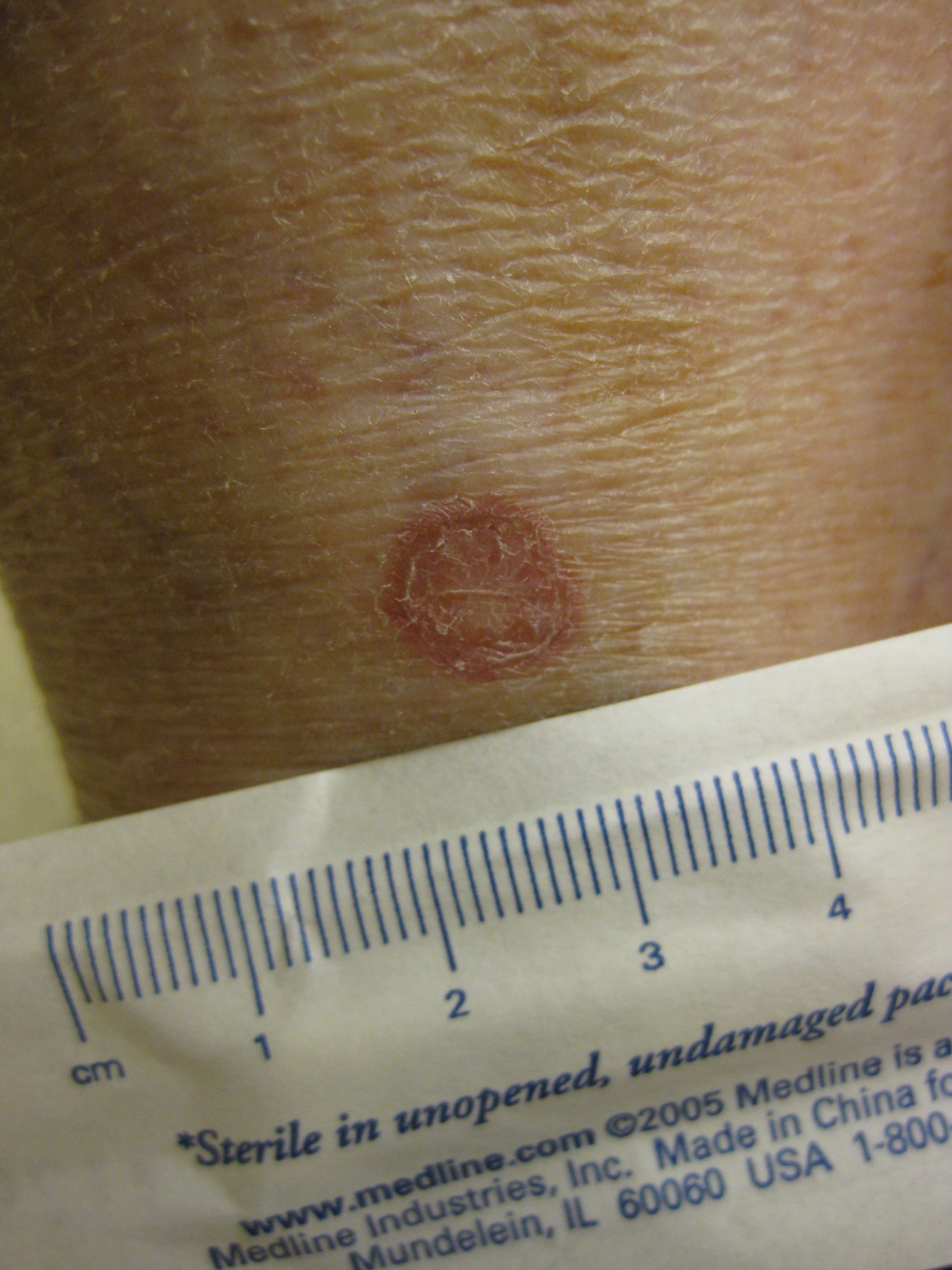
Porokeratosis
Wikipedia
Specialty Pediatrics , dermatology Porokeratosis is a specific disorder of keratinization that is characterized histologically by the presence of a cornoid lamella, a thin column of closely stacked, parakeratotic cells extending through the stratum corneum with a thin or absent granular layer. [1] : 532 Contents 1 Types 2 Genetics 3 Diagnosis 3.1 Pathology 4 Treatment 5 See also 6 References 7 External links Types [ edit ] Porokeratosis may be divided into the following clinical types: [1] : 532 Plaque-type porokeratosis (also known as "Classic porokeratosis" and "Porokeratosis of Mibelli" [2] ) is characterized by skin lesions that start as small, brownish papules that slowly enlarge to form irregular, annular, hyperkeratotic or verrucous plaques. [1] : 533 [3] : 566 Sometimes they may show gross overgrowth and even horn-like structures may develop. [4] Skin malignancy, although rare, is reported from all types of porokeratosis. ... This was the first report mentioning mucosal malignancy in any form of porokeratosis. [4] Disseminated superficial porokeratosis is a more generalized processes and involves mainly the extremities in a bilateral, symmetric fashion. [1] : 533 In about 50% of cases, skin lesions only develop in sun-exposed areas, and this is referred to as disseminated superficial actinic porokeratosis [1] : 533 Porokeratosis palmaris et plantaris disseminata is characterized by skin lesions that are superficial, small, relatively uniform, and demarcated by a distinct peripheral ridge of no more than 1mm in height. [1] : 534 [2] : 1668 [3] : 567 Linear porokeratosis is characterized clinically skin lesions are identical to those of classic porokeratosis, including lichenoid papules, annular lesions, hyperkeratotic plaques with central atrophy, and the characteristic peripheral ridge. [1] [2] : 1668 [3] : 567 Punctate porokeratosis is a skin condition associated with either classic porokeratosis or linear porokeratosis types of porokeratosis, and is characterized by multiple, minute, and discrete punctate, hyperkeratotic, seed-like skin lesions surrounded by a thin, raised margin on the palms and soles. [1] : 535 [2] : 1668 Porokeratosis plantaris discreta is a skin condition that occurs in adults, with a 4:1 female preponderance, characterized by a sharply marginated, rubbery, wide-based papules. [3] : 213 It is also known as "Steinberg's lesion". [5] It was characterized in 1970. [6] Genetics [ edit ] Linear porokeratosis has been associated with mutations in the PMVK and MVD genes. [7] The PMVK gene encodes the enzyme phosphomevalonate kinase and the MVD gene encodes the enzyme diphosphomevalonate decarboxylase . ... ISBN 0-07-138076-0 . ^ a b c d Rapini, Ronald P.; Bolognia, Jean L.; Jorizzo, Joseph L. (2007). Dermatology: 2-Volume Set . St. Louis: Mosby. p. 1668. ... "Porokeratosis plantaris discreta, a previously unrecognized dermatopathological entity". Int. J. Dermatol . 9 (2): 83–90. doi : 10.1111/j.1365-4362.1970.tb04584.x . ... S2CID 40802489 . ^ Atzmony L, Khan HM, Lim YH, Paller AS, Levinsohn JL, Holland KE, Mirza FN, Yin E, Ko CJ, Leventhal JS, Choate KA (2019) Second-Hit, Postzygotic PMVK and MVD Mutations in Linear Porokeratosis.
-
Lymphocele
Wikipedia
Lymphocele Specialty Surgery A lymphocele is a collection of lymphatic fluid within the body not bordered by epithelial lining . [1] It is usually a surgical complication seen after extensive pelvic surgery (such as cancer surgery) and is most commonly found in the retroperitoneal space . Spontaneous development is rare. [2] Contents 1 Signs and symptoms 2 Cause 3 Management 4 References 5 External links Signs and symptoms [ edit ] Many lymphoceles are asymptomatic. ... Smaller lymphoceles can be managed expectantly, and many lesions will regress over time. [2] For symptomatic lesions a number of approaches are available and include fine needle aspiration with US or CT guidance, catheter insertion and drainage (with possible use of sclerosants ), and surgical drainage. [2] [6] Sex and masturbation may cause the lymphocele to grow if it is in the genital area. ... "Clinical and experimental studies on so called lymphocyst which develops after radical hysterectomy in cancer of the uterine cervix". J Jpn Obstet Gynecol Soc . 2 : 178. ^ White M, Mueller PR, Ferrucci JT, et al. (1985). ... Ajr . 145 : 1065–1069. doi : 10.2214/ajr.145.5.1065 . ^ a b Kim JK, Jeong YY, Kim YH, Kim YC, Kang HK, Choi HS (1999).
-
Ornithophobia
Wikipedia
Ornithophobia , the abnormal and irrational fear of birds, is a type of specific phobia . [1] [2] The prefix ornitho- signifies "of or pertaining to birds", from Ancient Greek ὄρνις (órnis, "bird"). ... The Psychiatric Quarterly . 26 (1): 365–371. doi : 10.1007/BF01568473 . PMID 14949213 . ^ Irena Milosevic; Randi E.
-
Afterdepolarization
Wikipedia
Abnormal depolarizations of cardiac myocytes Afterdepolarizations are abnormal depolarizations of cardiac myocytes that interrupt phase 2, phase 3, or phase 4 of the cardiac action potential in the electrical conduction system of the heart . ... Early afterdepolarizations [ edit ] Early afterdepolarizations (EADs) occur with abnormal depolarization during phase 2 or phase 3, and are caused by an increase in the frequency of abortive action potentials before normal repolarization is completed. Phase 2 may be interrupted due to augmented opening of calcium channels , while phase 3 interruptions are due to the opening of sodium channels . ... They are due to elevated cytosolic calcium concentrations, classically seen with digoxin toxicity. [3] [4] The overload of the sarcoplasmic reticulum may cause spontaneous Ca 2+ release after repolarization, causing the released Ca 2+ to exit the cell through the 3Na + /Ca 2+ -exchanger. This results in a net depolarizing current. The classical feature is Bidirectional ventricular tachycardia .
-
Subungual Exostosis
Wikipedia
Subungual exostosis Other names Dupuytren subungual exostosis [1] Subungual exostosis (1/3), in a boy of 15 years old Specialty Orthopedic Subungual exostoses are bony projections which arise from the dorsal surface of the distal phalanx , most commonly of the hallux (the big toe). [2] Contents 1 Presentation 2 Diagnosis 3 Treatment 4 See also 5 References 6 External links Presentation [ edit ] They tend to be painful due to the pressure applied to the nail bed and plate. ... You can help by adding to it . ( September 2017 ) Treatment [ edit ] Surgical excision is common and is a very effective mode of treatment. [ citation needed ] Subungual exostosis (2/3) Subungual exostosis (3/3), after excision See also [ edit ] Sternal cleft List of cutaneous conditions References [ edit ] ^ "Dupuytren subungual exostosis | Genetic and Rare Diseases Information Center (GARD) – an NCATS Program" . rarediseases.info.nih.gov . Retrieved 11 October 2017 . ^ Rapini, Ronald P.; Bolognia, Jean L.; Jorizzo, Joseph L. (2007). Dermatology: 2-Volume Set . St. Louis: Mosby. ISBN 978-1-4160-2999-1 . ^ Suga H, Mukouda M (2005). ... Radiographics 20:1407-1434, 2000 ^ Lee SK, Jung MS, Lee YH, Gong HS, Kim JK, Baek GH (2007).

-
Chymosin Pseudogene
OMIM
In the process of secretion, preprochymosin, comprising 381 amino acids, is processed by the signal peptidase into an inactive 365-amino acid prochymosin. At low pH, prochymosin undergoes autocatalytic cleavage of 42 N-terminal amino acids, yielding active chymosin. ... The sequence showed a 1-bp deletion and a 2-bp deletion in the human sequence corresponding to bovine prochymosin exons 4 and 6, respectively.
-
Clanging
Wikipedia
This is associated with the irregular thinking apparent in psychotic mental illnesses (e.g. mania and schizophrenia ). [1] Gustav Aschaffenburg found that manic individuals generated these "clang-associations" roughly 10–50 times more than non-manic individuals. [2] Aschaffenburg also found that the frequency of these associations increased for all individuals as they became more fatigued. [3] Clanging refers specifically to behavior that is situationally inappropriate. ... "Formal thought disorder in schizophrenia: A factor analytic study". Comprehensive Psychiatry . 33 (2): 105–110. doi : 10.1016/0010-440X(92)90005-B . ... Livingstone. p. 32 . ^ Spitzer, Manfred (1999). The mind within the net: Models of learning, thinking, and acting .
-
Frigophobia
Wikipedia
This disorder has been linked to other psychological disorders such as hypochondriasis and obsessive-compulsive disorder . [ citation needed ] In a 1975 study among ethnic Chinese in Taiwan, it was noted that frigophobia may be culturally linked to koro . [ citation needed ] Where that disorder causes male sufferers to feel that their penis is retracting into the body due to an insufficiency of "male element" (or yang ), male frigophobia sufferers correlate coldness with an over-abundance of "female element" (or yin ). [1] Contents 1 Definition 2 Society and culture 2.1 China 3 See also 4 References 5 Further reading 6 External links Definition [ edit ] Frigophobia is defined as a persistent, abnormal, and unwarranted fear of coldness, despite conscious understanding by the phobic individual and reassurance by others that there is no danger. ... A case study of a 45-year-old Singaporean housewife with frigophobia concluded: frigophobia is closely related to, and strongly influenced by cultural beliefs. [2] Generally speaking, in therapy, treatments would consist of using low dose of anxiolytics and antidepressants , and psychological interventions. ... The Australian and New Zealand Journal of Psychiatry [1998, 32(4):582-585]. doi : 10.3109/00048679809068335 Further reading [ edit ] Chang YH, Rin H, Chen CC Frigophobia: a report of five cases.
-
Adenosine Deaminase 2 Deficiency
MedlinePlus
Adenosine deaminase 2 (ADA2) deficiency is a disorder characterized by abnormal inflammation of various tissues. ... Signs and symptoms that can occur with ADA2 deficiency include fevers that are intermittent, meaning they come and go; areas of net-like, mottled skin discoloration called livedo racemosa; an enlarged liver and spleen (hepatosplenomegaly); and recurrent strokes affecting structures deep in the brain that can start in the first few years of life. ... This gene provides instructions for making an enzyme called adenosine deaminase 2. Studies suggest that this enzyme plays an essential role in the growth and development of certain immune system cells, including macrophages , which are a type of white blood cell that plays a critical role in inflammation. ... Mutations in the ADA2 gene severely reduce or eliminate the activity of adenosine deaminase 2. Researchers do not fully understand how a shortage (deficiency) of this enzyme's activity leads to vasculitis and immune system abnormalities. ... Learn more about the gene associated with Adenosine deaminase 2 deficiency ADA2 Inheritance Pattern This condition is inherited in an autosomal recessive pattern , which means both copies of the gene in each cell have mutations.
-
Dogger Bank Itch
Wikipedia
Dogger Bank itch Specialty Dermatology Dogger Bank itch is a cutaneous condition characterized by a long-lasting dermatitis caused by exposure to the sea chervil , Alcyonidium diaphanum , a bryozoan . [1] The disease, common in fishermen who work in the North Sea , has been recognized by the Danish Workman's Compensation Act since 1939. [2] Contents 1 Pathogenesis 2 Treatment 3 Epidemiology 4 History 5 See also 6 References Pathogenesis [ edit ] The structural formula of the toxin responsible for Dogger Bank itch The rash is caused by a type of cell-mediated hypersensitivity reaction; this type of hypersensitivity normally occurs in people who become sensitized to volatile organic compounds . ... In Dogger Bank itch, sensitivity is acquired after repeated handling of the sea chervils that become entangled in fishing nets. [ citation needed ] The specific toxin responsible for the rash was determined to be the sulfur -bearing salt (2-hydroxyethyl) dimethylsulfoxonium chloride. [3] This salt is also found in some sea sponges and has potent in vitro activity against leukemia cells. [4] Treatment [ edit ] A study of two cases in 2001 suggests that the rash responds to oral ciclosporin . ... The sea chervil, abundant in the area, frequently came up with the fishing nets and had to be thrown back into the water. ... Allergy . 1 : 40–46. doi : 10.1111/j.1398-9995.1948.tb03301.x . ^ Carle JS, Christophersen C (1980). "Dogger Bank itch the allergen is 2-hydroxyethyldimethyl sulfonium ion". ... "Dogger Bank Itch revisited: isolation of (2-hydroxyethyl) dimethylsulfoxonium chloride as a cytotoxic constituent from the marine sponge Theonella aff. mirabilis ".
-
Zahn Infarct
Wikipedia
A Zahn infarct is a pseudo- infarction of the liver , consisting of an area of congestion with parenchymal atrophy but no necrosis , and usually due to obstruction of a branch of the portal vein . [1] [2] Zahn infarcts are unique in that there is collateral congestion of liver sinusoids that do not include areas of anoxia seen in most infarcts. ... Eponym [ edit ] The Zahn infarct is named for Friedrich Wilhelm Zahn . [1] References [ edit ] ^ a b Stegman, JK, ed. (2006), Stedman's Medical Dictionary (28th ed.), Baltimore, MD: Lippincott, Williams, & Wilkins ^ "GDPR page" . ^ Matsumoto T, Kuwabara N, Abe H, Fukuda Y, Suyama M, Fujii D, Kojima K, Futagawa S (1992), "Zahn infarct of the liver resulting from occlusive phlebitis in portal vein radicles", American Journal of Gastroenterology , 87 (3): 365–368, PMID 1539574 Reichelt HG (1985), "Partial Budd-Chiari syndrome with Zahn infarct of the liver in venous transmitted tumor thrombosis of a uterine cancer", Röntgen-Blätter (in German), 38 (11): 345–347, PMID 4081553 v t e Ischaemia and infarction Ischemia Location Brain ischemia Heart Large intestine Small intestine Infarction Types Anemic Hemorrhagic Location Heart Brain Spleen Limb Gangrene This article related to pathology is a stub .


