• FindZebra
  • About
  • Contact
  • Help
  • Login
Advanced
  • Retiform Parapsoriasis Wikipedia
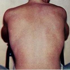
    Retiform parapsoriasis Specialty Dermatology Retiform parapsoriasis is a cutaneous condition, considered to be a type of large-plaque parapsoriasis . [1] It is characterized by widespread, ill-defined plaques on the skin, that have a net-like or zebra-striped pattern. [2] Skin atrophy , a wasting away of the cutaneous tissue , usually occurs within the area of these plaques. [1] See also [ edit ] Parapsoriasis Poikiloderma vasculare atrophicans List of cutaneous conditions References [ edit ] ^ a b Lambert WC, Everett MA (Oct 1981).
    • Poikiloderma Vasculare Atrophicans Wikipedia
      Poikiloderma vasculare atrophicans Other names Parapsoriasis variegata [1] or Parapsoriasis lichenoides [2] Typical skin changes and discoloration described as poikiloderma vasculare atrophicans Specialty Dermatology Poikiloderma vasculare atrophicans ( PVA ), is a cutaneous condition ( skin disease ) characterized by hypo- or hyperpigmentation (diminished or heightened skin pigmentation , respectively), telangiectasia and skin atrophy . [3] [4] [5] Other names for the condition include prereticulotic poikiloderma and atrophic parapsoriasis . [6] The condition was first described by pioneer American pediatrician Abraham Jacobi in 1906. [7] PVA causes areas of affected skin to appear speckled red and inflamed, yellowish and/or brown, gray or grayish-black, with scaling and a thinness that may be described as "cigarette paper". [3] On the surface of the skin, these areas may range in size from small patches, to plaques (larger, raised areas), to neoplasms (spreading, tumor-like growths on the skin). [3] [6] Mycosis fungoides, a type of skin lymphoma , may be a cause of PVA. The condition may also be caused by, associated with or accompany any of the following conditions or disorders: other skin lymphomas, dermatomyositis , lupus erythematosus , Rothmund–Thomson syndrome , Kindler syndrome , dyskeratosis congenita , and chronic radiodermatitis . [4] Rare causes include arsenic ingestion, and the condition can also be idiopathic . [1] [3] [5] PVA may be considered a rare variant of cutaneous T-cell lymphoma , a non-Hodgkin's form of lymphoma affecting the skin. [7] It may also be included among a number of similar conditions that are considered as precursors to mycosis fungoides . PVA is believed to be a syndrome closely associated with large-plaque parapsoriasis and its cohort retiform parapsoriasis ; including PVA, all three conditions fit within an updated view of the once ambiguous classification scheme known as parapsoriasis . [5] Contents 1 Presentation 2 Cause 3 Diagnosis 3.1 Classification 4 Management 5 See also 6 References 7 External links Presentation [ edit ] The layers of the epidermis (left). Melanocytes (rlght), located in the bottom epidermal layer, produce melanin. PVA can be characterized by speckled, combined hyper- and hypopigmentation in the plaques or patches of affected skin. [5] Hyperpigmentation is excess coloration, or darkening of the skin, [8] while hypopigmentation is a diminished or pallid coloring to the skin.
  • Epileptic Encephalopathy, Early Infantile, 45 OMIM
    In vitro functional studies in HEK293 cells showed that the mutation altered the kinetic properties of the channel, resulting in the net loss of GABAergic inhibition. In a boy with EIEE45, Lien et al. (2016) identified a de novo heterozygous missense mutation in the GABRB1 gene (T287I; 137190.0002).
    GABRB1
  • Maple Syrup Urine Disease GeneReviews
    Acute metabolic decompensation is corrected by treating the precipitating stress while delivering sufficient calories, insulin, free amino acids, isoleucine, and valine to achieve sustained net protein synthesis in tissues. Some centers use hemodialysis/hemofiltration to remove BCAAs from the extracellular compartment, but this intervention does not alone establish net protein accretion. ... Thus, leucine tolerance reflects a balance between unmeasured protein losses (e.g., sloughed skin, hair, and nails) and the net accretion of body protein, which in turn is linked to growth rate [Strauss et al 2010]. ... The risk for metabolic crisis in any ill person with MSUD depends on residual in vivo BCKD enzyme activity in relation to the net liberation of free leucine from protein catabolism. ... Following the neonatal period, acute metabolic intoxication (leucinosis) and neurologic deterioration can develop rapidly at any age as a result of net protein degradation precipitated by infection, surgery, injury, or psychological stress (see Figure 1). ... Plasma leucine levels rise predictably as a result of net protein catabolism provoked by a variety of physiologic stresses, including (more...)
    DBT, BCKDHB, BCKDHA, BCAT2, PPM1K, DLD, ARID4B, BDNF, CTSD, SERPINE1, TNS3, CACNA2D2, MKRN3, UMOD, SPN, NME1, PAH, NBN, MEA1, IL1B, GPR4, GLI2, F2, MECP2
    • Maple Syrup Urine Disease Wikipedia
      Metabolic disorder affecting branched-chain amino acids. It is one type of organic acidemia.[2] The condition gets its name from the distinctive sweet odor of affected infants' urine Maple syrup urine disease Other names Branched-chain ketoaciduria Isoleucine (pictured above), leucine , and valine are the branched-chain amino acids that build up in MSUD. Specialty Medical genetics Maple syrup urine disease ( MSUD ) is an autosomal recessive [1] metabolic disorder affecting branched-chain amino acids . It is one type of organic acidemia . [2] The condition gets its name from the distinctive sweet odor of affected infants' urine, particularly prior to diagnosis and during times of acute illness. [3] Contents 1 Classification 2 Signs and symptoms 2.1 Classic MSUD 2.2 Intermediate MSUD 2.3 Intermittent MSUD 2.4 Thiamine-response MSUD 2.5 Later onset 3 Causes 4 Pathophysiology 5 Diagnosis 6 Prevention 7 Treatment 7.1 Monitoring 7.2 Diet control 7.3 Liver transplantation 7.4 Pregnancy 8 Prognosis 9 Epidemiology 10 Research directions 10.1 Gene therapy 10.2 Phenylbutyrate therapy 11 See also 12 References 13 External links Classification [ edit ] Maple syrup urine disease can be classified by its pattern of signs and symptoms, or by its genetic cause. The most common and severe form of this disease is the classic type, which appears soon after birth, and as long as it remains untreated, gives rise to progressive and unremitting symptoms. Variant forms of the disorder may become apparent only later in infancy or childhood, with typically less severe symptoms that may only appear during times of fasting, stress or illness, but still involve mental and physical problems if left untreated.
    • Maple Syrup Urine Disease, Mild Variant OMIM
      A number sign (#) is used with this entry because of evidence that a mild variant of maple syrup urine disease (MSUDMV) is caused by homozygous mutation in the PPM1K gene (611065) on chromosome 4q22. One such family has been reported. Description The mild variant of MSUD is characterized by increased plasma levels of branched-chain amino acids (BCAA) apparent at birth. Treatment with a low-protein diet free of BCAA can result in normal psychomotor development and lack of metabolic episodes; however, plasma levels of BCAA may remain elevated (summary by Oyarzabal et al., 2013). For a general description and a discussion of genetic heterogeneity of maple syrup urine disease, see 248600. Clinical Features Oyarzabal et al. (2013) reported a 21-year-old woman with a mild variant of maple syrup urine disease.
    • Maple Syrup Urine Disease Orphanet
      A rare inherited disorder of branched-chain amino acid metabolism classically characterized by poor feeding, lethargy, vomiting and a maple syrup odor in the cerumen (and later in urine) noted soon after birth, followed by progressive encephalopathy and central respiratory failure if untreated. The four overlapping phenotypic subtypes are: classic, intermediate, intermittent and thiamine-responsive MSUD. Epidemiology The estimated prevalence is around 1/150,000 live births, from published and unpublished newborn screening data. Clinical description Classic MSUD presents in the first days of life with poor feeding and drowsiness followed by a worsening encephalopathy with lethargy, intermittent apnea, stereotypic movements ("fencing" and ''bicycling") and opisthotonus. Coma and central respiratory failure supervene 7 to 10 days after birth.
    • Maple Syrup Urine Disease GARD
      Maple syrup urine disease (MSUD) occurs when the body is unable to breakdown certain parts of proteins. This leads to the build-up of toxic substances that can cause organ and brain damage. There are several forms of MSUD. The most common is the classic or infantile form. Symptoms of the classic form of MSUD start in early infancy and include poor feeding, irritability, extra sleepiness, and muscle spasms. If untreated, respiratory failure (lack of oxygen getting to the blood) may occur.
  • Afterdepolarization Wikipedia
    They are due to elevated cytosolic calcium concentrations, classically seen with digoxin toxicity. [3] [4] The overload of the sarcoplasmic reticulum may cause spontaneous Ca 2+ release after repolarization, causing the released Ca 2+ to exit the cell through the 3Na + /Ca 2+ -exchanger. This results in a net depolarizing current. The classical feature is Bidirectional ventricular tachycardia .
  • Nephrolithiasis, Calcium Oxalate OMIM
    In vitro flux studies indicated that mice lacking Slc26a6 have a defect in intestinal oxalate secretion resulting in enhanced net absorption of oxalate. Jiang et al. (2006) concluded that the anion exchanger, SLC26A6, has a major constitutive role in limiting net intestinal absorption of oxalate, thereby preventing hyperoxaluria and calcium oxalate urolithiasis.
    SLC26A1, F2, FN1, AMBP, AHSG, HOGA1, HAO1, AGXT, SLC36A2, PAQR6, ADCY10, SLC6A20, OPLAH, GRHPR, SLC6A19, CD44, ABCB1, MSH3, CD9, SLC22A12, CDH1, SLC26A6, CHD1, GGCX, IL1B, IL1RN, TBC1D9, MDM4, ABCG2, ABCC1, KL, VDR, SPP1
    • Kidney Stones MedlinePlus
      Kidney stones (also called renal stones or urinary stones) are small, hard deposits that form in one or both kidneys; the stones are made up of minerals or other compounds found in urine. Kidney stones vary in size, shape, and color. To be cleared from the body (or "passed"), the stones need to travel through ducts that carry urine from the kidneys to the bladder (ureters) and be excreted. Depending on their size, kidney stones generally take days to weeks to pass out of the body. Kidney stones can cause abdominal or back pain (known as renal colic). Renal colic usually begins sporadically but then becomes constant and can lead to nausea and vomiting.
  • Steroid Diabetes Wikipedia
    Mechanism [ edit ] Glucocorticoids oppose insulin action and stimulate gluconeogenesis , especially in the liver , resulting in a net increase in hepatic glucose output.
  • Naegeli-Franceschetti-Jadassohn Syndrome/dermatopathia Pigmentosa Reticularis MedlinePlus
    Among the most common signs of NFJS/DPR is a net-like pattern of dark brown or gray skin coloring, known as reticulate hyperpigmentation.
    KRT14, TARDBP, C9orf72
    • Dermatopathia Pigmentosa Reticularis OMIM
      A number sign (#) is used with this entry because of evidence that dermatopathia pigmentosa reticularis (DPR) is caused by heterozygous mutation in the keratin-14 gene (KRT14; 148066) on chromosome 17q21. One such family has been reported. A closely related disorder, Naegeli-Franceschetti-Jadassohn syndrome (NFJS; 161000), is also caused by heterozygous mutation in the KRT14 gene. Description Dermatopathia pigmentosa reticularis is a rare heritable disorder consisting of a triad of cutaneous findings including reticulate hyperpigmentation, noncicatricial alopecia, and onychodystrophy. Variable features include adermatoglyphia, hypohidrosis or hyperhidrosis, and palmoplantar hyperkeratosis (Heimer et al., 1992). Clinical Features Heimer et al. (1992) described a family with 9 cases of dermatopathia pigmentosa reticularis distributed through 6 sibships of 4 generations.
    • Dermatopathia Pigmentosa Reticularis Orphanet
      A rare, genetic, ectodermal dysplasia characterized by a widespread, early-onset, reticulate hyperpigmentation that persists throughout life, mild, diffuse non-cicatricial alopecia, and onychodystrophy. There are no dental anomalies. Patients may also present with adermatoglyphia, palmoplantar hyperkeratosis, acral dorsal blistering, and hypohidrosis or hyperhidrosis.
    • Dermatopathia Pigmentosa Reticularis Wikipedia
      Dermatopathia pigmentosa reticularis Other names Dermatopathic pigmentosa reticularis [1] : 511 Dermatopathia pigmentosa reticularis has an autosomal dominant pattern of inheritance Specialty Medical genetics Dermatopathia pigmentosa reticularis is a rare, autosomal dominant [2] congenital disorder that is a form of ectodermal dysplasia . Dermatopathia pigmentosa reticularis is composed of the triad of generalized reticulate hyperpigmentation , noncicatricial alopecia , and onychodystrophy . [3] : 856 Contents 1 Presentation 2 Cause 3 Treatment 4 See also 5 References 6 External links Presentation [ edit ] Symptoms include lack of sweat glands, thin hair, brittle nails, mottled skin, and lack of fingerprints. [4] DPR is very similar to the related Naegeli-Franceschetti-Jadassohn syndrome . Both cause an affected person to lack fingerprints, have a lace-like pattern of hyperpigmentation and hyperkeratosis of the palms of the hands and soles of the feet. DPR is distinguished from NFJS by the duration of hyperpigmentation and lack of dental abnormalities. [5] Cause [ edit ] DPR is caused by a mutation in the keratin 14 gene. [6] Treatment [ edit ] This section is empty. You can help by adding to it . ( April 2017 ) See also [ edit ] List of cutaneous conditions caused by mutations in keratins References [ edit ] ^ Freedberg, et al. (2003).
  • Yellow Fever Orphanet
    Management and treatment As there is presently no antiviral drug available for YF, treatment is supportive, following the guidelines for treatment of severe septicemia. Insecticide-treated bed nets and/or room screens should be used in open-air settings to prevent further transmission.
    ERVK-32, ERVK-6, DDOST, PARTICL, TRIM56, CYP20A1, EMC3, TRAV12-2, BACE1, AMACR, TRPA1, TFPI, RPL19, RAF1, CRP, MRC1, IL10, IL6, IL1RN, IFNG, IFNAR1, GYPC, GPT, GLS, FCGR3B, FCGR3A, F10, MAP2K1
    • Yellow Fever Wikipedia
      Programmes for distribution of mosquito nets for use in homes produce reductions in cases of both malaria and yellow fever. ... Curtains and lids of water tanks can be sprayed with insecticides, but application inside houses is not recommended by the WHO. Insecticide-treated mosquito nets are effective, just as they are against the Anopheles mosquito that carries malaria. [4] Treatment [ edit ] As with other Flavivirus infections, no cure is known for yellow fever.
  • Dengue Fever Orphanet
    Management and treatment As there is presently no antiviral drug available for DF/DHF, treatment is supportive, following the guidelines for treatment of severe septicemia. Insecticide-treated bed nets, room screens and elimination of larval development sites should be used in open-air settings to prevent further transmission.
    CD209, PTPN11, SENP8, TNF, ERVK-6, IFNG, IVNS1ABP, IL10, ERVK-32, IFNA1, IFNA13, IL1B, PLAAT4, LSAMP, HLA-A, LAMP3, IFIH1, DDX58, HLA-DRB1, ROBO3, ERVW-1, IL6, PDR, ERVK-20, CLEC5A, CXCL10, CXCL8, IL4, CRP, GPT, RAF1, CCL5, MICB, VEGFA, CDK9, KRAS, RSAD2, MBL2, THBD, GP1BA, OAS3, RHD, ALB, FCGR2A, SARS1, PLCE1, HLA-C, FBXW7, SARS2, CCR5, SOAT1, RBM45, TNFSF10, TLR8, OAS1, MPZ, IFITM3, IFNAR2, CSPG5, IGHG3, CCL4L2, KDR, HPSE, EIF2AK3, KIR2DL1, PF4, SLC17A5, SPHK1, IL18, HAVCR1, MYOM2, KIR3DL1, KIR3DS1, TIMELESS, PMP22, IL1RN, ADAMTS13, PADI4, LGALS9, HSPA5, HLA-B, NLRP3, VDR, TAP2, MIR21, IFNL1, DHX9, ATN1, EGFR, CCL4, CCL3, CCL4L1, CCL2, ARHGEF5, FASN, THPO, RNASEL, RNASE1, CD40LG, STING1, TLR3, TAP1, GEM, PTGS2, MAVS, GZMB, TLR4, CFH, NR0B1, MBTPS1, CHST10, S1PR2, TXNRD1, TNFRSF10B, TTN, TSC2, HSP90B1, TPM4, IKBKE, TPI1, NUP153, SH2B3, VCAM1, P2RX6, PIWIL1, AURKB, PTPRU, HSD17B6, SQSTM1, USO1, MKNK1, APOL1, STAM, MGAM, HAP1, MOGS, SOCS3, CNBP, CLDN1, XBP1, VWF, VIM, IL1RL1, DNM1L, SERPINA3, RCL1, TIRAP, PIWIL4, NRSN1, TRIM69, EGFLAM, LRG1, CGAS, IL17F, GORASP1, IL33, HELZ2, MAGT1, DHX40, MAPKAP1, DDX50, RNF168, STT3B, MARCHF8, MIF-AS1, PRSS57, MIRLET7E, MIR146A, MIR378A, MIR484, NCF1, MIR744, KIR2DS2, LINC01672, MICA, P2RX5-TAX1BP3, PERCC1, MTCO2P12, WNK1, SMOC1, NOD1, TMED2, ARC, P2RX2, RAB18, MLXIP, CHP1, TPPP, LILRB1, GOLPH3, CERS1, OLFM4, AGPAT1, PRDX4, PROCR, NXF1, EXOC7, GRIP1, SEC14L2, SMUG1, NAAA, APEX2, MYLIP, NPC1L1, TRAT1, UBE2J1, SIRT6, RETREG1, SPHK2, AICDA, RPTOR, TRPV4, HPSE2, TP53, REL, TM7SF2, ILF2, GLS, GDF1, GC, GATA3, GAS6, GAPDH, GABPA, G6PD, IFI6, FLNB, FCGR3B, FCGR3A, FCGR2B, FCGR1A, F5, CXCR3, HGF, HLA-DQA1, IFNB1, IL6R, IL4R, IL3RA, IL2RA, IL2, IL1A, IFNAR1, HLA-E, ICAM3, HSP90AA1, HSF1, HP, HMGCR, HMGB1, ESR2, ESR1, ERN1, KLK3, CAT, BST2, BID, BCL2, BAK1, ARNTL, APRT, CD14, ANXA5, ANXA2, ANGPT1, AKT1, AHR, ADAR, CD9, CD33, ERBB4, CTLA4, ERBB2, ELAVL1, EIF4E, DDX6, DAXX, CYMD, CSF2, CD38, CLDN7, CP, CCR1, CEACAM8, CEBPB, CD69, IL11, ILF3, TLR2, INSR, PRNP, MAP2K1, PRKAB1, PRKAA2, PRKAA1, PPP2R5E, PLAT, PIK3CG, PIK3CD, PIK3CB, PIK3CA, SLC25A3, PAH, P4HB, P2RY2, RAB6A, ABO, REG1A, STAT3, TLR1, TGFB2, TGFB1, TCF7L2, SYT1, SYK, SSB, RELA, SPP1, SMARCB1, SI, SELE, CXCL11, SCD, P2RY1, P2RX7, P2RX5, KLRC1, MATK, CAPRIN1, LTA, TNPO1, KPNA2, KPNB1, KIR2DS5, CXCL9, KIR2DS1, KIR2DL3, ITGAX, IRF7, IRF1, INSRR, MFAP1, MMP2, P2RX4, NFKB1, P2RX3, P2RX1, OAS2, CNOT2, NOS3, NFYA, NFE2L2, MMP9, NCL, MYD88, COX2, COX1, MPL, MNT, H3P19
    • Dengue Fever Wikipedia
      The primary method of controlling A. aegypti is by eliminating its habitats . [35] This is done by getting rid of open sources of water, or if this is not possible, by adding insecticides or biological control agents to these areas. [35] Generalized spraying with organophosphate or pyrethroid insecticides, while sometimes done, is not thought to be effective. [22] Reducing open collections of water through environmental modification is the preferred method of control, given the concerns of negative health effects from insecticides and greater logistical difficulties with control agents. [35] People can prevent mosquito bites by wearing clothing that fully covers the skin, using mosquito netting while resting, and/or the application of insect repellent ( DEET being the most effective). [39] While these measures can be an effective means of reducing an individual's risk of exposure, they do little in terms of mitigating the frequency of outbreaks, which appear to be on the rise in some areas, probably due to urbanization increasing the habitat of A. aegypti . [7] The range of the disease also appears to be expanding possibly due to climate change. [7] Vaccine [ edit ] In 2016 a partially effective vaccine for dengue fever became commercially available in the Philippines and Indonesia. [4] [57] It has been approved for use by Mexico, Brazil, El Salvador, Costa Rica, Singapore, Paraguay, much of Europe, and the United States. [9] [57] [58] The vaccine is only recommended in individuals who have had a prior dengue infection or in populations where most (>80%) of people have been infected by age 9. [10] [59] In those who have not had a prior infection there is evidence it may worsen subsequent infections. [9] [10] [5] For this reason Prescrire does not see it as suitable for wide scale immunization, even in areas where the disease is common. [60] The vaccine is produced by Sanofi and goes by the brand name Dengvaxia . [61] It is based on a weakened combination of the yellow fever virus and each of the four dengue serotypes. [36] [62] Studies of the vaccine found it was 66% effective and prevented more than 80 to 90% of severe cases. [59] This is less than wished for by some. [63] In Indonesia it costs about US$207 for the recommended three doses. [57] Given the limitations of the current vaccine, research on vaccines continues, and the fifth serotype may be factored in. [7] One of the concerns is that a vaccine could increase the risk of severe disease through antibody-dependent enhancement (ADE). [64] The ideal vaccine is safe, effective after one or two injections, covers all serotypes, does not contribute to ADE, is easily transported and stored, and is both affordable and cost-effective. [64] Anti-dengue day [ edit ] A poster in Tampines , Singapore , notifying people that there are ten or more cases of dengue in the neighbourhood (November 2015).
    • Dengue Fever Mayo Clinic
      Permethrin can be applied to your clothing, shoes, camping gear and bed netting. You can also buy clothing made with permethrin already in it.
  • Antiphospholipid Syndrome Mayo Clinic
    Some people develop a red rash with a lacy, net-like pattern. Less common signs and symptoms include: Neurological symptoms. ... Do you have frequent headaches? Have you noticed a red, net-like rash on your wrists or knees?
    APOH, PPARG, FRMD4A, TSHR, F5, F3, SYCP2L, F2, PTPRO, GPI, ANXA5, TLR4, SH2B2, KLK3, ANXA2, TNF, AGER, CPB2, MTOR, MTHFR, F10, PLG, HT, TFPI, PLAT, SELPLG, ACR, SERPINE1, MOK, LRP8, VWF, HMGB1, VIM, SELP, RAB4A, CCL2, CXCL12, ATXN2, RO60, S100A10, TRIM21, ABCA1, SSB, THBD, TNFRSF1B, NR1I2, ADIPOQ, SH2B3, PROCR, ADAMTS13, TREX1, PTPN22, FOXP3, SLC52A1, IL21, ANXA8, ANXA8L1, PROS1, NOS3, PON1, PLSCR1, HLA-DPB1, GP1BA, GCY, FGA, FCGR2A, F2RL1, EMD, EDN1, DECR1, CRP, CD36, CD1D, CALR, B2M, SERPINC1, AQP4, AMH, HLA-DRB1, HRES1, IDS, MBL2, PF4, PC, SERPINB2, TNFRSF11B, MYD88, MSN, MPL, LPA, IFNG, LGALS9, LCT, CXCL10, CXCL8, IL1B, IGFBP1, IGF1, C20orf181
    • Antiphospholipid Syndrome Wikipedia
      Antiphospholipid syndrome Other names Hughes syndrome [1] Micrograph showing an advanced thrombotic microangiopathy , as may be seen in APLA syndrome. Kidney biopsy . PAS stain . Specialty Hematology , Rheumatology Antiphospholipid syndrome , or antiphospholipid antibody syndrome ( APS or APLS ), is an autoimmune , hypercoagulable state caused by antiphospholipid antibodies . APS provokes blood clots ( thrombosis ) in both arteries and veins as well as pregnancy-related complications such as miscarriage , stillbirth , preterm delivery , and severe preeclampsia . The diagnostic criteria require one clinical event (i.e. thrombosis or pregnancy complication) and two positive blood test results spaced at least three months apart that detect lupus anticoagulant , anti-apolipoprotein antibodies , or anti-cardiolipin antibodies . [2] Antiphospholipid syndrome can be primary or secondary. Primary antiphospholipid syndrome occurs in the absence of any other related disease.
  • Gerontophobia Wikipedia
    Part of a series on Discrimination General forms Age Class ( Caste ) Physical Disability Education Economic Employment Genetics Hair texture Height Housing Language Looks Race / Ethnicity / Nationality Rank Religion Sanity Sex Sexual orientation Size Skin color Specific forms Social Acephobia Adultism Amatonormativity Anti-albinism Anti-autism Anti-homelessness Anti-intellectualism Anti-intersex Anti-left handedness Anti-Masonry Antisemitism (Judeophobia) Aporophobia Audism Biphobia Clannism Cronyism Drug use Elitism Ephebiphobia Fatism Gerontophobia Heteronormativity Heterosexism HIV/AIDS stigma Homophobia Leprosy stigma Lesbophobia Misandry Misogyny Nepotism Pedophobia Perpetual foreigner Pregnancy Reverse Sectarianism Supremacism Black White Transphobia Non-binary Transmisogyny Vegaphobia Xenophobia Religious Ahmadiyya Atheism Baháʼí Faith Buddhism Catholicism Christianity post–Cold War era Druze Falun Gong Hinduism Persecution Islam Persecution Jehovah's Witnesses Judaism Persecution LDS or Mormon Neopaganism Eastern Orthodox Oriental Orthodox Copts Protestantism Rastafarianism Shi'ism Sufism Sunnism Zoroastrianism Ethnic/national African Albanian American Arab Armenian Australian Austrian Azerbaijani British Canadian Catalan Chechen Chilean Chinese Croat Dutch English Estonian European Filipino Finnish French Georgian German Greek Haitian Hazara Hispanic Hungarian Igbo Indian Indonesian Iranian Irish Israeli Italian Japanese Jewish Khmer Korean Kurdish Malay Manchu Mexican Middle Eastern Mongolian Montenegrin Pakistani Pashtun Polish Portuguese Quebec Romani Romanian Russian Scottish Serb Slavic Somali Soviet Tatar Thai Tibetan Turkish Ukrainian Venezuelan Vietnamese Western Manifestations Blood libel Bullying Compulsory sterilization Counter-jihad Cultural genocide Defamation Democide Disability hate crime Dog-whistle politics Eliminationism Ethnic cleansing Ethnic conflict Ethnic hatred Ethnic joke Ethnocide Forced conversion Freak show Gay bashing Gendercide Genital modification and mutilation Genocide examples Glass ceiling Hate crime Hate group Hate speech online Homeless dumping Indian rolling Lavender scare LGBT hate crimes Lynching Mortgage Murder music Occupational segregation Persecution Pogrom Purge Red Scare Religious persecution Religious terrorism Religious violence Religious war Scapegoating Segregation academy Sex-selective abortion Slavery Slut-shaming Trans bashing Victimisation Violence against women White flight White power music Wife selling Witch-hunt Policies Age of candidacy Blood purity Blood quantum Crime of apartheid Disabilities Catholic Jewish Ethnocracy Ethnopluralism Gender pay gap Gender roles Gerontocracy Gerrymandering Ghetto benches Internment Jewish quota Jim Crow laws Law for Protection of the Nation McCarthyism MSM blood donation restrictions Nonpersons Numerus clausus (as religious or racial quota) Nuremberg Laws One-drop rule Racial quota Racial steering Redlining Same-sex marriage (laws and issues prohibiting) Segregation age racial religious sexual Sodomy law State atheism State religion Ugly law Voter suppression Countermeasures Affirmative action Anti-discrimination law Cultural assimilation Cultural pluralism Diversity training Empowerment Feminism Fighting Discrimination Hate speech laws by country Human rights Intersex rights LGBT rights Masculism Multiculturalism Nonviolence Racial integration Reappropriation Self-determination Social integration Toleration Related topics Allophilia Anti-cultural, anti-national, and anti-ethnic terms Bias Christian privilege Civil liberties Cultural assimilation Dehumanization Diversity Ethnic penalty Eugenics Internalized oppression Intersectionality Male privilege Masculism Medical model of disability autism Multiculturalism Net bias Neurodiversity Oikophobia Oppression Police brutality Political correctness Polyculturalism Power distance Prejudice Prisoner abuse Racial bias in criminal news Racism by country Religious intolerance Second-generation gender bias Snobbery Social exclusion Social model of disability Social stigma Stereotype threat The talk White privilege v t e Gerontophobia is the fear of age-related self-degeneration (similar to Gerascophobia ), or a hatred or fear of the elderly due to memento mori . ... External links [ edit ] AGEISM AND AGING UP: A Q and A with Mariah MedFriendly Age Wave v t e Discrimination General forms Age Caste Class Disability Education Economic Employment Genetic Hair texture Height Housing Language Looks Race / Ethnicity / Nationality Rank Sanity Sex Sexual orientation Size Skin color Social Acephobia Adultism Amatonormativity Anti-albinism Anti-autism Anti-homelessness Anti-intellectualism Anti-intersex Anti-left handedness Anti-Masonry Antisemitism (Judeophobia) Aporophobia Audism Biphobia Clannism Cronyism Drug use Elitism Ephebiphobia Fatism Gerontophobia Heteronormativity Heterosexism HIV/AIDS stigma Homophobia Leprosy stigma Lesbophobia Misandry Misogyny Nepotism Pedophobia Perpetual foreigner Pregnancy Reverse Sectarianism Supremacism Black White Transphobia Non-binary Transmisogyny Vegaphobia Xenophobia Religious Ahmadiyya Atheism Baháʼí Faith Buddhism Catholicism Christianity post–Cold War era Falun Gong Hinduism Persecution Islam Persecution Jehovah's Witnesses Judaism Persecution LDS or Mormon Neopaganism Eastern Orthodox Oriental Orthodox Protestantism Rastafarianism Shi'ism Sufism Zoroastrianism Ethnic/National African Albanian American Arab Armenian Australian Austrian British Canadian Catalan Chilean Chinese Croat Dutch English Estonian European Filipino Finnish French Georgian German Greek Haitian Hazara Hindu Hispanic Hungarian Igbo Indian Indonesian Iranian Irish Israeli Italian Japanese Jewish Khmer Korean Kurdish Malay Manchu Mexican Middle Eastern Mongolian Pakistani Pashtun Polish Portuguese Quebec Romani Romanian Russian Scottish Serb Slavic Somali Soviet Tatar Thai Turkish Ukrainian Venezuelan Vietnamese Western Manifestations Blood libel Bullying Compulsory sterilization Counter-jihad Cultural genocide Defamation Democide Disability hate crime Dog-whistle politics Eliminationism Enemy of the people Ethnic cleansing Ethnic conflict Ethnic hatred Ethnic joke Ethnocide Forced conversion Freak show Gay bashing Gendercide Genital modification and mutilation Genocide examples Glass ceiling Hate crime Hate group Hate speech Homeless dumping Indian rolling Lavender scare LGBT hate crimes Lynching Mortgage Murder music Native American sports mascots Occupational segregation Persecution Pogrom Purge Red Scare Religious persecution Religious terrorism Religious violence Religious war Scapegoating Segregation academy Sex-selective abortion Slavery Slut-shaming Trans bashing Victimisation Violence against women White flight White power music Wife selling Witch-hunt Discriminatory policies Age of candidacy Blood purity Blood quantum Crime of apartheid Disabilities Catholic Jewish Ethnocracy Ethnopluralism Gender pay gap Gender roles Gerontocracy Gerrymandering Ghetto benches Internment Jewish quota Jim Crow laws Law for Protection of the Nation McCarthyism MSM blood donation restrictions Nonpersons Numerus clausus (as religious or racial quota) Nuremberg Laws One-drop rule Racial quota Racial steering Redlining Same-sex marriage (laws and issues prohibiting) Segregation age racial religious sexual Sodomy law State atheism State religion Ugly law Voter suppression Countermeasures Affirmative action Anti-discrimination law Cultural assimilation Cultural pluralism Diversity training Empowerment Feminism Fighting Discrimination Hate speech laws by country Human rights Intersex rights LGBT rights Masculism Multiculturalism Nonviolence Racial integration Reappropriation Self-determination Social integration Toleration Related topics Allophilia Anti-cultural, anti-national, and anti-ethnic terms Bias Christian privilege Civil liberties Cultural assimilation Dehumanization Diversity Ethnic penalty Eugenics Internalized oppression Intersectionality Male privilege Masculism Medical model of disability autism Multiculturalism Net bias Neurodiversity Oikophobia Oppression Police brutality Political correctness Polyculturalism Power distance Prejudice Prisoner abuse Racial bias in criminal news Racism by country Religious intolerance Second-generation gender bias Snobbery Social exclusion Social model of disability Social stigma Stereotype threat The talk White privilege Category
  • Rahman Syndrome OMIM
    The truncated proteins were predicted to have a reduced net charge compared to the wildtype protein, rendering them likely to be less effective in neutralizing negatively charged linker DNA.
    H1-4
    • Hist1h1e Syndrome GeneReviews
      Summary Clinical characteristics. The name HIST1H1E syndrome has been proposed as a mnemonic for the characteristic features of this emerging, recognizable phenotype: h ypotonia; i ntellectual disability with behavioral issues; s keletal; t estes (undescended) and t hyroid; h eart anomalies (most commonly atrial septal defect); and e ctodermal issues (including sparse hair, thin nails, and abnormal dentition). In the 47 affected individuals reported to date, predominant findings were intellectual disability (ranging from mild to profound) and behavioral problems (combinations of anxiety/phobias, obsessive behaviors, attention-deficit/hyperactivity disorder, and autistic spectrum disorder/traits among others). Skeletal involvement can include scoliosis and decreased bone mineral density. Other findings in some include seizures, craniosynostosis, and hearing loss. Life expectancy does not appear to be reduced in HIST1H1E syndrome. Diagnosis/testing.
    • Rahman Syndrome GARD
      Rahman syndrome is a genetic syndrome that includes mild to severe intellectual disability and an increase in height, weight, or head size (overgrowth). The overgrowth is more apparent in infancy and may lessen with time. Other symptoms may include curved fingers, eyes that may not line up in the same direction (strabismus), and facial features such as full cheeks and an increase in the distance between the eyes. The syndrome is caused by changes (mutations) in the HIST1H1E gene. The protein made from the HISTH1E gene helps control which genetic information is turned on (expressed) at any given time. Only one copy of the HIST1H1E gene needs to have a disease-causing genetic change to have Rahman syndrome, which is consistent with an autosomal dominant condition. However, most of the reported cases of Rahman syndrome have not been inherited from the parents, but have been caused by a genetic change that happens by mistake during the making of the egg or sperm (de novo).
  • Felty Syndrome Orphanet
    Neutrophil extracellular chromatin traps (NETs) containing deiminated histones, in complex with bacterial adjuvants, are the most likely antigenic trigger for the production of autoantibodies to deiminated histones.
    HLA-DRB1, RBM45, FAS, EEF1A1, HLA-A, HLA-C, HLA-DPB1, IL10, STAT3, TAP2, TRBV20OR9-2, TNF, B3GAT1
    • Felty's Syndrome GARD
      Felty's syndrome is a rare, potentially serious disorder that is defined by the presence of three conditions: rheumatoid arthritis (RA), an enlarged spleen ( splenomegaly ) and a decreased white blood cell count ( neutropenia ), which causes repeated infections. Although some individuals with Felty's syndrome are asymptomatic, others can develop serious and life-threatening infections. Symptoms of Felty's syndrome, in addition to those associated with the three conditions stated above, may include fatigue, fever, weight loss, discoloration of patches of skin, mild hepatomegaly (enlarged liver), lymphadenopathy (swelling of lymph nodes), Sjögren syndrome , vasculitis , lower-extremity ulcers, and other findings. The exact cause is unknown, but several risk factors have been proposed, including autoimmunity . A few familial cases of the condition have been reported. Treatment typically focuses on controlling the underlying RA; immunosuppressive therapy for RA may improve neutropenia and splenomegaly.
    • Felty's Syndrome Wikipedia
      Autoimmune disease Felty's syndrome Specialty Rheumatology Felty's syndrome , also called Felty syndrome , ( FS ) [1] is rare autoimmune disease characterized by the triad of rheumatoid arthritis , enlargement of the spleen and too few neutrophils in the blood . The condition is more common in those aged 50–70 years, specifically more prevalent in females than males, and more so in Caucasians than those of African descent. It is a deforming disease that causes many complications for the individual. [2] [3] Contents 1 Signs and symptoms 1.1 Complications 2 Causes 3 Mechanism 3.1 Rheumatoid Arthritis 3.2 Neutropenia 3.3 Splenomegaly 4 Diagnosis 4.1 Conditions of the Blood 4.2 Splenomegaly 4.3 Rheumatoid Arthritis 5 Treatment 6 Prognosis 7 History 8 See also 9 References 10 External links Signs and symptoms [ edit ] The symptoms of Felty's syndrome are similar to those of rheumatoid arthritis. Patients suffer from painful, stiff, and swollen joints, most commonly in the joints of the hands, feet, and arms. In some affected individuals, Felty's syndrome may develop during a period when the symptoms and physical findings associated with rheumatoid arthritis have subsided or are not present; in this case, Felty's syndrome may remain undiagnosed.
    • Felty Syndrome OMIM
      Blendis et al. (1976) described a mother and her son and daughter with Felty syndrome (rheumatoid arthritis, splenomegaly and neutropenia). Another sib had rheumatoid arthritis alone. Felty syndrome seems to be confined mainly to white males (Termini et al., 1979; Lewis, 1980). The low frequency in blacks may be related to the low frequency of HLA-DRw4, which shows association with Felty syndrome. Ditzel et al. (2000) identified eukaryotic elongation factor 1A-1 (EEF1A1; 130590) as an autoantibody in 66% of patients with Felty syndrome. Joints - Rheumatoid arthritis Inheritance - Autosomal dominant Misc - Confined mainly to white males - HLA-DRw4 association GI - Splenomegaly Heme - Neutropenia ▲ Close
  • Retinal Dystrophy, Reticular Pigmentary, Of Posterior Pole OMIM
    Variation in the presence of autofluorescent chromophores was observed, with the older sister and the unrelated boy exhibiting a milder and more punctiform hyperautofluorescence of the net, whereas the younger sister showed a more intense hyperautofluorescent pattern.
    RCBTB1
    • Reticular Dystrophy Of The Retinal Pigment Epithelium Orphanet
      A rare, patterned dystrophy of the retinal pigment epithelium, of progressive course, characterized by the presence of a bilateral hyperpigmented reticular pattern resembling a fishnet with knots, resulting in a slowly progressive loss of vision that often only becomes apparent in old age. This disorder is sometimes associated with scleral staphyloma, choroidal neovascularization, convergent strabismus, spherophakia with myopia and luxated lenses, and partial atrophy of the iris.
    • Retinal Dystrophy With Or Without Extraocular Anomalies OMIM
      A number sign (#) is used with this entry because of evidence that retinal dystrophy with or without extraocular anomalies (RDEOA) is caused by homozygous mutation in the RCBTB1 gene (607867) on chromosome 13q14. Clinical Features Coppieters et al. (2016) reported 6 families of varying ethnic backgrounds segregating autosomal recessive retinal dystrophy, in which affected individuals were homozygous for mutations in the RCBTB1 gene. In a consanguineous family of Turkish origin, 2 sisters and a maternal cousin exhibited severe retinal dystrophy with onset in the second decade of life that was deemed 'compatible with' retinitis pigmentosa (RP; see 268000). In addition, all 3 affected individuals had goiter, secondary amenorrhea, and mild intellectual disability, and the sisters' mother had goiter. Hormone analysis in the affected individuals was consistent with primary ovarian insufficiency.
    • Reticular Dystrophy Of Retinal Pigment Epithelium OMIM
      Description Reticular dystrophy is a disorder of protean manifestations occurring in the retinal pigment epithelium (RPE) with little or no involvement of the neurosensory retina. The disorder may be detected at an early age and may be slowly progressive, but the prognosis for visual acuity is good. Abnormalities of dark adaptation and nyctalopia may develop with time. Electrophysiologic testing may show a normal electroretinogram (ERG), subnormal electrooculogram (EOG), and subnormal results of dark adaptation studies (summary by Kingham et al., 1978). Clinical Features Hsieh et al. (1977) reported 2 sibs and their mother with patterned dystrophies of the RPE: the 21-year-old sister exhibited butterfly-shaped pigment dystrophy, her 23-year-old brother showed macroreticular dystrophy, and their 51-year-old mother had 'probable' reticular dystrophy.
  • Body Inflation Wikipedia
    ISBN 1-890451-03-7 . ^ More proof that the Net's a weird place: A new book assembles strange Web sites. , The Globe and Mail , 27 April 2000, page R7 ^ The Juicy, Round World of Blueberry Porn ^ a b Allerhand, Rhalou (2008).
  • Lichen Spinulosus Wikipedia
    PMID 2179296 . External links [ edit ] Derm Net NZ Emedicine Thehinhso v t e Diseases of the skin and appendages by morphology Growths Epidermal Wart Callus Seborrheic keratosis Acrochordon Molluscum contagiosum Actinic keratosis Squamous-cell carcinoma Basal-cell carcinoma Merkel-cell carcinoma Nevus sebaceous Trichoepithelioma Pigmented Freckles Lentigo Melasma Nevus Melanoma Dermal and subcutaneous Epidermal inclusion cyst Hemangioma Dermatofibroma (benign fibrous histiocytoma) Keloid Lipoma Neurofibroma Xanthoma Kaposi's sarcoma Infantile digital fibromatosis Granular cell tumor Leiomyoma Lymphangioma circumscriptum Myxoid cyst Rashes With epidermal involvement Eczematous Contact dermatitis Atopic dermatitis Seborrheic dermatitis Stasis dermatitis Lichen simplex chronicus Darier's disease Glucagonoma syndrome Langerhans cell histiocytosis Lichen sclerosus Pemphigus foliaceus Wiskott–Aldrich syndrome Zinc deficiency Scaling Psoriasis Tinea ( Corporis Cruris Pedis Manuum Faciei ) Pityriasis rosea Secondary syphilis Mycosis fungoides Systemic lupus erythematosus Pityriasis rubra pilaris Parapsoriasis Ichthyosis Blistering Herpes simplex Herpes zoster Varicella Bullous impetigo Acute contact dermatitis Pemphigus vulgaris Bullous pemphigoid Dermatitis herpetiformis Porphyria cutanea tarda Epidermolysis bullosa simplex Papular Scabies Insect bite reactions Lichen planus Miliaria Keratosis pilaris Lichen spinulosus Transient acantholytic dermatosis Lichen nitidus Pityriasis lichenoides et varioliformis acuta Pustular Acne vulgaris Acne rosacea Folliculitis Impetigo Candidiasis Gonococcemia Dermatophyte Coccidioidomycosis Subcorneal pustular dermatosis Hypopigmented Tinea versicolor Vitiligo Pityriasis alba Postinflammatory hyperpigmentation Tuberous sclerosis Idiopathic guttate hypomelanosis Leprosy Hypopigmented mycosis fungoides Without epidermal involvement Red Blanchable Erythema Generalized Drug eruptions Viral exanthems Toxic erythema Systemic lupus erythematosus Localized Cellulitis Abscess Boil Erythema nodosum Carcinoid syndrome Fixed drug eruption Specialized Urticaria Erythema ( Multiforme Migrans Gyratum repens Annulare centrifugum Ab igne ) Nonblanchable Purpura Macular Thrombocytopenic purpura Actinic/solar purpura Papular Disseminated intravascular coagulation Vasculitis Indurated Scleroderma / morphea Granuloma annulare Lichen sclerosis et atrophicus Necrobiosis lipoidica Miscellaneous disorders Ulcers Hair Telogen effluvium Androgenic alopecia Alopecia areata Systemic lupus erythematosus Tinea capitis Loose anagen syndrome Lichen planopilaris Folliculitis decalvans Acne keloidalis nuchae Nail Onychomycosis Psoriasis Paronychia Ingrown nail Mucous membrane Aphthous stomatitis Oral candidiasis Lichen planus Leukoplakia Pemphigus vulgaris Mucous membrane pemphigoid Cicatricial pemphigoid Herpesvirus Coxsackievirus Syphilis Systemic histoplasmosis Squamous-cell carcinoma This condition of the skin appendages article is a stub .
  • Diabetic Angiopathy Wikipedia
    Pathophysiology [ edit ] As insulin is required for glucose uptake, hyperglycemia in diabetes mellitus does not result in a net increase in intracellular glucose in most cells.
    NOS3, AGER, HP, THBS1, SERPINF1, MTHFR, ADCY3, HMOX1, CREM, CASP3, ASS1, FASLG, ADCY8, THBS2, ALB, RELA, VEGFA, FN1, PTK2B, NOS2, CCL2, SPARC, ICAM1, BDKRB1, MOK, EDN1, MPO, ADM, RENBP, AKR1B1, PPARG, PON1, SERPINE1, SMPD1, DPP4, SOD1, TFRC, STIP1, TLR2, ADAMTS13, MIR185, TGFBI, CIP2A, JTB, MIR200C, TCF7L2, SERP1, MIR216B, NOX4, SOD2, MIR146A, TNF, RNF10, MIR130A, TNFRSF1A, TRIB3, MIR126, NOP53, TRAF6, HPSE, VDR, CCDC8, FZD5, ARHGEF5, AOC3, SQSTM1, KLF4, OR10A4, PON2, SGK1, IAPP, GLP1R, GCG, FGF2, EGFR, E2F1, DUSP6, ACE, CMA1, CAT, CANX, ATF3, APOE, AKT1, AHR, AGT, HMGB1, IFIT3, SELL, IGF1, REN, PTEN, MAPK7, MAPK3, PRKCB, PPIA, PLG, TNFRSF11B, MSX2, LGALS3, KLK1, KDR, ITGAM, IRAK1, IGFBP7, H3P7
  • Megalencephaly-Capillary Malformation Syndrome MedlinePlus
    In other people with MCAP, the malformations appear as patches spread over the body or as a reddish net-like pattern on the skin (cutis marmorata).
    PIK3CA, PIK3R2, AKT3, AKT1, MMUT, MTOR, NF1, PIK3CB, PIK3CD, PIK3CG, CCR5, PTPN11, RASA1, CDC45, SYNGAP1, BRD4, CD274, MCM10
    • Megalencephaly-Capillary Malformation-Polymicrogyria Syndrome OMIM
      A number sign (#) is used with this entry because some cases of megalencephaly-capillary malformation-polymicrogyria syndrome (MCAP) have been found to have somatic mutations in the PIK3CA gene (171834) on chromosome 3q26. Description Megalencephaly-capillary malformation-polymicrogyria syndrome (MCAP) is characterized by a spectrum of anomalies including primary megalencephaly, prenatal overgrowth, brain and body asymmetry, cutaneous vascular malformations, digital anomalies consisting of syndactyly with or without postaxial polydactyly, connective tissue dysplasia involving the skin, subcutaneous tissue, and joints, and cortical brain malformations, most distinctively polymicrogyria (summary by Mirzaa et al., 2012). This disorder is also known as the macrocephaly-capillary malformation (MCM) syndrome (Conway et al., 2007). Mirzaa et al. (2012) suggested use of the term MCAP rather than MCM to reflect the very large brain size, rather than simply large head size, that characterizes this syndrome, and the importance and high frequency of perisylvian polymicrogyria. Clinical Features Moore et al. (1997) described 13 unrelated children with abnormalities of somatic growth, face, brain, and connective tissue including vasculature.
    • Megalencephaly-Capillary Malformation-Polymicrogyria Syndrome Orphanet
      A rare developmental defect during embryogenesis that is characterized by growth dysregulation with overgrowth of the brain and multiple somatic tissues, with capillary skin malformations, megalencephaly (MEG) or hemimegalencephaly (HMEG), cortical brain abnormalities (in particular polymicrogyria), typical facial dysmorphisms, abnormalities of somatic growth with asymmetry of the body and brain, developmental delay and digital anomalies. Epidemiology Over 200 patients have been reported without sex predominance. Clinical description Symptoms are usually recognizable at birth. Their severity varies widely among patients. Megalencephaly is a major clinical feature (MEG: occipitofrontal circumference [OFC] greater than or equal to 3 SD above the mean), which sometimes progresses to hydrocephaly, malformations of cortical development with polymicrogyria and Chiari malformation. Cutaneous capillary anomalies are often scattered over the limbs, palms, soles and trunk, are frequently pink/red and are aggravated by crying and emotions.
    • Macrocephaly-Capillary Malformation Wikipedia
      Macrocephaly-capillary malformation Other names Macrocephaly-cutis marmorata telangiectatica congenita syndrome, Megalencephaly-cutis marmorata telangiectatica congenita syndrome A newborn child with M-CM syndrome. A port-wine stain is visible under the nose. On the right side of a cheek, capillary malformations are present. Macrocephaly-capillary malformation ( M-CM ) is a multiple malformation syndrome causing abnormal body and head overgrowth and cutaneous , vascular , neurologic , and limb abnormalities. Though not every patient has all features, commonly found signs include macrocephaly , congenital macrosomia , extensive cutaneous capillary malformation ( naevus flammeus or port-wine stain type birthmark over much of the body; a capillary malformation of the upper lip or philtrum is seen in many patients with this condition), body asymmetry (also called hemihyperplasia or hemihypertrophy ), polydactyly or syndactyly of the hands and feet, lax joints, doughy skin, variable developmental delay and other neurologic problems such as seizures and low muscle tone . Contents 1 Genetics 2 Diagnosis 3 Treatment 4 Prognosis 5 History 6 References 7 External links Genetics [ edit ] Mosaic mutations in PIK3CA have been found to be the genetic cause of M-CM. [1] Other overgrowth conditions with distinct phenotypes have also been found to be caused by mosaic mutations in PIK3CA.
  • Distal Renal Tubular Acidosis Orphanet
    Distal renal tubular acidosis (dRTA) is a disorder of impaired net acid secretion by the distal tubule characterized by hyperchloremic metabolic acidosis.
    SLC4A1, ATP6V0A4, ATP6V1B1, WDR72, SLC4A4, CA2, GATA3, OSGEP, EPHA3, NCOA7, MRGPRF, SLC26A7, VAX2, RBFOX2, ALB, SLC26A4, AMH, GYPB, GYPA, FOXI1, CANX, ATP6AP1, GYPE
    • Distal Renal Tubular Acidosis Wikipedia
      Distal renal tubular acidosis Other names Type 1 renal tubular acidosis Radiograph of a rickets sufferer, a complication of both distal and proximal RTA. Distal renal tubular acidosis ( dRTA ) is the classical form of RTA, being the first described. Distal RTA is characterized by a failure of acid secretion by the alpha intercalated cells of the cortical collecting duct of the distal nephron . This failure of acid secretion may be due to a number of causes, and it leads to an inability to acidify the urine to a pH of less than 5.3. Contents 1 Symptoms 2 Causes 3 Diagnosis 4 Treatment 5 See also 6 References 7 External links Symptoms [ edit ] Because renal excretion is the primary means of eliminating acid from the body, there is consequently a tendency towards acidemia .
    • Autosomal Dominant Distal Renal Tubular Acidosis Orphanet
      A rare inherited form of distal renal tubular acidosis (dRTA) characterized by hyperchloremic metabolic acidosis often but not always associated with hypokalemia. Epidemiology The prevalence is unknown. Clinical description Disease onset occurs in adolescence or adulthood and initial manifestations can include polyuria, polydipsia, muscle weakness and fatigue. Osteomalacia or osteopenia can occur due to calcium salt loss from the bones. Hypercalciuria, nephrolithiasis and nephrocalcinosis may result from long term chronic metabolic acidosis. Renal failure has not been described. Etiology AD dRTA is due to mutations in the SLC4A1 gene (17q21.31) encoding the band 3 anion transport protein (AE1).
    • Slc4a1-Associated Distal Renal Tubular Acidosis MedlinePlus
      SLC4A1 -associated distal renal tubular acidosis is a kidney (renal) disorder that sometimes includes blood cell abnormalities. The kidneys normally filter fluid and waste products from the body and remove them in urine; however, in people with distal renal tubular acidosis, the kidneys are unable to remove enough acid from the body, and the blood becomes too acidic. This chemical imbalance is called metabolic acidosis. The inability to remove acids from the body often results in slowed growth and may also lead to softening and weakening of the bones, called rickets in children and osteomalacia in adults. This bone disorder is characterized by bone pain, bowed legs, and difficulty walking. In addition, most children and adults with SLC4A1 -associated distal renal tubular acidosis have excess calcium in the urine (hypercalciuria), calcium deposits in the kidneys (nephrocalcinosis), and kidney stones (nephrolithiasis).
    • Renal Tubular Acidosis, Distal, Autosomal Dominant OMIM
      A number sign (#) is used with this entry because autosomal dominant distal renal tubular acidosis is caused by heterozygous mutation in the SLC4A1 gene (109270) on chromosome 17q21. Clinical Features Randall and Targgart (1961) observed renal tubular acidosis in members of several successive generations. All affected members showed both acidosis and nephrocalcinosis. Randall (1967) provided follow-up of this family. The pedigree included 4 instances of male-to-male transmission. The features were nephrocalcinosis, fixed urinary specific gravity, fixed urinary pH of about 5.0, high serum chloride, low serum bicarbonate, osteomalacia, and hypocalcemia. Alkalinization was effective therapy. Seedat (1968) observed 18 affected persons in 3 generations.
  • Mutyh Polyposis GeneReviews
    A heterozygous MUTYH pathogenic variant was identified in two of 45 individuals with neuroendocrine tumors (NET) of the pancreas and eight of 160 individuals with adrenocortical carcinomas (ACC) [Pilati et al 2017, Scarpa et al 2017]. Two of 15 probands with familial NET of the small intestine and four of 215 individuals with nonfamilial NET of the small intestine were heterozygous for MUTYH pathogenic variant p.Gly396Asp [Dumanski et al 2017]. It is unclear if a heterozygous MUTYH pathogenic variant is a risk factor for NET or ACC, as the risk of NET or ACC in individuals with biallelic MUTYH pathogenic variants appears to be quite low.
  • ←
  • 1
  • 2
  • 3
  • 4
  • 5
  • 6
  • ...
  • 10
  • 11
  • →

FindZebra

contact@findzebra.com