• FindZebra
  • About
  • Contact
  • Help
  • Login
Advanced
  • Hypodysfibrinogenemia Wikipedia
    Functional fibrinogen below these levels should be treated preferably with fibrinogen concentrate or if not available, fibrinogen-rich cryoprecipitate or plasma to attain low risk levels of functional fibrinogen. [2] Antifibrinolytic drugs such as tranexamic acid or (ε-aminocaproic acid) may be considered as an alternative preventative or therapeutic treatments in cases of minor surgery, dental extractions, mucosal bleeding, or other episodes of mild bleeding. [2] [4] In individuals with a personal or family history of thrombosis, should be considered for long-term anticoagulation drugs such as low molecular weight heparin , coumadin , or rivaroxaban . [2] References [ edit ] ^ a b c d e f Casini A, Brungs T, Lavenu-Bombled C, Vilar R, Neerman-Arbez M, de Moerloose P (2017). "Genetics, diagnosis and clinical features of congenital hypodysfibrinogenemia: a systematic literature review and report of a novel mutation" . ... PMID 28211264 . ^ a b c d e f g h Casini A, de Moerloose P, Neerman-Arbez M (2016). "Clinical Features and Management of Congenital Fibrinogen Deficiencies". ... PMID 28550239 . ^ a b c d Neerman-Arbez M, de Moerloose P, Casini A (2016). ... PMID 27784620 . ^ Casini A, Neerman-Arbez M, Ariëns RA, de Moerloose P (2015).
    FGA, FGB, FGG, F2, F13A1, PLG
    • Familial Dysfibrinogenemia Orphanet
      Familial dysfibrinogenemia is a coagulation disorder characterized by a bleeding tendency due to a functional anomaly of circulating fibrinogen. Epidemiology Prevalence is unknown but dysfibrinogenemia is more frequent than afibrinogenemia which has a prevalence of 1/1,000,000. Clinical description Most patients with dysfibrinogenemia are asymptomatic. The others may have mild bleeding symptoms or even thrombosis. Etiology The deficiency is due to various mutations in the FGA , FGB , or FGG genes. Genetic counseling Transmission is mainly autosomal dominant.
    • Dysfibrinogenemia GARD
      Dysfibrinogenemia is a coagulation (clotting) disorder characterized by having an abnormal form of fibrinogen. Fibrinogen is a protein produced by the liver which helps control bleeding by helping blood clots to form. Having abnormal fibrinogen results in defective clot formation and can cause an increased or decreased ability to clot. Dysfibrinogenemias may be inherited (congenital) or acquired. Congenital dysfibrinogenemia is rare. About 40% of people with this form have no symptoms. About 50% have a bleeding disorder , and the remaining 10% have either a thrombotic disorder (excessive clotting) or both bleeding and thrombotic disorders.
    • Dysfibrinogenemia, Congenital OMIM
      A number sign (#) is used with this entry because congenital dysfibrinogenemia is caused by mutation in one of the fibrinogen genes: alpha (FGA; 134820), beta (FGB; 134830), or gamma (FGG; 134850). It is most often caused by heterozygous mutation, but can also be caused by homozygous or compound heterozygous mutation. Congenital hypodysfibrinogenemia is caused by heterozygous, homozygous, or compound heterozygous mutation in one of the fibrinogen genes. Description Inherited disorders of fibrinogen affect either the quantity (afibrinogenemia and hypofibrinogenemia; 202400) or the quality (dysfibrinogenemia) of the circulating fibrinogen, or both (hypodysfibrinogenemia). Patients with dysfibrinogenemia are frequently asymptomatic, but some patients have bleeding diathesis, thromboembolic complications, or both (summary by de Moerloose and Neerman-Arbez, 2009).
  • Stickler Syndrome Wikipedia
    . ^ a b c Richards AJ, Baguley DM, Yates JR, Lane C, Nicol M, Harper PS, Scott JD, Snead MP (2000). ... PMID 11007540 . ^ a b c Annunen S, Korkko J, Czarny M, Warman ML, Brunner HG, Kaariainen H, Mulliken JB, Tranebjaerg L, Brooks DG, Cox GF, Cruysberg JR, Curtis MA, Davenport SL, Friedrich CA, Kaitila I, Krawczynski MR, Latos-Bielenska A, Mukai S, Olsen BR, Shinno N, Somer M, Vikkula M, Zlotogora J, Prockop DJ, Ala-Kokko L (1999).
    COL9A2, COL9A1, COL9A3, COL2A1, COL11A1, COL11A2, LOXL3, BMP4, TECTA, ZAP70, WRN, LRP2, SULT2A1, RPS6KA3, PLXNA2, CGA, MYP10
    • Stickler Syndrome GeneReviews
      Summary Clinical characteristics. Stickler syndrome is a connective tissue disorder that can include ocular findings of myopia, cataract, and retinal detachment; hearing loss that is both conductive and sensorineural; midfacial underdevelopment and cleft palate (either alone or as part of the Robin sequence); and mild spondyloepiphyseal dysplasia and/or precocious arthritis. Variable phenotypic expression of Stickler syndrome occurs both within and among families; interfamilial variability is in part explained by locus and allelic heterogeneity. Diagnosis/testing. The diagnosis of Stickler syndrome is clinically based. At present, no consensus minimal clinical diagnostic criteria exist. Pathogenic variants in one of six genes ( COL2A1 , COL11A1 , COL11A2 , COL9A1 , COL9A2 , COL9A3 ) have been associated with Stickler syndrome; because a few families with features of Stickler syndrome are not linked to any of these six loci, pathogenic variants in other genes may also cause the disorder. Management. Treatment of manifestations: Management in a comprehensive craniofacial clinic when possible; tracheostomy as needed in infants with Robin sequence; mandibular advancement procedure to correct malocclusion for those with persistent micrognathia; correction of refractive errors with spectacles; standard treatment of retinal detachment and sensorineural and conductive hearing loss; symptomatic treatment for arthropathy.
    • Stickler Syndrome, Type Iv OMIM
      A number sign (#) is used with this entry because of evidence that Stickler syndrome type IV (STL4) is caused by homozygous mutation in the COL9A1 gene (120210) on chromosome 6q13. For a general phenotypic description and a discussion of genetic heterogeneity of Stickler syndrome, see 108300. Clinical Features Van Camp et al. (2006) described a consanguineous Moroccan family in which 4 of 10 sibs had features characteristic of Stickler syndrome, including moderate to severe sensorineural hearing loss, moderate to high myopia with vitreoretinopathy, and epiphyseal dysplasia. Nikopoulos et al. (2011) reported 2 sisters in a Turkish family and 1 boy in a Moroccan family with features of autosomal recessive Stickler syndrome. All 3 individuals had myopia, vitreous changes, sensorineural hearing loss, and epiphyseal dysplasia.
    • Stickler Syndrome Mayo Clinic
      Overview Stickler syndrome is a genetic disorder that can cause serious vision, hearing and joint problems. Also known as hereditary progressive arthro-ophthalmopathy, Stickler syndrome is usually diagnosed during infancy or childhood. Children who have Stickler syndrome often have distinctive facial features — prominent eyes, a small nose with a scooped-out facial appearance and a receding chin. They are often born with an opening in the roof of the mouth (cleft palate). While there is no cure for Stickler syndrome, treatments can help control symptoms and prevent complications.
    • Stickler Syndrome, Type V OMIM
      A number sign (#) is used with this entry because of evidence that Stickler syndrome type V (STL5) is caused by homozygous mutation in the COL9A2 gene (120260) on chromosome 1p34. One such family has been reported. For a general phenotypic description and a discussion of genetic heterogeneity of Stickler syndrome, see 108300. Clinical Features Baker et al. (2011) studied a large 5-generation consanguineous pedigree of Asian Indian origin segregating autosomal recessive Stickler syndrome. Affected family members had high myopia, vitreoretinal degeneration, retinal detachment, and mild to moderate sensorineural hearing loss. None of the family members was known to have cleft palate, and although there was short stature in childhood, adult height was thought to be appropriate for this family.
    • Stickler Syndrome Orphanet
      Stickler syndrome is an inherited vitreoretinopathy characterized by the association of ocular signs with more or less complete forms of Pierre-Robin sequence (see this term), bone disorders, and sensorineural deafness (10% of cases). Epidemiology Incidence at birth has been estimated at around 1/7,500. Clinical description Ocular disorders may include juvenile cataract, myopia, strabismus, vitreoretinal or chorioretinal degeneration, retinal detachment, and chronic uveitis. Bone anomalies include mild platyspondyly and large, often abnormal, epiphyses. Juvenile joint laxity is followed by early signs of arthrosis. Etiology Stickler syndrome type 1 is caused by mutations in the COL2A1 gene (12q13.11-q13.2), Stickler syndrome type 2 is caused by mutations in the COL11A1 gene (1p21) and Stickler syndrome type 3 (without ocular signs; see this term) is caused by mutations in the COL11A2 gene (6p21.3).
    • Stickler Syndrome GARD
      Stickler syndrome is a group of hereditary connective tissue disorders characterized by distinctive facial features, eye abnormalities, hearing loss, and joint problems. The symptoms of Stickler syndrome may vary but include near-sightedness ( myopia ), retinal detachment , underdevelopment of the middle of the face, and the development of arthritis at a young age. Stickler syndrome is caused by genetic changes (mutations or pathogenic variants) in one of six genes: COL2A1 , COL11A1 , COL11A2 , COL9A1 , COL9A2 , or COL9A3 . The syndrome can be inherited in an autosomal dominant or autosomal recessive manner. Stickler syndrome can be diagnosed when a doctor observes many symptoms consistent with the syndrome.
  • Arachnophobia Wikipedia
    However, having a disproportionate fear of spiders in comparison to other, potentially dangerous creatures [8] present during Homo sapiens' environment of evolutionary adaptiveness may have had drawbacks. [ citation needed ] In The Handbook of the Emotions (1993), psychologist Arne Öhman studied pairing an unconditioned stimulus with evolutionarily-relevant fear-response neutral stimuli ( snakes and spiders ) versus evolutionarily-irrelevant fear-response neutral stimuli ( mushrooms , flowers , and physical representation of polyhedra ) on human subjects and found that ophidiophobia and arachnophobia required only one pairing to develop a conditioned response while mycophobia, anthophobia, and phobias of physical representations of polyhedra required multiple pairings and went extinct without continued conditioning while the conditioned ophidiophobia and arachnophobia were permanent. [9] Psychiatrists Isaac Marks and Randolph M. Nesse and evolutionary biologist George C. ... In Lewis, Michael; Haviland, Jeannette M. (eds.). The Handbook of the Emotions (1st ed.). ... ISBN 978-0679746744 . ^ Nesse, Randolph M. (2005). "32. Evolutionary Psychology and Mental Health". In Buss, David M. (ed.). The Handbook of Evolutionary Psychology (1st ed.).
    BAAT, ELK3, EBPL
  • Blastoma Wikipedia
    Immunotherapy, as well as integrin signaling pathways inhibitors are also useful for its treatment, and the prognosis depends on the localization of the tumor, the degree of malignancy, genetic profile, proliferation rate and patient's age. [12] References [ edit ] ^ Alberts B, Johnson A, Lewis J, Raff M, Roberts K, Walter P (2008). Molecular Biology of the Cell (5th ed.). ... PMC 6113986 . PMID 30174723 . ^ Mlika M, El Mezni F (2019). Cancer, Pleuropulmonary Blastoma . ... Retrieved 2019-04-28 . ^ Aerts I, Lumbroso-Le Rouic L, Gauthier-Villars M, Brisse H, Doz F, Desjardins L (August 2006). ... PMID 16934146 . ^ Urbańska K, Sokołowska J, Szmidt M, Sysa P (2014). "Glioblastoma multiforme - an overview" .
    CTNNB1, ADRA1A, GPR42, CXCR6, LPAR2, TP53, SSTR4, NOTCH1, NCAM1, MPO, EDNRA, ADRA2B, ATN1, CD44, BRS3, BRAF, BMI1, ASCL1, ARSA, ACKR3
    • Embryoma Wikipedia
      Embryooma Other names Embryonal tumor Specialty Oncology Embryoma is a mass of rapidly growing cells believed to originate in embryonic ( fetal ) tissue. [1] Embryonal tumors may be benign or malignant , and include neuroblastomas and Wilms tumors . Also called embryoma. Embryomas have been defined as: "Adult neoplasms expressing one or more embryo-exclusive genes." [ citation needed ] Embryomas can appear in the lungs. [2] It is not a precise term, and it is not commonly used in modern medical literature. Embryomas have been defined as: "Adult neoplasms expressing one or more embryo-exclusive genes". References [ edit ] ^ " embryoma " at Dorland's Medical Dictionary ^ BARNARD WG (December 1952). "Embryoma of lungs" . Thorax . 7 (4): 299–301. doi : 10.1136/thx.7.4.299 .
  • Distal Renal Tubular Acidosis Wikipedia
    Lancet . 348 (9027): 578–80. doi : 10.1016/S0140-6736(96)03480-0 . PMID 8774570 . ^ Boton R, Gaviria M, Batlle DC (1987). "Prevalence, pathogenesis, and treatment of renal dysfunction associated with chronic lithium therapy". ... PMID 3314489 . ^ McCurdy DK, Frederic M, Elkinton JR (1968). "Renal tubular acidosis due to amphotericin B". ... PMID 5634966 . ^ Carlisle, E. J.; Donnelly, S. M.; Vasuvattakul, S.; Kamel, K. S.; Tobe, S.; Halperin, M.
    SLC4A1, ATP6V0A4, ATP6V1B1, WDR72, SLC4A4, CA2, GATA3, OSGEP, EPHA3, NCOA7, MRGPRF, SLC26A7, VAX2, RBFOX2, ALB, SLC26A4, AMH, GYPB, GYPA, FOXI1, CANX, ATP6AP1, GYPE
    • Distal Renal Tubular Acidosis Orphanet
      Distal renal tubular acidosis (dRTA) is a disorder of impaired net acid secretion by the distal tubule characterized by hyperchloremic metabolic acidosis. The classic form is often associated with hypokalemia whereas other forms of acquired dRTA may be associated with hypokalemia, hyperkalemia or normokalemia. Epidemiology Prevalence of dRTA is unknown but is often underreported. The hereditary forms of dRTA are more prevalent in areas of high consanguinity (Arabic peninsula and North Africa) whereas acquired dRTA has been reported more frequently in Western countries. Clinical description Disease onset can occur at any age, depending on cause.
    • Autosomal Dominant Distal Renal Tubular Acidosis Orphanet
      A rare inherited form of distal renal tubular acidosis (dRTA) characterized by hyperchloremic metabolic acidosis often but not always associated with hypokalemia. Epidemiology The prevalence is unknown. Clinical description Disease onset occurs in adolescence or adulthood and initial manifestations can include polyuria, polydipsia, muscle weakness and fatigue. Osteomalacia or osteopenia can occur due to calcium salt loss from the bones. Hypercalciuria, nephrolithiasis and nephrocalcinosis may result from long term chronic metabolic acidosis. Renal failure has not been described. Etiology AD dRTA is due to mutations in the SLC4A1 gene (17q21.31) encoding the band 3 anion transport protein (AE1).
    • Slc4a1-Associated Distal Renal Tubular Acidosis MedlinePlus
      SLC4A1 -associated distal renal tubular acidosis is a kidney (renal) disorder that sometimes includes blood cell abnormalities. The kidneys normally filter fluid and waste products from the body and remove them in urine; however, in people with distal renal tubular acidosis, the kidneys are unable to remove enough acid from the body, and the blood becomes too acidic. This chemical imbalance is called metabolic acidosis. The inability to remove acids from the body often results in slowed growth and may also lead to softening and weakening of the bones, called rickets in children and osteomalacia in adults. This bone disorder is characterized by bone pain, bowed legs, and difficulty walking. In addition, most children and adults with SLC4A1 -associated distal renal tubular acidosis have excess calcium in the urine (hypercalciuria), calcium deposits in the kidneys (nephrocalcinosis), and kidney stones (nephrolithiasis).
    • Renal Tubular Acidosis, Distal, Autosomal Dominant OMIM
      A number sign (#) is used with this entry because autosomal dominant distal renal tubular acidosis is caused by heterozygous mutation in the SLC4A1 gene (109270) on chromosome 17q21. Clinical Features Randall and Targgart (1961) observed renal tubular acidosis in members of several successive generations. All affected members showed both acidosis and nephrocalcinosis. Randall (1967) provided follow-up of this family. The pedigree included 4 instances of male-to-male transmission. The features were nephrocalcinosis, fixed urinary specific gravity, fixed urinary pH of about 5.0, high serum chloride, low serum bicarbonate, osteomalacia, and hypocalcemia. Alkalinization was effective therapy. Seedat (1968) observed 18 affected persons in 3 generations.
  • Amyloidosis Wikipedia
    TTR is also deposited in the heart in wild-type transthyretin amyloidosis , also known as senile systemic amyloidosis. [23] Also found in leptomeningeal amyloidosis . 105210 Aβ 2 M β 2 microglobulin Not to be confused with Aβ, β 2 m is a normal serum protein, part of major histocompatibility complex (MHC) Class 1 molecules. ... Retrieved 19 December 2015 . ^ a b c d e f g h i j k l m n o Hazenberg BP (May 2013). "Amyloidosis: a clinical overview" (PDF) . ... PMID 23425518 . ^ a b c d e f g h i j k l m n o Falk RH, Comenzo RL, Skinner M (September 1997). ... PMID 9302305 . ^ a b c d e f g h i j Ebert EC, Nagar M (March 2008). "Gastrointestinal manifestations of amyloidosis". ... S2CID 4762107 . ^ a b c Rosenzweig M, Landau H (November 2011). "Light chain (AL) amyloidosis: update on diagnosis and management" .
    TTR, PSEN1, GSN, APP, ACHE, APOE, HMOX1, APOC3, ZDHHC13, APOC2, TNFRSF1A, CCND1, POLA1, B2M, BCHE, BDNF, NLRP3, PRNP, OSMR, CASP3, MME, CSF2, CST3, SAA2, MEFV, MAPT, LYZ, LIG4, LAMC2, IL6, IL1B, TREM2, IDE, BACE1, IAPP, SAA1, GFAP, APCS, TNF, AGER, SNCA, APOA1, NEFL, TGFBI, CLU, ADAM10, IL1A, SUCLA2, DPYD, IL4, NGF, NFE2L2, APOA2, LPAL2, ABCB1, BECN1, PPARG, NRGN, TARDBP, BIN1, BCL2, TLR4, MMP9, ABCA7, CTSD, DCLRE1B, LRP1, TYROBP, SIRT1, TGFB1, LECT2, CHI3L1, MOK, RELN, PSEN2, S100A9, TSPO, LEP, CTSB, SOD1, SYP, ALOX5, KHDRBS1, DLG4, MCIDAS, CAT, CRP, IGF1, FGA, SMUG1, NUP62, DCTN4, HP, S100B, GTF2H1, GSK3B, SOD2, AQP4, CHRNA4, VEGFA, PIK3CD, ABCB6, CDK5, PLG, SQSTM1, PIK3CG, PIK3CB, MAPK3, ITM2B, PIK3CA, SORL1, IL10, LPA, VSNL1, ROS1, RXRA, PTPRC, EGR1, PARP1, ALB, CD40, STS, TLR2, PLD3, CD36, GABPA, HTT, SDC1, PYCARD, SH2D1A, LINC01672, ACE, MAPK8, CNR2, MAP3K5, CYP46A1, TSHZ1, CD55, PINK1, SERPINA3, ADIPOQ, GRM5, HTRA1, MAPK1, TRPC6, DYSF, TYRP1, RNR2, UBQLN1, QPCT, NCAM1, NFIB, MMP2, HLA-DRB1, NFIA, HCRT, PPARA, HDAC3, UCHL1, ABCG1, MAP2, PTGS2, PPARGC1A, PDB1, LDLR, NFIC, LBP, TXN, DKK1, INSR, SYNM, NCSTN, IL17A, IL13, GH1, P2RX7, RBP4, IL1RN, MFAP1, PDE5A, NFIX, A2M, CD40LG, FYN, NGB, DDIT3, CRYAB, CD38, APLP2, C4BPA, MIR200A, ARG1, ESCO1, EPO, DDR1, CYLD, CALCA, FCGRT, FAP, DECR1, LRRK2, APRT, CR1, GCG, DYRK1A, CHRM3, GDNF, ECE1, EDN1, ACTB, DRD1, IGAN1, TNMD, CHAT, DPYSL2, BAX, MFT2, PCSK9, GCSAM, LRRTM3, STOX1, CDK5R1, SUMO4, GOLGA6A, OSTN, ANO5, LOC390714, ARHGEF7, DUOX2, CD163, UBR1, FSIP1, SV2A, SNAP91, MAGI2, ZEB2, KEAP1, UCN3, TOMM20, PCLAF, CLSTN3, HDAC9, CD109, ACTRT1, CARTPT, MAD2L1BP, BCAR1, GDF15, TRIM69, TMC4, GRAP2, TICAM1, RMDN2, DAB2IP, CRADD, MSC, SLC2A12, RAB11A, MIR137, MIR107, RIPK1, VWF, DEFB103A, VTN, DNM1P33, LOC643387, SCFV, VIP, SFTPA1, VDR, VCAM1, UTRN, NR1H2, C20orf181, BACE1-AS, OCLN, TYRO3, MICA, TMX2-CTNND1, LOC102723407, LOC102724971, LINC02210-CRHR1, UPK3B, HSP90B2P, TRAF6, LOC105379528, WNT1, XBP1, XK, AGPS, MIR132, URI1, WDR1, EIF3A, MIR15B, MIR181C, UNC5C, DENR, DEGS1, MIR29A, MIR29B1, CST7, LRP8, ULK1, AXIN1, PICALM, MIR29C, BAS, FXR1, PSCA, AIMP2, TFEB, PLA2G7, NPHS2, DBA2, PPIF, DGCR2, RAB21, TP53, HSPA14, DCDC2, GDE1, DUOX1, GSAP, DAPK2, TLR9, TREM1, QPCTL, DDAH1, ODAM, SLC25A38, RMDN3, RCBTB1, SYBU, SIRT3, USE1, DEFB103B, CRTC1, CTNNBL1, ARC, SLC17A7, ARL6IP1, TBC1D9, PRDX5, BACE2, RNF19A, ADIPOR1, SNX12, EEF2K, SNX8, FLVCR1, TMEM176B, F11R, IGKV2-29, IGKV3-20, ASCC1, PRLH, GAL, IGKV1D-8, SH2B1, IGKV3D-15, IGHV3-69-1, IGHV3OR16-7, APH1A, HTRA2, VPS4A, RMDN1, HSPB8, POLDIP2, SDF4, CHMP2B, KIF1B, SHANK2, HDAC5, ATF6, GPNMB, OLFM1, PRMT5, PHF6, RTN3, COL25A1, LILRB2, CALCOCO2, MFSD2A, ABI2, NAV3, CHRFAM7A, BMF, NLRP12, BMS1P20, OPTN, PRRT2, MBL3P, NR1H3, DNM1L, CDCA5, SH2D3C, TOM1, BCL2L11, PDCD6IP, CIB1, MAP1LC3B, KAT5, FERMT2, NLGN1, NLRP1, HPS5, CENPK, KLK8, WDHD1, TPPP, ABCG4, RIPK3, HHIP, SDS, STIP1, SLC8B1, CLEC7A, UBE2Z, EDAR, CARD14, MMEL1, CLDN16, PAGR1, TXNIP, EHMT1, AHSA1, ATG7, TPSG1, PPP3R1, CLDN5, EREG, NR5A1, MTOR, FPR1, FN1, FOXO1, FGF2, FCN2, EFEMP1, FAT1, FANCD2, F10, F3, ESR2, ESR1, EPHB2, TLR5, EPHA3, EPHA1, ELN, SERPINB1, EHHADH, E2F1, DUSP6, DPP4, DPEP1, DNMT3A, DNMT1, DNM1, DLX4, DLG2, GAST, FUS, G6PD, GALNS, HSPA9, HSPA4, HSF1, HSD11B1, FOXA2, NR4A1, HMGB1, HMBS, HLA-DMA, NRG1, HFE, HDAC2, GSK3A, CXCL2, GRM3, GRM2, GRIN2B, GRIN1, GRN, GRIA1, GPX1, GNA12, GEM, GDF2, GCHFR, GC, GATA6, GAS6, GAPDH, DES, DCX, DCN, C1QB, C1QBP, BSG, BRCA2, BGN, BCR, TNFRSF17, AVP, ATF4, SERPINC1, ARSA, ABCC6, FAS, APOA4, APLP1, BIRC3, APBB2, APAF1, ANXA5, ANXA1, ALOX15, ALOX12, AKT1, AIF1, AGTR1, AGT, ACAN, ACAT1, ABCA4, ABCA1, C1QA, C1QC, CYBB, C3, CTNND1, CSF3, CSE1L, MAPK14, CRMP1, CRK, CRHR1, CRH, CPB2, COL11A2, CLCN3, CHIT1, AKR1C4, CETN1, CEBPA, CDR1, CD74, CD68, CD33, CD19, CD14, RUNX1T1, CAV1, CASR, CASP1, CAMK2G, CAMK4, CACNA1C, C4BPB, HSP90AA1, HSPD1, HSPG2, MAPK12, S100A8, S100A6, SORT1, S100A1, RPL29, RHD, RET, RARB, PVALB, PTX3, PTGS1, MAP2K6, MAP2K1, PRKCB, PRKCA, PPY, PTPA, PPID, PPARD, POR, POLB, PMP22, PLK1, PLA2G4A, PITX3, PIN1, SERPINB9, SERPINB6, SLC25A3, SAA4, ATXN7, PAWR, SCD, TIMP1, TGFBR2, PRDX2, TAT, SYT1, SYN1, VAMP1, STK11, ST13, SST, TRIM21, SPRR2A, SPP1, SOAT1, SNCG, SMN2, SMN1, SLC6A3, SLC5A5, SLC1A3, PMEL, SH3GL2, SGCG, SRSF2, SEMG1, SELPLG, CXCL12, CCL5, SCT, SERPINF1, REG3A, HTR1B, MAZ, MAOA, MAG, TACSTD2, LTB, LNPEP, LMNB1, LIMK1, LCN2, LAIR1, KRAS, KLC1, KNG1, KIR3DL1, ITIH4, ITGAM, IRS1, PDX1, INS, CXCL10, ING1, IL18, IL12A, CXCR2, CXCL8, IL2, IFNG, IFNB1, ID2, HTR2A, MATN1, MBL2, SERPINE1, MBP, PAEP, P2RY2, P2RX4, OCA2, NTRK2, YBX1, SLC11A2, NPTX1, NPPA, NPC1, NPY, NOTCH1, NOS3, NFKB1, NEFH, NCF2, NAGLU, MYOC, COX1, MT3, ABCC1, MPZ, MPV17, MNAT1, MMP3, AFF1, MFGE8, DNAJB9, CHST6, H3P40
    • Amyloidosis Mayo Clinic
      Overview Amyloidosis (am-uh-loi-DO-sis) is a rare disease that occurs when a protein called amyloid builds up in organs. This amyloid buildup can make the organs not work properly. Organs that may be affected include the heart, kidneys, liver, spleen, nervous system and digestive tract. Some types of amyloidosis occur with other diseases. These types may improve with treatment of the other diseases. Some types of amyloidosis may lead to life-threatening organ failure. Treatments may include chemotherapy with strong drugs used to treat cancer.
    • Amyloidosis Orphanet
      A vast group of diseases defined by the presence of insoluble protein deposits in tissues. Amyloidoses are classified according to clinical signs and biochemical type of amyloid protein involved. Clinical description Most amyloidoses are multisystemic, 'generalize' or 'diffuse'. Mainly affected are the kidneys, heart, GI tract, liver, skin, peripheral nerves and eyes, but any organ can be affected. Progression is usually severe, as affected organs are destroyed. There are a few forms of localized amylosis.
  • Vitiligo Wikipedia
    The longevity of the repigmentation differed from person to person. [43] References [ edit ] ^ a b c d e f g h i j k l m n o p q r s t u v w x y z aa ab ac ad ae af Ezzedine, K; Eleftheriadou, V; Whitton, M; van Geel, N (4 July 2015). ... S2CID 208791128 . ^ a b c d e f g h i j k l m "Questions and Answers about Vitiligo" . ... Retrieved 11 August 2016 . ^ a b c Whitton, M; Pinart, M; Batchelor, JM; et al. ... PMID 26022363 . S2CID 20038695 . ^ Lamkanfi M, Vande Walle L, Kanneganti TD (2011). ... S2CID 205222684 . ^ Al Aboud, Daifallah M.; Gossman, William (2019). "Woods Light (Woods Lamp)".
    PTPN22
    • Vitiligo-Associated Multiple Autoimmune Disease Susceptibility 6 OMIM
      For a phenotypic description and a discussion of genetic heterogeneity of vitiligo susceptibility, see VAMAS1 (606579). Mapping Arcos-Burgos et al. (2002) collected pedigree data on vitiligo from a set of 56 multigeneration families belonging to the Paisa community in Antioquia, Colombia, with the goal of applying the unified model of complex segregation and linkage disequilibrium analyses to test the hypotheses of the existence of a major gene predisposing to vitiligo and that allelic or haplotype polymorphisms of microsatellite loci at 6p21.4-p21.3 spanning HLA are associated with this predisposition. Among the 15 models of complex segregation used, the one that best fit the data was that of a major dominant gene and the existence of strong environmental effects acting on the recessive genotype. The penetrance and risk estimations discriminated 2 sets of vitiligo patients: those with early onset of vitiligo cosegregating with a dominant mode of inheritance without environmental effects, and those with late onset of vitiligo cosegregating with the recessive genotype and being influenced by environmental effects. After establishing the normal distribution of allelic frequencies and performing correction for multiple comparisons, the linkage disequilibrium analysis suggested that a major genetic factor could be located at 6p21.3, because significant case-control differences for allele 122 at marker D6S265 and significant linkage disequilibrium between D6S276 and D6S273 were detected in cases but not in controls.
  • Pornography Addiction Wikipedia
    In the DSM-5, the term addiction is synonymous with the classification of severe substance-use disorder. ^ a b c d Twohig, M. P.; Crosby, J. M. (2010). "Acceptance and Commitment Therapy as a Treatment for Problematic Internet Pornography Viewing" . ... PMID 27114191 . ^ Grant, J. E.; Potenza, M. N.; Weinstein, A.; Gorelick, D. A. (2010). ... PMC 3164585 . PMID 20560821 . ^ Kafka, M. P. (2009). "Hypersexual Disorder: A Proposed Diagnosis for DSM-V" (PDF) . ... Sexual Addiction and Compulsivity . 7 (1–2): 5–29. doi : 10.1080/10720160008400205 . S2CID 144124065 . ^ Twohig, M. P.; Crosby, J. M.; Cox, J. M. (2009). ... S2CID 144495292 . ^ Weinstein, A.; Lejoyeux, M. (2010). "Internet Addiction or Excessive Internet Use".
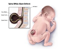
  • Neural Tube Defect Wikipedia
    Pediatrics . 1 (2): 113. doi : 10.3171/PED/2008/1/2/113 . PMID 18352777 . ^ a b Molloy, A. M.; Kirke, P. N.; Troendle, J. F.; Burke, H.; Sutton, M.; Brody, L. ... PMID 19706381 . ^ Brouwer, I. A.; Van Dusseldorp, M.; West, C. E.; Meyboom, S.; Thomas, C. M.; Duran, M.; Van Het Hof, K. H.; Eskes, T. ... BMJ 2005;330:571 ^ Wein, TN; Pike, E; Wisløff, T; Staff, A; Smeland, S; Klemp, M (12 January 2012). "Cancer risk with folic acid supplements: a systematic review and meta-analysis" . ... ISSN 1871-403X . PMID 27155922 . ^ Wang, M; Wang, ZP; Gong, R; Zhao, ZT (January 2014).
    VANGL1, VANGL2, FUZ, MTHFD1, MTHFR, PAX3, FOLR1, GRHL3, BHMT, CECR2, FOLR2, ZIC2, PTK7, SHROOM3, ZIC5, CYP1A2, SPINT2, NAT2, CCL2, TBXT, GLI3, CSF2, INS, IFNG, GHRL, SKI, NPY1R, PRSS8, PYY, RRM1, RFC1, CDH1, AFP, PGPEP1, MTRR, MTR, PRCP, SETBP1, BMP1, CBSL, SLC19A1, CBS, TP53, MTHFD1L, PDGFRA, FOLH1, PAX1, TCN2, PARD3, SHMT1, CASP8, GLDC, TYMS, FZD3, CELSR1, UCP2, LMNB1, LRP6, IGF2, SOX3, ALDH1A2, SCRIB, ZIC3, DVL2, DHFR, COMT, CASP9, GCLC, PPARGC1A, MARCKS, ALDH1L1, MGMT, MSX2, MMUT, NFKB1, NOS2, PCMT1, TRAF4, CASP3, NOG, PRKACB, PRICKLE1, PTCH1, BMP4, SHH, TBX1, FZD6, ALX1, UGCG, CUBN, LRP2, RAB11FIP3, FOXO3, RAB23, HLA-A, F2RL2, GRHL2, MARCKSL1, EPHA4, INPP5E, DVL1, NHLRC2, H4C14, H4C8, H4C2, EED, MIR206, PTF1A, H4C5, H4C13, MIR197, FOXN1, TAGLN2, MIR129-2, MIR30B, CYP26B1, SELENOH, CUL4B, TRADD, MIRLET7G, ZGPAT, H4C3, H4C11, MALL, LST1, PERCC1, CDH23, GORASP1, CRPPA, POU5F1P4, CSRP3, DVL1P1, ARID1A, H4C9, POU5F1P3, SALL4, H4C1, H4C4, H4C6, H4C12, H4C15, TNFSF12, PCSK9, SLC19A2, PEMT, SEC24B, SLC46A1, PRRT2, WDR20, TRIM4, EMG1, CARM1, NKX2-8, CITED2, TCTN3, TXN2, TUBGCP2, SLC22A16, FKBP8, KDM2B, KCNQ1OT1, ANKRD6, DLC1, RXYLT1, WNK1, SHROOM1, SIRT1, TRPM6, NOL3, RIN2, FTO, PPIG, SUFU, DACT1, CD320, TNIP1, H4-16, LIN28A, TUBA3D, PRX, SLC40A1, KEAP1, WLS, ZEB2, GPR161, NAT1, ZIC1, GOLGA4, EZH2, FASN, FGF8, GPC5, FOXO1, FUT2, GCH1, GCKR, CBLIF, GJA1, GLI2, GNAS, CFHR1, TRDMT1, HHEX, HIF1A, HLA-B, HLA-C, HMOX1, HOXB7, HOXD@, ID1, ID2, IGF1, IL10, ITGA3, DVL3, SARDH, XPC, ATRX, ACTB, ADA, AHR, AKT2, ALX3, AMT, ANXA5, ANXA11, APAF1, APOB, SHROOM2, ASCL1, BCHE, DLX5, C5, C5AR1, CALCA, CD6, CDC25C, CLDN3, CRABP2, CTNNA1, CYP1B1, DAPK3, DDIT3, DIO3, ITGB1, ITPK1, JARID2, TERC, RELA, S100B, SALL2, CXCL6, SLC2A2, SNAI2, SMARCC1, SOX2, SULT1A1, SYT1, TCF7L2, TCN1, TFAP2A, KCNQ1, TGFB3, TGIF1, TNF, TRIP6, TUBA4A, TUBA3C, TULP3, TXN, KDM6A, VCL, WNT7B, WNT2B, MAPK8, PRKCB, PRKCA, PRKACA, KRT1, LAMC2, LEP, LEPR, LGALS1, LIFR, LIG3, MAB21L1, MAP3K5, MLH1, MRC1, ABCC1, MSH2, MUC2, MYH2, MYLK, NAP1L2, NCAM1, SEPTIN2, NFE2L2, NOS1, NOS3, POU3F1, POU5F1, PPBP, RN7SL263P
  • Scleroderma Wikipedia
    PMID 21315153 . ^ Arnson Y, Amital H, Agmon-Levin N, Alon D, Sánchez-Castañón M, López-Hoyos M, Matucci-Cerinic M, Szücs G, Shapira Y, Szekanecz Z, Shoenfeld Y (June 2011). ... PMID 23541012 . ^ a b Kowal-Bielecka O, Bielecki M, Kowal K (2013). "Recent advances in the diagnosis and treatment of systemic sclerosis" (PDF) . ... Retrieved 27 November 2013 . [ full citation needed ] ^ a b Sticherling M (October 2012). "Systemic sclerosis-dermatological aspects. ... PMID 29573101 . S2CID 4230441 . ^ Nikpour M, Stevens WM, Herrick AL, Proudman SM (December 2010). ... PMC 4553970 . PMID 23793108 . ^ a b c d Lidar M, Langevitz P (May 2012). "Pregnancy issues in scleroderma".
    LMNA, TNF, CSF3, XYLT2, LBR, PAH, SLC29A3, POLD1, CCN2, CLCNKB, UROD, SMAD3, HFE, SLC12A3, UROS, LEMD3, TGFB1, WRN, COL1A2, HLA-DRB1, SS18L1, CCL2, MMP1, TGFBR1, FBN1, SPARC, IL6, HGF, RNPC3, FLI1, EDN1, FN1, MAPK1, IFNG, SMAD7, COL1A1, IL13, PDGFRB, CYCS, IL1B, HLA-DQB1, RBM45, TLR4, ICAM1, CAV1, POLR3A, TNFSF14, KL, POLDIP2, MYBL2, DKK1, CCN1, GZMA, IL10, ITGA5, BTG3, GRAP2, TNC, HLA-G, HLA-DQA1, HLA-DPB1, RNF19A, VEGFA, CSF1, FGF2, STAT4, SRY, TGFBI, TGFBR2, MIR21, MIR196A1, THBS1, HDAC5, TIMP1, CD34, CENPA, SMAD2, CRK, MAPK14, IL2, DBA2, TP53, AIMP2, MMP3, PLAU, ESR1, FBL, EPHB2, ENG, POSTN, EGR1, EDNRB, AHSA1, UBE4A, PPIG, GDF15, PLAA, TSIX, FGF23, KLF4, YY1, TG, THY1, TIMP3, TNFAIP3, TNFRSF1A, TPO, CRISP2, TTN, TXN, VDR, CEBPZ, COPB2, LTBP4, RTCA, PDE5A, TNFSF10, TNFRSF14, TNFRSF10B, ASAP2, EIF2B4, EIF2B2, EIF2S2, EBI3, ACR, ZMPSTE24, PRSS55, TRPV4, FBRS, GORASP1, SMURF2, WNK1, COL18A1, TSSK1B, SPZ1, TSLP, PRRT2, CARD16, SLCO6A1, IL23R, DNAAF3, UBE4B, GSTK1, MIR130B, MIR150, MIR155, MIR17, MIR206, MIR29A, MIR17HG, MIR135B, MIR202, CCR2, C20orf181, KIR2DS2, SPG16, MRTFA, SMURF1, IFT122, TNIP1, EMG1, TNFSF13B, UTP14A, MRPS30, PAPOLA, RNPS1, SPIN1, PDAP1, TUSC2, NID2, TAC1, SMG1, SIRT1, RBFOX2, POLR1A, ERAL1, DNAI1, DLL1, TBX21, ASAP1, CSAD, SCLY, KRT20, BANK1, MTPAP, CARMIL1, TAL1, EXOSC10, SYT1, STAT6, EGR3, EIF2B1, EPHA1, ETS1, EZH2, PTK2B, FCGR2B, FKBP1A, FOXC1, FOXO3, FLII, FOLR2, FPR2, ACKR1, GEM, GLB1, GTF2I, GYPA, HDC, HIF1A, HLA-A, HLA-B, HLA-DRB5, IFI16, IFNA1, IFNA13, IFNAR1, IL1A, IL3, EGR2, EGFR, EGF, CD247, ADA, AKT1, ALDH3A2, ALOX5, ALOX5AP, ABCC6, BCL2, BCL6, BLM, BMP6, BMPR2, CAT, SERPINH1, MS4A1, DNMT1, CD44, CD69, CENPC, LTB4R, COL3A1, COMP, CSF2, NKX2-5, CTLA4, CYP2B6, DECR1, DNAH5, DNASE1L3, IL4, IL13RA1, IL16, PRKCD, PGM1, SERPINE2, PIK3CA, PIK3CB, PIK3CD, PIK3CG, PLAUR, PLOD2, ACP3, POMC, PPARG, PRKCA, PRKCB, MAP2K7, PDGFA, PRS, PRTN3, PSMB6, PTGS2, RELA, S100A9, CCL5, SLC25A1, SP1, SPG7, SRF, SRPK2, STAT3, ENPP2, PCM1, IL17A, MET, ITGAV, ITGB3, KIR2DL2, KIR3DL1, KNG1, KRAS, KRT7, LGALS3, LOX, LOXL2, SMAD4, MBD1, MECP2, KITLG, PCBD1, MIF, MPO, MT3, COX2, MYB, MYC, NOS3, DDR2, OXCT1, P2RX7, SERPINE1, PAK3, REG3A, MTCO2P12
    • Scleroderma Orphanet
      Women are predominantly affected (F/M sex ratio around 4:1). Clinical description Localized scleroderma is the cutaneous form of scleroderma characterized by fibrosis of the skin causing cutaneous plaques (morphea) or strips (linear scleroderma).
    • Scleroderma Mayo Clinic
      Overview Scleroderma (sklair-oh-DUR-muh), also known as systemic sclerosis, is a group of rare diseases that involve the hardening and tightening of the skin. It may also cause problems in the blood vessels, internal organs and digestive tract. Scleroderma is often categorized as "limited" or "diffuse," which refers only to the degree of skin involvement. Both types can involve any of the other vascular or organ problems. Localized scleroderma, also known as morphea, affects only the skin. While there is no cure for scleroderma, treatments can ease symptoms, slow progression and improve quality of life.
  • Infections Associated With Diseases Wikipedia
    PMID 23974921 . ^ Kolehmainen P, Koskiniemi M, Oikarinen S, et al. (September 2013). ... S2CID 12678568 . ^ Negro, F; Alaei, M (2009). "Hepatitis C virus and type 2 diabetes" . ... E. P.; Arendrup, M. C.; Nielsen, H. V.; Mølbak, K. (2009). ... S2CID 29730230 . ^ Arcari, Christine M.; Gaydos, Charlotte A.; Nieto, F. ... PMID 23313649 . ^ Muller, N; Riedel, M; Blendinger, C; Oberle, K; Jacobs, E; Abelehorn, M (2004).
  • Food Intolerance Wikipedia
    Dig Dis Sci . 54 (1): 8–14. doi : 10.1007/s10620-008-0331-x . PMID 18594978 . ^ Maurer M, Hanau A, Metz M, Magerl M, Staubach P (February 2003). ... Retrieved 14 April 2009 . ^ Woods RK, Abramson M, Raven JM, Bailey M, Weiner JM, Walters EH (January 1998). ... PMID 9012205 . ^ Feinle-Bisset C, Horowitz M (August 2006). "Dietary factors in functional dyspepsia". ... PMID 7460264 . S2CID 12346266 . ^ Montalto M, Santoro L, D'Onofrio F, et al. (2008). ... PMC 5492024 . PMID 28445436 . ^ Heyman M (December 2005). "Gut barrier dysfunction in food allergy".
    ELN, ALG3, MLXIPL, COG8, FOXO3, IL15, SLC7A7, ATG16L1
  • Benzodiazepine Overdose Wikipedia
    PMID 9926062 . ^ a b Ngo AS, Anthony CR, Samuel M, Wong E, Ponampalam R (July 2007). ... PMID 7572872 . ^ Reidenberg MM, Levy M, Warner H, Coutinho CB, Schwartz MA, Yu G, Cheripko J (April 1978). ... PMID 14705844 . ^ Rumpl E, Prugger M, Battista HJ, Badry F, Gerstenbrand F, Dienstl F (December 1988). ... PMID 1611650 . ^ Marchant B, Wray R, Leach A, Nama M (September 1989). "Flumazenil causing convulsions and ventricular tachycardia" . ... PMID 16439763 . ^ Carlsten A, Waern M, Holmgren P, Allebeck P (2003). "The role of benzodiazepines in elderly suicides".
  • Alcohol Withdrawal Syndrome Wikipedia
    S2CID 2139796 . ^ a b c d e f g Bayard M, McIntyre J, Hill KR, Woodside J (March 2004). ... PMID 18365930 . S2CID 25872623 . ^ Hornyak M; Haas P; Veit J; Gann H; Riemann D (November 2004). ... PMID 19249388 . S2CID 20197292 . ^ Heilig M, Egli M, Crabbe JC, Becker HC (April 2010). ... PMID 2597811 . ^ Amato L, Minozzi S, Vecchi S, Davoli M (2010). Amato L (ed.). "Benzodiazepines for alcohol withdrawal". ... S2CID 19857498 . ^ a b Minozzi, S.; Amato, L.; Vecchi, S.; Davoli, M.; Minozzi, Silvia (2010). Minozzi, Silvia (ed.).
    AR, LEP, MPDZ, CCK, NGF, CYP19A1, KDM4C, GSTM1, NQO1, MALAT1, GLUL, SORCS2, SLC6A3, BDNF, CRH, SLC6A4, DRD2, FKBP5, DBH, GRIN1, GRIN2B, NPY, CCL2, APOE, POMC, PPARA, PPARD, PPARG, SORT1, SLC6A2, COMT, KCNQ4, SLC18A1, SNCA, PIK3CG, ADIPOQ, BMS1, C1D, PPARGC1A, AKT1, TMEM97, PNPLA3, PNOC, NPY5R, PIK3CD, HTR2C, CCKAR, CAT, GAD1, GATA4, GH1, NR3C1, GRM5, TSPO, HDAC2, IL6, PIK3CB, KRT18, LBP, MAOA, ADH1B, NQO2, NPY1R, NPY2R, CFP, PIK3CA, ACACA
  • Compartment Syndrome Wikipedia
    Retrieved 25 July 2017 . ^ Peitzman AB, Rhodes M, Schwab CW (2008). The Trauma Manual: Trauma and Acute Care Surgery . ... PMID 25050098 . ^ a b Via AG, Oliva F, Spoliti M, Maffulli N (2015-03-27). "Acute compartment syndrome" . ... PMID 25878982 . ^ a b Chandwani D, Varacallo M (2020). "Exertional Compartment Syndrome" . ... Retrieved 2020-01-22 . ^ a b c d e f g h i j k l m n o Via AG, Oliva F, Spoliti M, Maffulli N (2015). ... PMID 17992173 . ^ a b c d Cetinus E, Uzel M, Bilgiç E, Karaoguz A, Herdem M (April 2004).
    HPX, MB, F3, TNF
  • Drug Reaction With Eosinophilia And Systemic Symptoms Wikipedia
    PMID 24388810 . ^ Sauvetre G, MahÉvas M, Limal N, Guillaud C, Khellaf M, Bierling P, Languille L, Delbos F, Noizat-Pirenne F, Michel M, Godeau B (October 2015). ... PMID 18256392 . ^ Konvinse KC, Trubiano JA, Pavlos R, James I, Shaffer CM, Bejan CA, Schutte RJ, Ostrov DA, Pilkinton MA, Rosenbach M, Zwerner JP, Williams KB, Bourke J, Martinez P, Rawandamuriye F, Chopra A, Watson M, Redwood AJ, White KD, Mallal SA, Phillips EJ (2019). ... PMID 30776417 . ^ a b Hoetzenecker W, Nägeli M, Mehra ET, Jensen AN, Saulite I, Schmid-Grendelmeier P, Guenova E, Cozzio A, French LE (2016). ... The Journal of Dermatology . 43 (7): 758–66. doi : 10.1111/1346-8138.13430 . PMID 27154258 . ^ Lerch M, Mainetti C, Terziroli Beretta-Piccoli B, Harr T (2017). ... S2CID 46796285 . ^ Chong HY, Mohamed Z, Tan LL, Wu DB, Shabaruddin FH, Dahlui M, Apalasamy YD, Snyder SR, Williams MS, Hao J, Cavallari LH, Chaiyakunapruk N (2017).
    ALK, HLA-B, HLA-A, HSPG2, IL1B, IL1A, CYP2C9, CCL17, GPT, TNF, IRF3, LCT, PPARG, TRIM27, TPO, IL10, TSHR, GNLY, IL22, SNX14, ZBP1, TSLP, IL33, EMB, IL13, NAT2, IL6, EBM, ANK1, BRAF, TPP1, CRP, CYP2C19, CYP2C18, FDXR, IL5, FPR2, GZMB, HMGB1, IFNG, AKR1B1, IL4, SYT14
    • Drug Rash With Eosinophilia And Systemic Symptoms Orphanet
      A rare hypersensitivity reaction characterized by a generalized skin rash, fever, eosinophilia, lymphocytosis and visceral involvement (hepatitis, nephritis, pneumonitis, pericarditis and myocarditis) and, in some patients, reactivation of human herpes virus 6. Epidemiology Prevalence is unknown. Etiology Onset usually occurs 2-6 weeks after administration of the causal medication. DRESS syndrome is most frequently associated with anticonvulsants and sulfonamides but other medications (allopurinol, cyclosporine, azathioprine, gold salts and antiviral agents) have also been implicated. The manifestations may persist for several weeks after withdrawal of the causative medication.
  • Lumbar Spinal Stenosis Wikipedia
    PMID 20227646 . ^ a b c d e f g h i j k l m Djurasovic M, Glassman SD, Carreon LY, Dimar JR (April 2010). ... PMID 17549525 . ^ Takahashi K, Kitahara H, Yamagata M, Murakami M, Takata K, Miyamoto K, Mimura M, Akahashi Y, Moriya H (November 1990). ... PMC 3591818 . PMID 14534849 . ^ Benoist M (October 2003). "Natural history of the aging spine" . ... PMC 3591827 . PMID 12961079 . ^ Szpalski M, Gunzburg R (October 2003). "Lumbar spinal stenosis in the elderly: an overview" . ... PMID 25901233 . ^ a b c Hicks, Gregory E.; Gaines, Jean M.; Shardell, Michelle; Simonsick, Eleanor M. (2008-09-15).
    CAPN1, EHMT1, IL6, ELN, CRNKL1, EOS, LAP, LTBP4, CRLF1, ANGPTL2, AMH, CLQTL1, VEGFA, MIR155, MIR21, MIR221, MIR29A, GOLPH3, THBS2, VDR, TGFB1, PTGS2, NHS, COX2, MMP9, MMP2, MAP6, LTBP2, LSS, LEP, LPAR1, COL9A2, CAT, MTCO2P12
    • Spinal Stenosis Wikipedia
      Retrieved 19 December 2017 . ^ Boos N, Aebi M (2008). Spinal Disorders: Fundamentals of Diagnosis and Treatment . ... P., Lambing, C. L., Malanga, G. A., Maurer, P. M., Ralph R., R. (2010). Algorithmic approach to the management of the patient with lumbar spinal stenosis.
  • Hellp Syndrome Wikipedia
    Retrieved 5 October 2018 . ^ a b c d e f g h i j k l m Haram K, Svendsen E, and Abildgaard U (February 2009). ... PMID 16579935 . ^ Strand S, Strand D, Seufert R, Mann A, Lotz J, Blessing M, Lahn M, Wunsch A, Broering DC, Hahn U, Grischke EM, Rogiers X, Otto G, Gores GJ, Galle PR (March 2004). ... Eur J Hum Genet . 9 (10): 758–64. doi : 10.1038/sj.ejhg.5200706 . PMID 11781687 . ^ a b Habli M, Eftekhari N, Wiebracht E, Bombrys A, Khabbaz M, How H, Sibai B (October 2009). ... PMC 2270909 . PMID 18382655 . ^ a b Geary M (August 1997). "The HELLP syndrome" . ... South Med J . 93 (8): 768–71. doi : 10.1097/00007611-200093080-00005 . PMID 10963506 . ^ Haeger M, Unander M, Norder-Hansson B, Tylman M, Bengtsson A (January 1992).
    CD46, CFH, CFI, HELLPAR, FASLG, FAS, PGF, F5, LEP, HADHA, F2, TNF, HPGDS, LGALS13, FLT1, VEGFA, MTHFR, MAPK14, TLR4, AIMP2, TLR2, MAPK3, TPBG, VEGFC, TGFB3, VWF, MAPK1, ABCG2, TFPI2, IL18R1, GRAP2, EBI3, AHSA1, ADAMTS13, SIRT4, RNF19A, POLDIP2, SLC17A5, ERVW-1, MBL3P, AHSP, NOD2, POTEF, SERPINE2, ACTB, SERPINE1, PAH, APC, CFB, CA9, CD40LG, CD59, CDKN1C, COX8A, CP, CRK, ENG, EPHX1, GAPDH, GNB3, GPT, NR3C1, GSTM1, GSTT1, HSPA4, HSPG2, IFNG, IL1B, IL1RN, CXCL8, IL10, LEPR, LNPEP, ADM, NOS3, PAEP, MBL2
    • Hellp Syndrome Orphanet
      A rare hemorrhagic disorder due to an acquired platelet anomaly characterized by hemolysis, elevated liver enzymes and thrombocytopenia that affects pregnant or post-partum women, and is frequently associated with severe preeclampsia. Symptoms are variable, typically including right upper quadrant or epigastric abdominal pain, nausea, vomiting, excessive weight gain, generalized edema, hypertension, general malaise, right shoulder pain, backache, and/or headache. Hepatic hemorrhage and rupture, renal failure, and pulmonary edema can result in maternal and/or fetal death.
    • Hellp Syndrome GARD
      HELLP syndrome is a life-threatening condition that can potentially complicate pregnancy. It is named for 3 features of the condition: H emolysis, E levated L iver enzyme levels, and L ow P latelet levels. It typically occurs in the last 3 months of pregnancy (the third trimester) but can also start soon after delivery. A wide range of non-specific symptoms may be present in women with HELLP syndrome. Symptoms may include fatigue; malaise; fluid retention and excess weight gain; headache; nausea and vomiting; pain in the upper right or middle of the abdomen; blurry vision; and rarely, nosebleed or seizures.
  • Emergent Virus Wikipedia
    PMC 3291398 . PMID 16494711 . ^ a b Eidson M. "Zoonotic disease" . Britannica . ... Global Biosecurity . 1 (3). doi : 10.31646/gbio.43 . ISSN 2652-0036 . ^ Woolhouse M, Scott F, Hudson Z, Howey R, Chase-Topping M (October 2012). ... PMC 1539106 . PMID 16847084 . ^ Woolhouse M, Gaunt E (2007). "Ecological origins of novel human pathogens". ... PMID 16447497 . ^ a b c d e Bolles M, Donaldson E, Baric R (December 2011). ... PMID 25474536 . ^ Farag E, Sikkema RS, Vinks T, Islam MM, Nour M, Al-Romaihi H, et al. (December 2018).
  • Progressive Supranuclear Palsy Wikipedia
    PMC 6183005 . PMID 30363831 . ^ Gearing M, Olson DA, Watts RL, Mirra SS (June 1994). ... PMID 12802605 . S2CID 20275104 . ^ Hattori M, Hashizume Y, Yoshida M, Iwasaki Y, Hishikawa N, Ueda R, Ojika K (August 2003). ... S2CID 25741692 . ^ Komori T, Arai N, Oda M, Nakayama H, Mori H, Yagishita S, et al. ... Current Opinion in Neurology . 23 (4): 394–400. doi : 10.1097/WCO.0b013e32833be924 . PMID 20610990 . ^ Kanazawa M, Tada M, Onodera O, Takahashi H, Nishizawa M, Shimohata T. (2013). ... Retrieved 19 February 2019 . ^ Litvan I, Campbell G, Mangone CA, Verny M, McKee A, Chaudhuri KR, et al. (January 1997).
    MAPT, STX6, MOBP, EIF2AK3, SRSF2, TRA2B, SLCO1A2, TRIM11, SP1, PSPH, REG1A, SNCA, RIDA, STXBP3, MSMB, PSPN, TPO, CD8B, ASAP1, RUNX2, BPIFA2, PIK3C2G, IRF4, APOE, SLC6A3, TARDBP, LRRK2, NEFL, SOD1, CIT, C9orf72, CSF2, MAOB, GRN, LAMC2, DCTN1, STH, SMUG1, PRKN, UBB, PYCARD, NPC1, APP, TYMS, CRHR1, TH, TGM2, SLC25A38, ATXN2, GFAP, IGLON5, CST3, NPEPPS, VEGFA, RAB35, YWHAE, OGA, CXCR4, PICALM, NPC2, SNCAIP, BSN, MAP3K14, OPN1MW3, DUSP10, ARL17B, ROCK2, SCRN1, MAP4K4, NF1P1, UNC13A, DNAJB1P1, FLAD1, UBASH3B, SPECC1, FOXP2, RMDN2, ASXL1, MCIDAS, SETX, MIR132, MIR518E, GGTLC5P, GGTLC3, GGT2, OPN1MW2, CTNNBL1, SYBU, PSPC1, RMDN3, LRRC37A4P, TET2, TMEM106B, TREM2, LCMT1, PPME1, RMDN1, GGTLC4P, PSAT1, TBK1, CSDC2, LMOD1, SF3B1, MINK1, NAT1, TPI1, OPN1MW, FMR1, MTOR, FUS, GABPA, GABRG2, GBA, GGT1, EGFR, GLDC, GSTM1, NRG1, HSPA4, DNAJB1, IFNG, ERBB4, DLX1, IGFALS, CASP3, AP2A2, ANXA6, KLK3, BDNF, BNIP1, BRCA1, CBS, ACE, CDK5, CHI3L1, CLU, CRP, CTSS, CYP2D6, IGF1, IL2, TP53BP1, MAP2K4, PSEN2, PTEN, PTPRC, RAPSN, ROCK1, ATXN8OS, NAT2, PROS1, SPOCK1, SPP1, TCOF1, TGFB1, TGM1, TNF, PSEN1, PRNP, IL6, NR4A2, IRS1, MUSK, NFE2L2, NGF, NOS1, NSF, PAEP, PTPA, PAFAH1B1, PDK1, PIN1, PLAG1, PLCG2, PLXNA2, ATXN2-AS
    • Parkinson-Dementia Syndrome OMIM
      A number sign (#) is used with this entry because of evidence that a form of atypical supranuclear palsy is caused by mutation in the microtubule-associated protein tau gene (MAPT; 157140). Clinical Features Mata et al. (1983) described 2 brothers and a sister with a 'new' Parkinson-dementia syndrome. The disorder, characterized also by ophthalmoparesis and pyramidal signs, came on in the third decade and progressed for several years. Kyphoscoliosis was present in all 3 sibs. Examination of the brain in the sister, who died at age 31 years, showed neurofibrillary degeneration of the hippocampus, basal ganglia and brainstem nuclei. The parents were not related. The authors suggested that the disorder most closely resembled the Parkinson-dementia complex of Guam (105500) but could be distinguished by the lack of Chamorro descent (a dubious argument) and the earlier age of onset.
    • Progressive Supranuclear Palsy Mayo Clinic
      Overview Progressive supranuclear palsy is an uncommon brain disorder that causes serious problems with walking, balance and eye movements, and later with swallowing. The disorder results from deterioration of cells in areas of your brain that control body movement, coordination, thinking and other important functions. Progressive supranuclear palsy is also called Steele-Richardson-Olszewski syndrome. Progressive supranuclear palsy worsens over time and can lead to life-threatening complications, such as pneumonia and swallowing problems. There's no cure for progressive supranuclear palsy, so treatment focuses on managing the signs and symptoms.
    • Progressive Supranuclear Palsy Orphanet
      A rare late-onset neurodegenerative disease characterized by supranuclear gaze palsy, postural instability, progressive rigidity, and mild dementia. Epidemiology Prevalence is conservatively estimated at about 1/16,600. Clinical description PSP usually manifests during the sixth or seventh decade of life. Five clinical variants have been described with clinicopathological correlations: Classical PSP (Richardson's syndrome), and four atypical variants of PSP including PSP-Parkinsonism (PSP-P), PSP-Pure akinesia with gait freezing (PSP-PAGF), PSP-corticobasal syndrome (PSP-CBS), and PSP-progressive non fluent aphasia (PSP-PNFA) (see these terms). Richardson's syndrome is the most common clinical variant and manifests with a lurching gait, falls due to postural instability, cognitive impairment and slowing of vertical saccadic eye movements.
    • Supranuclear Palsy, Progressive, 3 OMIM
      For a phenotypic description and a discussion of genetic heterogeneity of progressive supranuclear palsy (PSP), see PSNP1 (601104). Mapping Using a pooling-based genomewide association study of more than 500,000 SNPs in 288 patients with PSP and 344 age- and sex-matched controls, Melquist et al. (2007) identified candidate SNPs with large differences in allelic frequency by ranking all SNPs by their probe-intensity difference between cohorts. The MAPT H1 haplotype (see 157140) was strongly detected by this approach as was a second major locus (PSNP3) on chromosome 11p12-p11 that showed evidence of association at allelic (p less than 0.001), genotypic (p less than 0.001), and haplotypic (p less than 0.0001) levels and was narrowed to a single haplotype block containing the DNA damage-binding protein-2 (DDB2; 600811) and lysosomal acid phosphatase-2 (ACP2; 171650) genes. Since DNA damage and lysosomal dysfunction have been implicated in aging and neurodegenerative processes, Melquist et al. (2007) considered both genes to be viable candidates for conferring risk of disease.
    • Supranuclear Palsy, Progressive, 2 OMIM
      For a phenotypic description and a discussion of genetic heterogeneity of progressive supranuclear palsy (PSP), see PSNP1 (601104). Clinical Features De Yebenes et al. (1995) studied a 5-generation Spanish family in which progressive supranuclear palsy was transmitted as an autosomal dominant trait. The proband had the classic presentation of this disorder beginning with axial rigidity, slowness of movement, and gait difficulty. Over the course of 2 years he progressed to complete vertical gaze palsy, axial dystonia, and retrocollis, as well as generalized severe akinesia. Postmortem examination demonstrated neuronal loss and atrophy of the brainstem, cerebellum, and diencephalon.
    • Supranuclear Palsy, Progressive, 1 OMIM
      A number sign (#) is used with this entry because of evidence that progressive supranuclear palsy-1 (PSNP1) is caused by heterozygous mutation in the gene encoding microtubule-associated protein tau (MAPT; 157140) on chromosome 17q21. Description Progressive supranuclear palsy (PSP) is the second most frequent cause of degenerative parkinsonism. In addition to parkinsonism, the clinical symptoms include early postural instability, supranuclear gaze palsy, and cognitive decline. Neuropathologically, the disorder is characterized by abundant neurofibrillary tangles, which differ in both distribution and composition from those associated with Alzheimer disease. In progressive supranuclear palsy, the tangles are primarily localized to subcortical regions and are found in both neurons and glia, whereas in Alzheimer disease they are more widespread, largely cortical, and limited to neurons.
    • Progressive Supranuclear Palsy GARD
      Progressive supranuclear palsy (PSP) is a degenerative neurologic disease due to damage to nerve cells in the brain. Signs and symptoms vary but may include loss of balance; blurring of vision; problems controlling eye movement; changes in mood, behavior and judgment; cognitive decline; and slowing and slurred speech. PSP is often misdiagnosed as Parkinson disease due to similar symptoms. Onset is usually after age 60 but may occur earlier. Most cases of PSP appear to be sporadic , but familial cases have been reported. Some cases have been found to be caused by a mutation in the MAPT gene, and other genetic factors are being studied.
    • Classic Progressive Supranuclear Palsy Syndrome Orphanet
      Classical progressive supranuclear palsy, also known as Richardson's syndrome, is the most common clinical variant of progressive supranuclear palsy (PSP; see this term), a rare late-onset neurodegenerative disease characterized by postural instability, progressive rigidity, supranuclear gaze palsy and mild dementia. Epidemiology The prevalence is conservatively estimated at about 1/16,600. Clinical description PSP usually manifests during the sixth or seventh decade of life with postural instability and falls, slowing of vertical saccadic eye movements and cognitive slowing. Progressively patients develop other eye abnormalities (dry and red eyes, blurred vision, spontaneous involuntary eyelid closure, photophobia), a dysexecutive cognitive syndrome with impulsivity, problems in speech (slow speech) and a supranuclear gaze palsy and difficulties in swallowing. The disease is characterized neuropathologically by gliosis with astrocytic plaques, accumulation of tau-immunoreactive neurofibrillary tangles and neuronal loss in specific brain areas, especially in the subthalamic nucleus and the substantia nigra.
  • ←
  • 1
  • 2
  • ...
  • 78
  • 79
  • 80
  • 81
  • 82
  • 83
  • 84
  • ...
  • 297
  • 298
  • →

FindZebra

contact@findzebra.com