• FindZebra
  • About
  • Contact
  • Help
  • Login
Advanced
  • Papillary Renal Cell Carcinoma Wikipedia
    PMID 31867475 . ^ a b c Couvidat C, Eiss D, Verkarre V, Merran S, Corréas JM, Méjean A, Hélénon O (November 2014). ... PMID 19448113 . ^ a b c d e Bourlon MT, Meneses-Medina M, Vázquez-Manjarrez S, Bustamante-Romero FM, Gallegos-Garza AC, Lam ET (November 2015). ... PMID 26185343 . ^ Weidner N, Cote RJ, Suster S, Weiss LM (2009-07-08). Modern Surgical Pathology E-Book . ... PMID 28153029 . ^ Ravaud, A.; Oudard, S.; De Fromont, M.; Chevreau, C.; Gravis, G.; Zanetta, S.; Theodore, C.; Jimenez, M.; Sevin, E.; Laguerre, B.; Rolland, F.; Ouali, M.; Culine, S.; Escudier, B. (2015-06-01). ... S2CID 205659715 . ^ Kamai T, Abe H, Arai K, Murakami S, Sakamoto S, Kaji Y, Yoshida KI (March 2016).
    MET, MITF, PRCC, VHL, TFE3, KRT7, ERBB2, GSTP1, SLC2A1, ELOC, POMC, PIK3CA, ADIPOQ, RPL14, INPP4B, FPGT, TNFSF10, CRADD, UNC5C, TAGLN2, BAP1, KDM5C, PEBP1, MLLT10, PAK1, TSC1, TP53, TGM2, PDHB, HNF1B, HNF1A, NF2, SOD2, SLC5A3, PGK1, ACY1, RYR1, RELA, PVALB, PTGS2, PTEN, ZNF536, ACHE, KEAP1, AHNAK, LMAN2L, CARD11, SLC49A4, NAV3, ZNF765, NLRP12, ZNF804A, CSMD3, LRRK2, ASB15, DNHD1, FAAH2, FLCN, OR4C13, VMO1, MSGN1, FAM111B, BIRC7, SPTBN4, CUL7, FMN2, NDRG1, CPQ, BTG3, AKAP13, RNF139, PDXDC1, SYNE2, PRAME, YIPF3, PNKD, SETD2, SHANK1, LRP1B, TET2, PBRM1, PIDD1, AP5M1, OGG1, SFRP2, APRT, MUC4, IL6, ALDH1A1, ALK, ALOX5, IFNA2, HSPD1, HSPB1, HSPA9, ALOX12B, ANXA4, APAF1, BIRC5, HARS1, EEF2, BCHE, GSTT1, GSTM1, GRB7, GJB1, EPAS1, MTOR, IL6R, IL4R, IL13, KRT32, CNN2, SCARB1, CASP2, CAPG, M6PR, ALAD, LDHB, L1CAM, CRABP1, KRT8, KCNMA1, DAPK1, CTSD, CTSB, CRYAB, FLT1, CDC73, HABP2, LMNA, FOXE1, MINPP1, VEGFA, AMACR, KDR, CD38, FH, MME, RASSF1, CA9, MYC, KIT, SNORD14D, SNORD14C, CD47, MIR1180, TWSG1, MIR1293, MIR379, KMT2C, ACE2, FBXO47, EIF2AK4, MIR34A, MIR200C, MIR181C, MIR134, MIR127, FBXO11, AKT1, DDR1, RAB37, APC, CDC34, SETDB2, BRAF, SNORD35B, RIOX2, BSG, BUB1, SNORD14E, SNORD14B, MEG3, DHCR24, LARP6, CDH1, HIF1A, TOP2A, TIMP3, TIMP1, UBE2K, SYP, IGFBP7, SLC16A1, SFPQ, RRM2, SNORD15A, CD82, PYCR1, PTK7, CFP, PAX2, MUC1, TBC1D25, OAT, NONO, NFE2L2, XRCC1, PAX8, FZD1, KDM4C, CDR2, CMA1, APH1A, CD274, CLDN7, MCTS1, MCAT, UBE2S, HAVCR1, ACE, CUL3, RALBP1, EGF, EGFR, ELAVL2, ETFA, NAPSA, FGFR2, FOLH1, GABPA, MIR3199-2
    • Papillary Renal Cell Carcinoma GARD
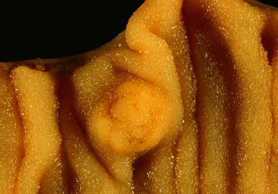
      It accounts for about 10-15% of all renal cell carcinomas. R enal cell carcinoma s are a type of kidney cancer that develop in the lining of very small tubes (tubules) in the kidney.The term "papillary" describes the finger-like projections that can be found in most of the tumors.
    • Papillary Renal Cell Carcinoma Orphanet
      Papillary renal cell carcinoma is a rare subtype of renal cell carcinoma, arising from the renal tubular epithelium and showing a papillary growth pattern, which typically manifests with hematuria, flank pain, palpable abdominal mass or nonspecific symptoms, such as fatigue, weight loss or fever. Symptoms related to metastatic spread, such as bone pain or persistent cough, are frequently associated since early diagnosis is not common. It is typically multifocal, bilateral, and in most cases sporadic, although different hereditary syndromes, such as Hereditary leiomyoma renal cell carcinoma, Birt-Hogg-Dubé syndrome and Tuberous sclerosis, may predispose to the development of papillary renal cell carcinoma.
  • Thiopurine S Methyltranferase Deficiency GARD
    Thiopurine S-methyltransferase deficiency is an autosomal recessive disorder that affects the body's ability to metabolize thiopurine drugs. Thiopurine S-methyltransferase (TPMT) is an enzyme that the body uses to break down thiopurine drugs. Thiopurine S-methyltransferase deficiency patients have a mutation in either one or both copies of the TPMT gene that causes reduced enzyme activity and difficulties breaking down thiopurine drugs.
    TPMT, ABCC4, NUDT15
    • Thiopurine S-Methyltransferase Deficiency MedlinePlus
      Thiopurine S-methyltransferase (TPMT) deficiency is a condition characterized by significantly reduced activity of an enzyme that helps the body process drugs called thiopurines. ... Learn more about the gene associated with Thiopurine S-methyltransferase deficiency TPMT Inheritance Pattern The activity of the TPMT enzyme is inherited in a pattern described as autosomal codominant .
    • Thiopurines, Poor Metabolism Of, 1 OMIM
      INHERITANCE - Autosomal recessive HEMATOLOGY - Myelosuppression develops on standard doses of thiopurine drugs - Hematopoietic toxicity develops on standard doses of thiopurine drugs LABORATORY ABNORMALITIES - Decreased activity of thiopurine S-methyltransferase - Decreased metabolism of thiopurine drugs - Increased toxic thioguanine nucleotides (TGNs) on standard doses of thiopurine drugs MISCELLANEOUS - Increased susceptibility to toxic effects of treatment with 6-mercaptopurine (6MP), 6-thioguanine (6TG), and azathioprine (AZA) - Heterozygotes may also show increased susceptibility to toxic effects of thiopurine treatment MOLECULAR BASIS - Caused by mutation in the thiopurine S-methyltransferase gene (TPMT, 187680.0001 ) ▲ Close
  • Carcinoid Wikipedia
    Contents 1 Signs and symptoms 1.1 Gastrointestinal 1.2 Lung 1.3 Other sites and metastases 1.4 Goblet cell carcinoid 2 Cause 3 Treatment 4 History 5 See also 6 References 7 External links Signs and symptoms [ edit ] Primary site of a carcinoid cancer of gut While most carcinoids are asymptomatic through the natural lifetime and are discovered only upon surgery for unrelated reasons (so-called coincidental carcinoids ), all carcinoids are considered to have malignant potential. ... Occasionally, haemorrhage or the effects of tumor bulk are the presenting symptoms. The most common originating sites of carcinoid is the small bowel, particularly the ileum; carcinoid tumors are the most common malignancy of the appendix. ... The next most common affected area is the respiratory tract , with 28% of all cases—per PAN-SEER data (1973–1999). The rectum is also a common site. Gastrointestinal [ edit ] Main article: Small intestine neuroendocrine tumor Carcinoid tumors are apudomas that arise from the enterochromaffin cells throughout the gut. ... Main article: Typical lung carcinoid tumor Carcinoid tumors are also found in the lungs . Other sites and metastases [ edit ] Metastasis of carcinoid can lead to carcinoid syndrome .
    MIR409, MIR155, MIR222, MIR369, MIR154, MIR193A, MIR127, MIR224, MIR30A, MIR34B, MIR34C, MIR302D, MIR136, MIR370, MIR10A, MIR146A, MIR410, MIR432, MIR487B, MIR494, MIR938, MIR511, MEN1, CDKN1B, CDKN1A, BRCA2, CHGA, SST, CDKN2C, CDKN2B, POMC, TP53, KIF16B, GAST, SCLC1, SYP, ASCL1, MTA1, CTNNB1, EGFR, SLC6A2, BCL2, OTP, TGFB1, ELK3, TTF1, RAF1, EPHB1, CD44, PIK3CA, IGF1, INSM1, RASSF1, PAX5, MGMT, NKX2-1, CD274, DLL3, PYY, SSTR2, RET, PIK3CG, PIK3CD, PIK3CB, KRT19, KIT, SMUG1, VEGFA, PDGFRA, SDHD, SIM2, NOTCH1, NME1, MYC, APC, TTR, SCTR, AKT1, TERT, CDKN2A, CCN2, MAP2K1, MTOR, MAPK3, GRP, CCKBR, SSTR1, HDC, DAD1, SOX2, TAC1, SSTR5, HMGA2, KMT2D, FZD7, SPHK1, VIM, CXCR4, WT1, TGFA, STC1, TGFB2, TGFB3, TIMP3, UVRAG, UCHL1, TP63, ACTB, GPRC5A, TMED7, RTN4, INTS2, MARCKSL1, WLS, TMPRSS13, LMLN, CDHR1, SPECC1, PIK3IP1, OR51E1, IPMK, NUTM1, TICAM2, MIR100, MIR21, MIR431, MIR497, MIR885, TMED7-TICAM2, LOC105373985, H3P23, BCOR, HPGDS, CLDN2, MYCBP, CLDN1, ATG12, ZFYVE9, NAPSA, CARTPT, KEAP1, BCL2L11, PRMT5, CIB1, ZNRD2, IGF2BP3, DCTN6, POLQ, MAGED2, CKAP4, NLRP1, TBC1D9, SMS, CRTC1, RAB38, PRKD2, SATB2, POU4F1, SLC18A1, FGF3, CPE, CLDN4, CLDN3, DDC, DES, DIO3, EGF, EPHB2, ERBB2, ERBB4, ETS1, FHIT, HES1, FOS, FOSB, GABPA, GCG, GHRH, GIP, GLI1, GNAS, GSK3B, GSTP1, HIF1A, CLU, CHRNA7, CFTR, CDX2, AMY1A, AMY1B, AMY1C, AMY2A, AMY2B, APLP1, APP, AQP4, STS, ATP1A2, ATP4A, BAAT, CEACAM1, BRAF, CA2, CALCA, CASP8, CCNA2, CCNE1, CDH1, CDH13, CDK6, CDKN2D, HOXC6, HSP90AA1, SLC3A2, PRKD1, PAK3, PCNA, PDGFA, PDGFRB, ABCB1, PIP, PITX1, PLAU, PLCB3, PLG, POU3F2, PSMD9, HTR2B, PTEN, PTH, PTHLH, PTPRN, RAD51C, RARB, RNASE3, RPE65, RTN1, SCT, SLC2A1, SERPINE1, NTS, NMB, NGF, IFI27, IL12A, ISL1, JUN, JUNB, JUND, LUM, SMAD4, MAP2, MCAM, CD99, MKI67, MLH1, MMP2, CD200, ABCC1, MSH2, MSMB, MST1, NEUROD1, NF1, NFE2L2, NFKB1, H3P10
    • Carcinoid Tumor GARD
      A carcinoid tumor is a type of neuroendocrine tumor that usually develops in the digestive (GI) tract (such as the stomach or intestines) or in the lungs. In some cases, a carcinoid tumor develops in another part of the body, such as the pancreas, testicle (in men), or ovary (in women). It is a slow-growing tumor that typically does not cause symptoms in the early stages, so a person may have the tumor for years before being diagnosed. In later stages, symptoms may vary depending on where the tumor is located. Symptoms of a GI carcinoid tumor may only develop if the tumor has spread to the liver.
    • Carcinoid Tumors Mayo Clinic
      Overview Carcinoid tumors are a type of slow-growing cancer that can arise in several places throughout your body. Carcinoid tumors, which are one subset of tumors called neuroendocrine tumors, usually begin in the digestive tract (stomach, appendix, small intestine, colon, rectum) or in the lungs. Carcinoid tumors often don't cause signs and symptoms until late in the disease. Carcinoid tumors can produce and release hormones into your body that cause signs and symptoms such as diarrhea or skin flushing. Treatment for carcinoid tumors usually includes surgery and may include medications.
  • Diabetes Mellitus, Insulin-Dependent, 2 OMIM
    They found that HLA-DR4-positive diabetics showed an increased risk associated with common variants at polymorphic sites in a 19-kb segment spanned by the 5-prime INS VNTR and the third intron of the gene for insulinlike growth factor II (IGF2; 147470). ... By 'cross-match' haplotype analysis and linkage disequilibrium mapping, Bennett et al. (1995) mapped the IDDM2 mutation to a site within the VNTR locus itself. Other polymorphisms were systematically excluded as primary disease determinants. ... The ILPR is located in the proximal promoter of the insulin gene, 365 bp from the transcription start site. From this location, it was initially thought that the ILPR might be an important transcriptional regulatory region. ... The ILPR contains numerous high-affinity binding sites for the transcription factor Pur-1 (PUR1; 600473), and transcriptional activation of Pur-1 is modulated by naturally occurring sequences in the ILPR. ... In search of a more plausible mechanism for the dominant effect of class III alleles, Vafiadis et al. (1997) analyzed expression of insulin in human fetal thymus, a critical site for tolerance induction to self proteins.
    INS, IGF2, INS-IGF2
  • Plasma Cell Leukemia Orphanet
    Neoplastic plasma cells may also be found in extramedullary sites, such as the liver or spleen, among others.
    IL6, CDKN2A, MYC, TP53, CCND1, IGH, FGFR3, RASSF1, RB1, HLA-A, H3P10, CDKN2B, CD40, CKS1B, SOCS3, TMSB4X, BCR, VEGFA, NSD2, CXCR4, BCL10, PKD2L1, SFRP5, MAFB, NES, BCL2, PHF19, MUC16, RBM45, TIMP3, SFRP1, FLT4, SAT1, BRAF, PTEN, PSMB5, NCAM1, MLH1, MGMT, MDM2, KRAS, CD40LG, IL3, CD79A, ICAM1, FRZB, PLIN2
    • Plasma Cell Leukemia GARD
      Plasma cell leukemia (PCL) is a rare and aggressive form of multiple myeloma that involves high levels of plasma cells circulating in the peripheral blood. The signs and symptoms of PCL include aggressive clinical features, such as extramedullary disease, bone marrow failure, advanced stage disease and expression of distinct immunophenotypic markers. Different types of treatments are available for patients with PCL. Some treatments are standard (the currently used treatment), and some are being tested in clinical trials. For detailed information on the available treatment options, please visit the following link. http://www.cancer.gov/cancertopics/pdq/treatment/myeloma/Patient/page4
    • Plasma Cell Leukemia Wikipedia
      American Journal of Hematology / Oncology . 13 (3). ^ a b c d Joseph NS, Gentili S, Kaufman JL, Lonial S, Nooka AK (2017). ... PMID 23303035 . ^ a b c d Cifola I, Lionetti M, Pinatel E, Todoerti K, Mangano E, Pietrelli A, Fabris S, Mosca L, Simeon V, Petrucci MT, Morabito F, Offidani M, Di Raimondo F, Falcone A, Caravita T, Battaglia C, De Bellis G, Palumbo A, Musto P, Neri A (2015). ... PMID 28466550 . ^ a b Barbieri M, Manzoni M, Fabris S, Ciceri G, Todoerti K, Simeon V, Musto P, Cortelezzi A, Baldini L, Neri A, Lionetti M (2016). ... PMID 26456599 . ^ Robillard N, Jego G, Pellat-Deceunynck C, Pineau D, Puthier D, Mellerin MP, Barillé S, Rapp MJ, Harousseau JL, Amiot M, Bataille R (1998). ... PMID 24957143 . ^ Bommannan K, Sachdeva MU, Malhotra P, Kumar N, Sharma P, Naseem S, Ahluwalia J, Das R, Varma N, Prakash G, Khadwal A, Srinivasan R, Varma S (2016).
  • Acute Myeloid Leukemia With T(6;9)(P23;q34) Orphanet
    Frequently associated with multilineage bone marrow dysplasia, it usually presents with anemia, thrombocytopenia (often pancytopenia), and other nonspecific symptoms related to ineffective hematopoesis (fatigue, bleeding and bruising, recurrent infections, bone pain) and/or extramedullary site involvement (gingivitis, splenomegaly).
  • Epstein-Barr Virus-Positive Diffuse Large B-Cell Lymphoma Of The Elderly Orphanet
    Epstein-Barr virus-positive diffuse large B-cell lymphoma of the elderly is a rare form of diffuse large B-cell lymphoma occurring most commonly in patients over the age of 50 (usually between 70-75 years of age), without overt immunodeficiency, and presenting with nodal and extranodal involvement (in sites such as the stomach, lung, skin and pancreas) and B symptoms (fever, night sweats, weight loss).
  • Poliomyelitis GARD
    The prognosis depends on the form of the disease (subclinical, nonparalytic, or paralytic) and the site affected.
    PVR, AFP, NECTIN2, PIK3CA, NECTIN4, LSM2, CPVL, PART1, RACK1, MIA, TLR5, MMUT, AIC, MRC1, MEFV, CD46, ICAM1, FCGR3A, ELF4, CST3, CCR5, CHAT, VHLL
    • Polio Wikipedia
      Most patients with CNS involvement develop nonparalytic aseptic meningitis , with symptoms of headache, neck, back, abdominal and extremity pain, fever, vomiting, lethargy , and irritability. [12] [13] About one to five in 1000 cases progress to paralytic disease, in which the muscles become weak, floppy and poorly controlled, and, finally, completely paralyzed; this condition is known as acute flaccid paralysis . [14] Depending on the site of paralysis, paralytic poliomyelitis is classified as spinal, bulbar , or bulbospinal. ... Poliovirus can survive and multiply within the blood and lymphatics for long periods of time, sometimes as long as 17 weeks. [33] In a small percentage of cases, it can spread and replicate in other sites, such as brown fat , the reticuloendothelial tissues, and muscle. [34] This sustained replication causes a major viremia, and leads to the development of minor influenza-like symptoms. ... It was produced by the repeated passage of the virus through nonhuman cells at sub physiological temperatures. [55] The attenuated poliovirus in the Sabin vaccine replicates very efficiently in the gut, the primary site of wild poliovirus infection and replication, but the vaccine strain is unable to replicate efficiently within nervous system tissue. [56] A single dose of Sabin's oral polio vaccine produces immunity to all three poliovirus serotypes in about 50 percent of recipients. ... Please update this article to reflect recent events or newly available information. ( October 2020 ) The Poliovirus Antivirals Initiative was launched in 2007 with the aim of developing antiviral medications for polio, but while several promising candidates were identified, none have progressed beyond Phase II clinical trials. [155] [156] Pocapavir (a capsid inhibitor ) and V-7404 (a protease inhibitor ) may speed up viral clearance and are being studied for this purpose. [157] References ^ a b c d e f g h i j k l m n o p q r s t u v w x y z aa ab ac ad ae af ag Hamborsky J, Kroger A, Wolfe C, eds. (2015), "Poliomyelitis" , Epidemiology and Prevention of Vaccine-Preventable Diseases (The Pink Book) (13th ed.), Washington DC: Public Health Foundation, (chap. 18), archived from the original on 30 December 2016 . ^ a b "Post-Polio Syndrome Fact Sheet" . ... ISBN 978-0-8385-8529-0 . ^ Katz SL, Gershon AA, Krugman S, Hotez PJ (2004). Krugman's infectious diseases of children .
    • Poliomyelitis Orphanet
      Poliomyelitis is a viral infection caused by any of three serotypes of human poliovirus, which is part of the family of enteroviruses. Epidemiology Progress in global poliomyelitis eradication, since its beginning in 1988, has been remarkable. In 1988, 125 countries were endemic for poliomyelitis and an estimated 1000 children were being paralyzed every day by wild poliovirus. By the end of 2003, six polio-endemic countries remained (Afghanistan, Egypt, India, Niger, Nigeria, Pakistan), and less than 3 children per day were being paralyzed by the poliovirus. The Global Poliomyelitis Eradication Initiative is ongoing. There remain only 4 endemic countries (Pakistan, Afghanistan, India, and Nigeria) and just 2000 reported cases globally in 2006.
    • Polio Mayo Clinic
      Common side effects are pain and redness at the injection site. IPV can cause an allergic reaction in some people.
  • Gross Pathology Wikipedia
    It is vital to systematically explain the gross appearance of a pathological state, for example, a malignant tumor, noting the site, size, shape, consistency, presence of a capsule and appearance on cut section whether well circumscribed or diffusely infiltrating, homogeneous or variegated, cystic, necrotic, hemorrhagic areas, as well as papillary projections.
  • Post-Transplant Lymphoproliferative Disease Orphanet
    Patients may have more than one type of PTLD in a single or in different locations. The most commonly involved sites are lymph nodes, gastrointestinal tract, lungs, and liver, although the disease may occur almost anywhere in the body.
    KRT20, MS4A1, ABO, PIK3CB, RTEL1, PDLIM7, TP53, TNF, TGFB1, PIK3CG, PIK3CD, PIK3CA, XIAP, NCAM1, IL10, HLA-B, FCGR3B, FCGR3A, CRP, CDKN2A, CD19, H3P10
    • Post-Transplant Lymphoproliferative Disorder Wikipedia
      Similarly, CMV mismatching (with a CMV negative recipient from a CMV positive donor) increases the risk of PTLD. References [ edit ] ^ Gottschalk S, Rooney CM, Heslop HE (2005). "Post-transplant lymphoproliferative disorders".
  • Ascariasis Wikipedia
    References [ edit ] ^ a b c d e f g h i j k l m n o p q r s t u Dold C, Holland CV (July 2011). ... PMID 22291577 . ^ a b Fung IC, Cairncross S (March 2009). "Ascariasis and handwashing". ... PMID 18789465 . ^ Jia TW, Melville S, Utzinger J, King CH, Zhou XN (2012). ... PMID 22325616 . ^ a b Lozano R, Naghavi M, Foreman K, Lim S, Shibuya K, Aboyans V, et al. (December 2012). ... Image (warning, very graphic): Image 1 CDC DPDx Parasitology Diagnostic Web Site Classification D ICD - 10 : B77 ICD - 9-CM : 127.0 OMIM : 604291 MeSH : D001196 DiseasesDB : 934 External resources MedlinePlus : 000628 eMedicine : article/212510 v t e Parasitic disease caused by helminthiases Flatworm/ platyhelminth infection Fluke/trematode ( Trematode infection ) Blood fluke Schistosoma mansoni / S. japonicum / S. mekongi / S. haematobium / S. intercalatum Schistosomiasis Trichobilharzia regenti Swimmer's itch Liver fluke Clonorchis sinensis Clonorchiasis Dicrocoelium dendriticum / D. hospes Dicrocoeliasis Fasciola hepatica / F. gigantica Fasciolosis Opisthorchis viverrini / O. felineus Opisthorchiasis Lung fluke Paragonimus westermani / P. kellicotti Paragonimiasis Intestinal fluke Fasciolopsis buski Fasciolopsiasis Metagonimus yokogawai Metagonimiasis Heterophyes heterophyes Heterophyiasis Cestoda ( Tapeworm infection ) Cyclophyllidea Echinococcus granulosus / E. multilocularis Echinococcosis Taenia saginata / T. asiatica / T. solium (pork) Taeniasis / Cysticercosis Hymenolepis nana / H. diminuta Hymenolepiasis Pseudophyllidea Diphyllobothrium latum Diphyllobothriasis Spirometra erinaceieuropaei Sparganosis Diphyllobothrium mansonoides Sparganosis Roundworm/ Nematode infection Secernentea Spiruria Camallanida Dracunculus medinensis Dracunculiasis Spirurida Filarioidea ( Filariasis ) Onchocerca volvulus Onchocerciasis Loa loa Loa loa filariasis Mansonella Mansonelliasis Dirofilaria repens D. immitis Dirofilariasis Wuchereria bancrofti / Brugia malayi / | B. timori Lymphatic filariasis Thelazioidea Gnathostoma spinigerum / G. hispidum Gnathostomiasis Thelazia Thelaziasis Spiruroidea Gongylonema Strongylida ( hookworm ) Hookworm infection Ancylostoma duodenale / A. braziliense Ancylostomiasis / Cutaneous larva migrans Necator americanus Necatoriasis Angiostrongylus cantonensis Angiostrongyliasis Metastrongylus Metastrongylosis Ascaridida Ascaris lumbricoides Ascariasis Anisakis Anisakiasis Toxocara canis / T. cati Visceral larva migrans / Toxocariasis Baylisascaris Dioctophyme renale Dioctophymosis Parascaris equorum Rhabditida Strongyloides stercoralis Strongyloidiasis Trichostrongylus spp.
    GOT2, IFNG, STAT6
    • Ascariasis Mayo Clinic
      This technology uses sound waves to create images of internal organs. computed tomography (CT) scans or Magnetic resonance imaging (MRI)s. Both types of tests create detailed images of the internal structures, which can help your doctor detect worms that are blocking ducts in the liver or pancreas.
  • Oral And Maxillofacial Pathology Wikipedia
    Atlas of oral & maxillofacial surgery . Tiwana, Paul S. St. Louis, Mo. ISBN 9781455753284 . OCLC 912233495 . ^ a b c M., Balaji, S. (2007). Textbook of oral and maxillofacial surgery . ... ISBN 9780198564898 . OCLC 61756542 . ^ Elad S, Zadik Y, Hewson I, et al. (August 2010). ... Retrieved on 2010-02-01 ^ Women's Oral Health and Overall ^ Elad S, Zadik Y, Zeevi I, et al. (December 2010). ... Retrieved on 2010-02-01 ^ Zadik Y, Drucker S, Pallmon S (Aug 2011). "Migratory stomatitis (ectopic geographic tongue) on the floor of the mouth".
    BRCA1, S100A12, CYP26B1, DDIT4, HSPA14, CHAF1A, GDF15, CHAF1B, PTGS2, DNAJA1, GSS, FANCG, CRP, FGFR1, P2RX5, MEFV, P2RX5-TAX1BP3
  • Berylliosis Wikipedia
    PMID 25398119 . ^ Frome, Edward L; Newman, Lee S; Cragle, Donna L; Colyer, Shirley P; Wambach, Paul F (2003-02-01). ... I.; Latza, U.; Groneberg, D.; Letzel, S. (2012-10-01). "Systematic review: progression of beryllium sensitization to chronic beryllium disease" . ... M.; Zhen, B.; Martyny, J. W.; Newman, L. S. (1993-10-01). "Epidemiology of beryllium sensitization and disease in nuclear workers". ... ISSN 0003-0805 . PMID 8214955 . ^ Newman, Lee S.; Mroz, Margaret M.; Balkissoon, Ronald; Maier, Lisa A. (2005-01-01). ... K.; Cumro, D.; Deubner, D. D.; Kent, M. S.; McCawley, M.; Kreiss, K. (2001-04-01).
    HLA-DPB1, TNF, TGFB1, NAT2, IL9, BTNL2, SLC27A5, UCHL1, TRBV20OR9-2, PMP22, ITGB2, ITGAL, IL9R, IL1A, CEL, HLA-DRB3, HLA-DRB1, HLA-DPA1, GSTT1, FOLH1, FGF2, FCGR3B, FCGR3A, CTLA4, CCR5, PARP9
  • Acute Myeloid Leukemia With Inv(3)(Q21q26.2) Or T(3;3)(Q21;q26.2) Orphanet
    Patients typically present with leukocytosis, anemia, variable platelet counts and a variety of nonspecific symptoms related to ineffective hematopoesis (fatigue, bleeding, bruising, recurrent infections, bone pain) and/or extramedullary site involvement (gingivitis, splenomegaly).
  • Acute Myeloid Leukemia And Myelodysplastic Syndromes Related To Topoisomerase Type 2 Inhibitor Orphanet
    This subgroup of t-MN is typically associated with overt leukemia, without preceding myelodysplastic syndrome, developing 2-3 years after exposure, presenting with non-specific symptoms related to ineffective hematopoesis (fatigue, bleeding and bruising, recurrent infections, bone pain) and/or extramedullary site involvement.
  • Clear Cell Sarcoma Of Kidney Orphanet
    Metastatic spread to lymph nodes, bones, lungs, retroperitoneum, brain and liver is common at time of diagnosis and therefore bone pain, cough or neurological compromise may be associated. Metastasis to unusual sites, such as the scalp, neck, nasopharynx, axilla, orbits and epidural space, have been reported.
    BCOR, YWHAE, NUTM2B, TERT, IRX2, NUTM2E, TP53, CCND1, CCNB3, EGFR, TCF21, IGF2, NGFR, PAX8, SLC12A9, MAML3, OSR1, SATB2, TBC1D9, THBS1, WT1, CDKN1A, SNRPN, ABCB1, PAX2, CITED1, MDM2, KIT, FOXD1, EZH2, TARID
    • Clear-Cell Sarcoma Of The Kidney Wikipedia
      Clear-cell sarcoma of the kidney Specialty Oncology , nephrology Clear cell sarcoma of the kidney ( CCSK ) is an extremely rare type of kidney cancer comprising 3% of all pediatric renal tumours. [1] Clear cell sarcoma of the kidney can spread from the kidney to other organs, most commonly the bone , but also including the lungs , brain, and soft tissues of the body. Despite the similarities in names, clear cell sarcoma of the kidney is unrelated to clear cell sarcoma of soft tissue, also known as malignant melanoma of soft parts. [2] Contents 1 Signs and symptoms 2 Cause 3 Diagnosis 4 Treatment 5 References 6 External links Signs and symptoms [ edit ] Typical gross features include large size (mean diameter 11.3 cm), a mucoid texture, foci of necrosis, and prominent cyst formation. [3] Cause [ edit ] Research in 2005 showed that CCSK can arise within a renal mesenchymal cell that shows a wide variety of neural markers. [4] Typical presentation is between 1 and 4 years of age, and a 2:1 male-to-female ratio is observed. [5] Diagnosis [ edit ] This section is empty. You can help by adding to it . ( February 2018 ) Treatment [ edit ] 2004 research showed that CCSK patients exhibit an improved relapse-free survival from a longer course of therapy when using vincristine , doxorubicin , and dactinomycin , but their long-term survival is unchanged compared with patients receiving 6 months of therapy. [6] References [ edit ] ^ Sharma SC, Menon PA, Clear cell sarcoma of the kidney , Journal of Postgraduate Medicine , 2001, vol. 47, p. 206, jpgmonline.com. PMID 11832627 . Accessed 2012-5-21. ^ Poppe B, Forsyth R, Dhaene K, Speleman F., Soft tissue tumors: Clear cell sarcoma , Atlas Genet Cytogenet Oncol Haematol , November 2002. ^ Argani, Pedram M.D.; Perlman, Elizabeth J. M.D.; Breslow, Norman E. Ph.D.; Browning, Nancy G. M.S.; Green, Daniel M.
  • Ocular Cicatricial Pemphigoid GARD
    If left untreated, OCP can progress to severe conjunctiva scarring and vision loss. Involvement of other mucosal sites and the skin may also occur in OCP.
    PTGER3, HLA-DQB1, SERPINH1, HLA-DRB1, TGFB1, BSG, CSF1, IL4, SGCA, TNF, AKR1C3, KLRK1, RBM45, KLRC4-KLRK1, LOC110013312
  • Scorpion Envenomation Orphanet
    Scorpion envenomation is a rare intoxication caused by a scorpion sting which typically manifests with localized pain, edema, erythema, and paresthesias at the site of the sting and, when severe, progresses to produce systemic symptoms of variable severity that include respiratory difficulties, abnormal systemic blood pressure, cardiac arrhythmia, and a combination of parasympathetic (i.e. excessive salivation and lacrimation, diaphoresis, miosis, frequent urination, diarrhea, vomiting, priapism) and sympathetic (e.g. hyperthermia, hyperglycemia, mydriasis) manifestations.
    • Scorpion Sting Mayo Clinic
      Symptoms Signs and symptoms at the site of a scorpion sting may include: Pain, which can be intense Numbness and tingling Slight swelling Warmth Signs and symptoms related to widespread (systemic) venom effects usually occur in children who are stung and may include: Difficulty breathing Muscle twitching or thrashing Unusual head, neck and eye movements Drooling Sweating Nausea and vomiting High blood pressure (hypertension) Accelerated heart rate (tachycardia) Restlessness or excitability, or inconsolable crying in children As with other stinging insects, such as bees and wasps, it is possible for people who have previously been stung by scorpions to have allergic reactions with subsequent stings.
  • Rhabdoid Tumor Predisposition Syndrome GeneReviews
    If ultrasound is not sufficient consider MRI at least every two to three months for affected site and ultrasound for all other sites. ... Variants may include small intragenic deletions/insertions and missense, nonsense, and splice site variants; typically, exon or whole-gene deletions/duplications are not detected. ... The absence of a clinical and family history of rhabdoid tumor(s) distinguishes these individuals from those with RTPS. ... If ultrasound is not sufficient consider MRI at least every two to three months for affected site and ultrasound for all other sites.
    SMARCB1, SMARCA4, SMARCA1
  • Autosomal Dominant Nocturnal Frontal Lobe Epilepsy Wikipedia
    PMID 7550350 . ^ a b Matsushima N, Hirose S, Iwata H, Fukuma G, Yonetani M, Nagayama C, Hamanaka W, Matsunaka Y, Ito M, Kaneko S, Mitsudome A, Sugiyama H (2002). ... J Neurosci . 17 (23): 9035–47. doi : 10.1523/JNEUROSCI.17-23-09035.1997 . PMID 9364050 . ^ a b Bertrand S, Weiland S, Berkovic S, Steinlein O, Bertrand D (1998). ... PMID 9831911 . ^ a b c d Bertrand D, Picard F, Le Hellard S, Weiland S, Favre I, Phillips H, Bertrand S, Berkovic S, Malafosse A, Mulley J (2002). ... PMID 12754307 . ^ Steinlein O, Magnusson A, Stoodt J, Bertrand S, Weiland S, Berkovic S, Nakken K, Propping P , Bertrand D (1997). ... PMID 11062464 . ^ Phillips H, Favre I, Kirkpatrick M, Zuberi S, Goudie D, Heron S, Scheffer I, Sutherland G, Berkovic S, Bertrand D, Mulley J (2001).
    CHRNA4, CHRNB2, CHRNA2, KCNT1, CRH, DEPDC5, CABP4, ENFL2, IGBP1, FGFR3, KCNQ2, KCNQ3, SCN1A, CHRNA3, CHRNA7, CHRNA5, CHRFAM7A
    • Epilepsy, Nocturnal Frontal Lobe, 1 OMIM
      A number sign (#) is used with this entry because of evidence that nocturnal frontal lobe epilepsy-1 (ENFL1) is caused by heterozygous mutation in the gene encoding the neuronal nicotinic acetylcholine receptor (nAChR) alpha-4 subunit (CHRNA4; 118504) on chromosome 20q13. Description Autosomal dominant nocturnal frontal lobe epilepsy (ENFL, ADNFLE) is a partial epilepsy with frontal lobe seizure semiology. It is characterized by childhood onset of frequent violent and brief motor seizures occurring at night. The disorder may be misdiagnosed as night terrors, nightmares, hysteria, or paroxysmal nocturnal dystonia. The condition usually persists through adult life (Scheffer et al. (1994, 1995)).
    • Autosomal Dominant Nocturnal Frontal Lobe Epilepsy Orphanet
      A rare seizure disorder characterized by intermittent dystonia and/or choreoathetoid movements that occur during sleep. The clusters of nocturnal motor seizures are often stereotyped and brief. Epidemiology Over 100 families have been described in the literature to date. Males and females are affected equally. Clinical description The age of onset varies between 3 and 47 years (usually < 20 years, with a peak during childhood). Autosomal dominant nocturnal frontal lobe epilepsy (ADNFLE) is defined by different motor events of increasing complexity and duration, arising during non-rapid eye movement sleep (NREM), including short-lasting (2-4 sec) stereotyped movements involving the limbs, axial musculature, and/or the head; paroxysmal arousals characterized by sudden and brief arousals (5-10 sec) sometimes accompanied by stereotyped movements, vocalization, and fear; and major attacks (20-30 sec), featuring asymmetric tonic or dystonic posturing, or complex movements (pelvic thrusting, pedaling, choreoathetoid, and ballistic movements of the limbs).
    • Epilepsy, Nocturnal Frontal Lobe, 3 OMIM
      A number sign (#) is used with this entry because of evidence that nocturnal frontal lobe epilepsy-3 (ENFL3) is caused by heterozygous mutation in the gene encoding the beta-2 nicotinic acetylcholine receptor (nAChR) subunit (CHRNB2; 118507) on chromosome 1q21. Clinical Features For a general phenotypic description of autosomal dominant nocturnal frontal lobe epilepsy (ADNFLE, ENFL), see 600513. Clustered attacks of epileptic episodes originating from the frontal lobe during sleep represent the main manifestation of ADNFLE. Mapping Gambardella et al. (2000) reported a large Italian family with ADNFLE. Eight members were affected and 5 were asymptomatic, suggesting incomplete penetrance of the disorder.
    • Epilepsy, Nocturnal Frontal Lobe, 2 OMIM
      Description Nocturnal frontal lobe epilepsy-2 (ENFL2) is an autosomal dominant focal epilepsy syndrome characterized by childhood onset of clusters of motor seizures during sleep. Some patients may develop behavioral or psychiatric manifestations (summary by Derry et al., 2008). For a general description and a discussion of genetic heterogeneity of ENFL, see ENFL1 (600513). Clinical Features Derry et al. (2008) reported a large multigenerational family of English descent (family A) with nocturnal frontal lobe epilepsy associated in some with psychiatric disorders and cognitive impairment. The family had previously been reported as family C by Scheffer et al. (1995) and family M by Phillips et al. (1998).
    • Epilepsy, Nocturnal Frontal Lobe, 4 OMIM
      A number sign (#) is used with this entry because nocturnal frontal lobe epilepsy-4 (ENFL4) is caused by heterozygous mutation in the gene encoding the neuronal nicotinic cholinergic receptor alpha-2 subunit (CHRNA2; 118502) on chromosome 8p21. Description Nocturnal frontal lobe epilepsy is a childhood-onset focal epilepsy that displays clusters of sleep-related hypermotor seizures (summary by Aridon et al., 2006). Some patients with CHRNA2 mutations may have a slightly different phenotype that is more consistent with a clinical diagnosis of benign familial infantile seizures (BFIS6) (Trivisano et al., 2015). For a general phenotypic description and a discussion of genetic heterogeneity of nocturnal frontal lobe epilepsy, see ENFL1 (600513). For a general phenotypic description and a discussion of genetic heterogeneity of benign familial infantile seizures, see BFIS1 (601764).
    • Autosomal Dominant Nocturnal Frontal Lobe Epilepsy GARD
      Autosomal dominant nocturnal frontal lobe epilepsy (ADNFLE) is an uncommon, inherited form of epilepsy . People with ADNFLE have seizures that usually occur at night during sleep. Some people with ADNFLE also have seizures during the day. These seizures can last from a few seconds to a few minutes, and can vary from causing simple arousal from sleep, to dramatic muscle spasms and movements. The onset of ADNFLE ranges from infancy to adulthood, but most cases begin in childhood. Episodes tend to become milder and less frequent with age. It is diagnosed based on symptoms and the results of tests such as an EEG.
    • Autosomal Dominant Nocturnal Frontal Lobe Epilepsy GeneReviews
      Pathogenic variants may include small intragenic deletions/insertions and missense, nonsense, and splice site variants; typically, exon or whole-gene deletions/duplications are not detected.
    • Epilepsy, Nocturnal Frontal Lobe, 5 OMIM
      A number sign (#) is used with this entry because nocturnal frontal lobe epilepsy-5 (ENFL5) is caused by heterozygous mutation in the KCNT1 gene (608167) on chromosome 9q34. Description Nocturnal frontal lobe epilepsy-5 is an autosomal dominant focal epilepsy syndrome characterized by childhood onset of clusters of motor seizures during sleep. Some patients may develop behavioral or psychiatric manifestations and/or intellectual disability. The phenotype is more severe than observed in other genetic forms of ENFL (summary by Heron et al., 2012). For a general description and a discussion of genetic heterogeneity of ENFL, see ENFL1 (600513).
  • ←
  • 1
  • 2
  • ...
  • 60
  • 61
  • 62
  • 63
  • 64
  • 65
  • 66
  • ...
  • 400
  • 401
  • →

FindZebra

contact@findzebra.com