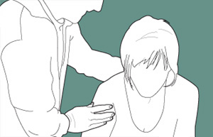
These panic attacks are less comprehensive, with fewer than four bodily symptoms being experienced. [9] It is not unusual to experience only one or two symptoms at a time, such as vibrations in their legs, shortness of breath, or an intense wave of heat traveling up their bodies, which is not similar to hot flashes due to estrogen shortage.
Furthermore, contrary to common belief, and in contrast to diving conditions, the role of facial barotrauma in the cause of in-flight barodontalgia is only minor (about one-tenth of cases). [14] Classification [ edit ] The Fédération dentaire internationale describes 4 classes of barodontalgia. [1] The classes are based on signs and symptoms.
When laryngospasm is coincident with a cold or flu, it may be helpful for some sufferers to take acid reflux medication to limit the irritants in the area. [ citation needed ] If a cough is present, then treat a wet cough; but limit coughing whenever possible, as it is only likely to trigger a spasm. [ citation needed ] Drink water or tea to keep the area from drying up. [ citation needed ] Saline drops also help to keep the area moist. [ citation needed ] Pseudoephederine may also help to clear any mucus that may cause coughing and thereby triggering more spasms. [ citation needed ] Treatment [ edit ] Minor laryngospasm will generally resolve spontaneously in the majority of cases. [1] Laryngospasm in the operating room is treated by hyperextending the patient's neck and administering assisted ventilation with 100% oxygen .
Please improve it by verifying the claims made and adding inline citations . Statements consisting only of original research should be removed. ( October 2013 ) ( Learn how and when to remove this template message ) ( Learn how and when to remove this template message ) GEFS+ Specialty Neurology Generalized epilepsy with febrile seizures plus ( GEFS+ ) is a syndromic autosomal dominant disorder where afflicted individuals can exhibit numerous epilepsy phenotypes. [1] GEFS+ can persist beyond early childhood (i.e., 6 years of age).
This process takes place over months to years and eventually causes disabling arthritis , particularly of the femoral head (hip). [2] Dysbaric osteonecrosis lesions are typically bilateral and usually occur at both ends of the femur and at the proximal end of the humerus . Symptoms are usually only present when a joint surface is involved, which typically does not occur until a long time after the causative exposure to a hyperbaric environment.
Between agencies, current tolerable upper limit guidelines vary from 10 mg per day to 100 mg per day. [37] Daily vitamin B 6 tolerable upper limits for adults as established by agency Agency Upper limit Notes Reference National Health Service (NHS) United Kingdom 10 mg/day [40] Norwegian Scientific Committee for Food and Environment (VKM) 25 mg/day In 2017 VKM proposed to raise this to 25 mg/day, it was previously 4.2 mg/day. [37] Netherlands Food and Consumer Product Safety Authority [ nl ] (NVWA) 25 mg/day Supplements may only contain dosages of 21 mg/day. [41] European Food Safety Authority 25 mg/day [42] National Health and Medical Research Council (NHMRC) Australia 50 mg/day [43] U.S.
In tests of the effect of hydrogen narcosis, where divers dived to 500 m (1,600 ft) with a hydrogen–helium–oxygen ( Hydreliox ) mixture containing 49% hydrogen, it was found that while the narcotic effect of hydrogen was detectable, the neurological symptoms of high-pressure nervous syndrome were only moderate. [2] [3] See also [ edit ] Nitrogen narcosis – Reversible narcotic effects of respiratory nitrogen at elevated partial pressures References [ edit ] ^ Taylor, Larry "Harris".
A single layer of cells are surrounding the basal membrane. [13] In 2014, PRCC was first acknowledged as a renal tumor subtype by the World Health Organization (WHO) considering its distinct genetic, molecular and histologic characteristics. [10] It is further divided into type 1 and type 2 based on morphological features. [14] Type 1 Papillary Renal Cell Carcinoma [ edit ] A micrograph of Type 2 PRCC, showing cells containing abundant eosinophilic granular cytoplasm with prominent nucleoli. [13] Type 1 PRCC, also known as a renal tumor caused by a genetic predisposition of hereditary papillary renal cancer syndrome, compromises approximately 25% of all PRCCs. [15] [16] In the perspective of immunochemistry, it has a profile of strong CK7 and alpha-methyl acyl-CoA racemase (AMACR) expression at most focal CA-IX expression. [17] Histologically, its epithelium is composed of relatively small-sized simple cuboidal cells lined in a single layer. [18] These cells are well-characterized by basophilic cytoplasm. [19] Due to its solid growth, an extremely compact papillary architecture is often observed. [17] Other morphological characteristics include intracellular hemosiderin and foamy macrophages placed inside of papillary fibrovascular cores or psammoma bodies. [20] In general, the nuclei of type 1 PRCC belong to grade 1-2 of the Fuhrman system. [17] Type 2 Papillary renal cell carcinoma [ edit ] Accounting for 25% of PRCCs, type 2 PRCC is the pathological subtype that is most commonly associated with hereditary leiomyomatosis and renal cell carcinoma (HLRCC) syndrome. [15] [16] When compared to type 1, it shows more variation in protein expression mostly by loss of CK7. [17] In a gross examination, it shows papillae covered by large cells abundant in eosinophilic cytoplasm. [19] Its large spherical nuclei on papillary cores are arranged in a pseudo-stratified manner. [17] Unlike type 1 PRCC, foamy macrophages and psammoma bodies are less common in case of type 2. [16] The majority of type 2 PRCC has high Fuhrman grade nuclei with prominent nucleoli. [17] Signs and Symptoms [ edit ] Due to its asymptomatic nature, PRCC is often undetectable, and the majority of cases are incidentally diagnosed during the radiological workup of unrelated diseases. [3] [8] Its clinical manifestations are similar to those of clear cell RCC , which are the classical triad of renal cell carcinoma ( hematuria , flank pain and palpable abdominal mass ; only 6-10% of patients) or even nonspecific symptoms including fatigue , weight loss , fever , and anorexia . [4] [10] [21] Since early diagnosis is relatively uncommon, PRCC patients may experience symptoms caused by the metastatic spread to secondary sites.
Papillary renal cell carcinoma (PRCC) is a type of cancer that occurs in the kidneys. It accounts for about 10-15% of all renal cell carcinomas. R enal cell carcinoma s are a type of kidney cancer that develop in the lining of very small tubes (tubules) in the kidney.The term "papillary" describes the finger-like projections that can be found in most of the tumors. PRCC can be divided into two types: type 1, which is more common and usually grows more slowly and type 2, which are usually more aggressive .Though the exact cause of papillary renal cell carcinoma is unknown, smoking, obesity, and genetic predisposition conditions (such as hereditary leiomyomatosis and renal cell cancer ) may contribute to the development of this type of cancer. Treatment often begins with surgery to remove as much of the cancer as possible, and may be followed by radiation therapy, chemotherapy, biological therapy, or targeted therapy .
Papillary renal cell carcinoma is a rare subtype of renal cell carcinoma, arising from the renal tubular epithelium and showing a papillary growth pattern, which typically manifests with hematuria, flank pain, palpable abdominal mass or nonspecific symptoms, such as fatigue, weight loss or fever. Symptoms related to metastatic spread, such as bone pain or persistent cough, are frequently associated since early diagnosis is not common. It is typically multifocal, bilateral, and in most cases sporadic, although different hereditary syndromes, such as Hereditary leiomyoma renal cell carcinoma, Birt-Hogg-Dubé syndrome and Tuberous sclerosis, may predispose to the development of papillary renal cell carcinoma.
DTI), [2] and drug screening assays, such as stem cell -derived hepatocyte-like cells, that are capable of detecting toxicity early in the drug development process. [3] Chemicals often cause subclinical injury to the liver, which manifests only as abnormal liver enzyme tests .