The cycloid psychosis is therefore only partially described in the diagnostic classification systems used. ... Because some dietary supplements may also induce psychosis or mania, but cannot be ruled out with laboratory tests, a psychotic individual's family, partner, or friends should be asked whether the patient is currently taking any dietary supplements. [110] Common mistakes made when diagnosing people who are psychotic include: [108] Not properly excluding delirium, Not appreciating medical abnormalities (e.g., vital signs), Not obtaining a medical history and family history, Indiscriminate screening without an organizing framework, Missing a toxic psychosis by not screening for substances and medications, Not asking their family or others about dietary supplements, Premature diagnostic closure, and Not revisiting or questioning the initial diagnostic impression of primary psychiatric disorder. Only after relevant and known causes of psychosis are excluded, a mental health clinician may make a psychiatric differential diagnosis using a person's family history, incorporating information from the person with psychosis, and information from family, friends, or significant others. ... Classification [ edit ] The word was also used to distinguish a condition considered a disorder of the mind, as opposed to neurosis , which was considered a disorder of the nervous system. [136] The psychoses thus became the modern equivalent of the old notion of madness , and hence there was much debate on whether there was only one (unitary) or many forms of the new disease. [137] One type of broad usage would later be narrowed down by Koch in 1891 to the 'psychopathic inferiorities'—later renamed abnormal personalities by Schneider . [132] The division of the major psychoses into manic depressive illness (now called bipolar disorder ) and dementia praecox (now called schizophrenia ) was made by Emil Kraepelin , who attempted to create a synthesis of the various mental disorders identified by 19th-century psychiatrists , by grouping diseases together based on classification of common symptoms. ... Hippocrates writes: Men ought to know that from the brain, and from the brain only, arise our pleasures, joys, laughter, and jests, as well as our sorrows, pains, griefs and tears.
Typical treatment for chronic iron overload requires subcutaneous injection over a period of 8–12 hours daily. [ citation needed ] Two newer iron-chelating drugs that are licensed for use in patients receiving regular blood transfusions to treat thalassaemia (and, thus, who develop iron overload as a result) are deferasirox and deferiprone . [25] [26] Chelating polymers [ edit ] A nover experimental approach to the hereditary haemochromatosis treatment is the maintenance therapy with polymeric chelators . [27] [28] [29] These polymers or particles have a negligible or null systemic biological availability and they are designed to form stable complexes with Fe 2+ and Fe 3+ in the GIT and thus limiting their uptake and long-term accumulation. Although this method has only a limited efficacy, unlike small-molecular chelators , the approach has virtually no side effects in sub-chronic studies. [29] Interestingly, the simultaneous chelation of Fe 2+ and Fe 3+ increases the treatment efficacy. [29] Prognosis [ edit ] In general, provided there has been no liver damage, patients should expect a normal life expectancy if adequately treated by venesection.
They concluded 'no' because the benefit exceeds 1 year only if the prevalence of aneurysm exceeds 30%, the surgical complication rate is 1% or less, and the patient is under 25 years of age. ... The Caucasian family studied by Vinet et al. (1989) had the leftward type of deletion alpha(+)-thalassemia which, except for 1 case in a Mediterranean population (Troungos et al., 1984), had been described only in Asiatic populations (Winichagoon et al., 1984). ... Hannig et al. (1992) found that of 5,026 renal transplants done in 1988, 390 (7.8%) involved a PKD1 recipient. Only 7% of these 390 transplants used a living, related donor compared to the 20% rate reported for all renal transplants. DNA linkage studies were not used by any of the centers surveyed and only 29% reported provision of risk counseling. ... Prenatal Diagnosis Breuning et al. (1990) recommended that prenatal diagnosis of PKD by chorionic villus sampling should be attempted only after the linkage phase of the DNA markers has been established by haplotyping the index family.
Polycystic kidney disease is a disorder that affects the kidneys and other organs. Clusters of fluid-filled sacs, called cysts, develop in the kidneys and interfere with their ability to filter waste products from the blood. The growth of cysts causes the kidneys to become enlarged and can lead to kidney failure. Cysts may also develop in other organs, particularly the liver. Frequent complications of polycystic kidney disease include dangerously high blood pressure (hypertension), pain in the back or sides, blood in the urine (hematuria), recurrent urinary tract infections, kidney stones, and heart valve abnormalities. Additionally, people with polycystic kidney disease have an increased risk of an abnormal bulging (an aneurysm ) in a large blood vessel called the aorta or in blood vessels at the base of the brain.
A rare, genetic, renal tubular disease characterized by progressive outgrowths of fluid-filled cysts from the renal epithelium, which can manifest with hematuria, urinary tract infections, hypertension, and abdominal or flank pain. The slowly progressive loss of kidney function may evolve to end stage kidney disease (ESKD). Epidemiology Autosomal dominant polycystic kidney disease (ADPKD) is the most common inherited renal disease, with estimated prevalence of 1/2,500 in Europe. Clinical description Symptoms typically manifests in adulthood; however, approximately 2-5% of cases present before 15 years of age. The renal phenotype ranges from patients in advanced age with preserved kidney function to rare cases of enlarged kidneys that are detected in utero.
Sequence analysis of TBC1D24 is performed first and followed by gene-targeted deletion/duplication analysis if only one or no pathogenic variant is found.
A family history of more than one individual with a pituitary adenoma Note: (1) A germline AIP pathogenic variant is identified in approximately 20% of families with FIPA [Chahal et al 2010] and in 40% of families in which somatotropinomas are the only tumor type observed. (2) To date, AIP pathogenic variants have not been identified in families with two adults with microprolactinomas (prolactin secreting tumors <10 mm in diameter); therefore, the probability of identifying an AIP pathogenic variant in such a family is low.
The incidence of deaths due to silicosis declined by 84% between 1968 and 1999, and only 187 deaths in 1999 had silicosis as the underlying or contributing cause. [24] Additionally, cases of silicosis in Michigan, New Jersey, and Ohio are highly correlated to industry and occupation. [25] Although silicosis has been known for centuries, the industrialization of mining has led to an increase in silicosis cases.
Silicosis is a respiratory disease caused by breathing in (inhaling) silica dust. There are three types of silicosis: Simple chronic silicosis, the most common type of silicosis, results from long-term exposure (usually more than 20 years) to low amounts of silica dust. Simple chronic silicosis may cause people to have difficulty breathing. Accelerated silicosis occurs after 5 to 15 years of exposure of higher levels of silica. Swelling of the lungs and other symptoms occur faster in this type of silicosis than in the simple chronic form.
In most cases, relatives at risk need only be tested for the family-specific germline pathogenic variant, except in the following situations: Individuals of Ashkenazi Jewish heritage should consider testing for all three founder germline pathogenic variants because of the high population frequency of these founder pathogenic variants as well as reports of the coexistence of more than one founder germline pathogenic variant in some families.
Alcohol related brain damage is not only due to the direct toxic effects of alcohol; alcohol withdrawal, nutritional deficiency, electrolyte disturbances, and liver damage are also believed to contribute to alcohol-related brain damage. [98] Cognition and dementia [ edit ] Excessive alcohol intake is associated with impaired prospective memory . ... In cases of severe liver disease, the only treatment option may be a liver transplant from alcohol abstinent donors. ... However, the benefits of drinking occurred only among nonsmokers. [...] Although alcohol consumption did not influence risk of clinical illness for smokers, moderate alcohol consumption was associated with decreased risk for nonsmokers." [184] Another study concluded, "Findings suggest that wine intake, especially red wine, may have a protective effect against common cold. ... A study conducted in Germany in 2016 found the economic burden for those dependent on alcohol was 50% higher than those who were not. [211] In the study, over half of the economic cost was due to lost productivity, and only 6% was due to alcohol treatment programs.
However, these loci are estimated to only account for 25-44% of the genetic component of insulin resistance. [26] Pathophysiology [ edit ] In normal metabolism, the elevated blood glucose instructs beta (β) cells in the Islets of Langerhans , located in the pancreas , to release insulin into the blood.
A number sign (#) is used with this entry because of evidence that estrogen resistance (ESTRR) is caused by homozygous mutation in the alpha estrogen receptor gene (ESR1; 133430) on chromosome 6q25. Description Estrogen resistance (ESTRR) is characterized by absence of puberty with elevated estradiol and gonadotropic hormones, as well as markedly delayed bone maturation. Female patients show absent breast development, small uterus, and enlarged multicystic ovaries; male patients may show small testes (Bernard et al., 2017). Some patients exhibit continued growth into adulthood (Smith et al., 1994). Clinical Features Smith et al. (1994) described a 28-year-old man with estrogen resistance.
Estrogen resistance syndrome is a rare, genetic endocrine disease characterized by estrogen-receptor insensitivity to estrogens and the presence of elevated estrogen and gonadotropin serum levels. Clinical manifestations include absent breast development and primary amenorrhea in association with multicystic ovaries and/or hypoplastic uterus in female patients, normal or abnormal gonadal development in male patients and markedly delayed bone maturation, persistence of open epiphyses, reduced bone mineral density, and variable tall stature in both sexes. Glucose intolerance, hyperinsulinemia and lipid abnormalities may also be present.
The RNA from the expanded trinucleotide repeat region forms intranucleoplasmic hairpin loops due to the extensive hydrogen bonding between C-G base pairs, and it has been demonstrated that these sequester the splicing regulator MBNL1 to form distinctive foci. [11] DM2 [ edit ] DM2 is caused by a defect of the CNBP gene on chromosome 3 . [12] The specific defect is a repeat of the cytosine-cytosine-thymine-guanosine (CCTG) tetranucleotide in the CNBP gene. [12] As it involves the repeat of four nucleotides, it is not a trinucleotide repeat disorder , but rather a tetranucleotide repeat disorder. [13] [14] The repeat expansion for DM2 is much larger than for DM1, ranging from 75 to over 11,000 repeats. [12] Unlike in DM1, the size of the repeated DNA expansion in DM2 does not appear to make a difference in the age of onset or disease severity. [9] Anticipation appears to be less significant in DM2 and most current reviews only report mild anticipation as a feature of DM2. [ citation needed ] Diagnosis [ edit ] This section needs more medical references for verification or relies too heavily on primary sources .
Myotonic dystrophy is a disease that affects the muscles and other body systems. It is the most common form of muscular dystrophy that begins in adulthood, usually in a person’s 20s or 30s. This disease is characterized by progressive muscle loss and weakness. Myotonic dystrophy may be further classified into two types, and the two types may affect different muscles. People with myotonic dystrophy usually have prolonged muscle tensing ( myotonia ) and are not able to relax certain muscles after use.
These symptoms include the inability to be intimate ( intimacy anorexia ), depression and bipolar disorders. [51] The resulting hypersexuality may have an impact in the person's social and occupational domains if the underlying symptoms have a large enough systemic influence. [52] [53] As a disorder [ edit ] Main articles: Sexual addiction and Hypersexual disorder As of 2010 [update] , a proposal to add Sexual Addiction to the Diagnostic and Statistical Manual of Mental Disorders (DSM) system has failed to get support of the American Psychiatric Association (APA). [54] [55] [56] The DSM does include an entry called Sexual Disorder Not Otherwise Specified (Sexual Disorder NOS) to apply to, among other conditions, "distress about a pattern of repeated sexual relationships involving a succession of lovers who are experienced by the individual only as things to be used". [57] The International Statistical Classification of Diseases and Related Health Problems (ICD-10) of the World Health Organization (WHO), includes two relevant entries.
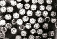
There are several licensed test kits on the market which are sensitive, specific and detect all serotypes of rotavirus A . [40] Other methods, such as electron microscopy and PCR , are used in research laboratories. [18] Reverse transcription-polymerase chain reaction ( RT-PCR ) can detect and identify all species and serotypes of human rotavirus. [41] Prevention [ edit ] Because improved sanitation does not decrease the prevalence of rotaviral disease, and the rate of hospitalisations remains high, despite the use of oral rehydrating medicines, the primary public health intervention is vaccination. [2] Two rotavirus vaccines against Rotavirus A infection are safe and effective in children: [13] Rotarix by GlaxoSmithKline [42] and RotaTeq by Merck . [43] Both are taken orally and contain attenuated live virus. [13] Rotavirus vaccines are licensed in more than 100 countries, but only 17 countries have introduced routine rotavirus vaccination. [44] Following the introduction of routine rotavirus vaccination in the US in 2006, the health burden of rotavirus gastroenteritis "rapidly and dramatically reduced" despite lower coverage levels compared to other routine infant immunizations. [45] Clinical trials of the Rotarix rotavirus vaccine in South Africa and Malawi, found that the vaccine significantly reduced severe diarrhoea episodes caused by rotavirus, and that the infection was preventable by vaccination. [46] A 2019 Cochrane systematic review of 55 clinical trials that included 216,480 participants concluded RV1 (Rotarix), RV5 (RotaTeq), and Rotavac and are effective vaccines. [47] Additional rotavirus vaccines are under development. [48] The World Health Organization (WHO) recommends that rotavirus vaccine be included in all national immunisation programmes. [49] The incidence and severity of rotavirus infections has declined significantly in countries that have acted on this recommendation. [12] [13] The Rotavirus Vaccine Program is a collaboration between PATH , the (WHO), and the U.S. ... External links [ edit ] WHO Rotavirus web page CDC About Rotavirus Classification D ICD - 10 : A08.0 ICD - 9-CM : 008.61 MeSH : D012400 DiseasesDB : 11667 External resources MedlinePlus : 000252 eMedicine : emerg/401 v t e Infectious diseases – viral systemic diseases Oncovirus DNA virus HBV Hepatocellular carcinoma HPV Cervical cancer Anal cancer Penile cancer Vulvar cancer Vaginal cancer Oropharyngeal cancer KSHV Kaposi's sarcoma EBV Nasopharyngeal carcinoma Burkitt's lymphoma Hodgkin lymphoma Follicular dendritic cell sarcoma Extranodal NK/T-cell lymphoma, nasal type MCPyV Merkel-cell carcinoma RNA virus HCV Hepatocellular carcinoma Splenic marginal zone lymphoma HTLV-I Adult T-cell leukemia/lymphoma Immune disorders HIV AIDS Central nervous system Encephalitis / meningitis DNA virus Human polyomavirus 2 Progressive multifocal leukoencephalopathy RNA virus MeV Subacute sclerosing panencephalitis LCV Lymphocytic choriomeningitis Arbovirus encephalitis Orthomyxoviridae (probable) Encephalitis lethargica RV Rabies Chandipura vesiculovirus Herpesviral meningitis Ramsay Hunt syndrome type 2 Myelitis Poliovirus Poliomyelitis Post-polio syndrome HTLV-I Tropical spastic paraparesis Eye Cytomegalovirus Cytomegalovirus retinitis HSV Herpes of the eye Cardiovascular CBV Pericarditis Myocarditis Respiratory system / acute viral nasopharyngitis / viral pneumonia DNA virus Epstein–Barr virus EBV infection / Infectious mononucleosis Cytomegalovirus RNA virus IV : Human coronavirus 229E / NL63 / HKU1 / OC43 Common cold MERS coronavirus Middle East respiratory syndrome SARS coronavirus Severe acute respiratory syndrome SARS coronavirus 2 Coronavirus disease 2019 V , Orthomyxoviridae : Influenza virus A / B / C / D Influenza / Avian influenza V, Paramyxoviridae : Human parainfluenza viruses Parainfluenza Human orthopneumovirus hMPV Human digestive system Pharynx / Esophagus MuV Mumps Cytomegalovirus Cytomegalovirus esophagitis Gastroenteritis / diarrhea DNA virus Adenovirus Adenovirus infection RNA virus Rotavirus Norovirus Astrovirus Coronavirus Hepatitis DNA virus HBV ( B ) RNA virus CBV HAV ( A ) HCV ( C ) HDV ( D ) HEV ( E ) HGV ( G ) Pancreatitis CBV Urogenital BK virus MuV Mumps v t e Diseases of the digestive system Upper GI tract Esophagus Esophagitis Candidal Eosinophilic Herpetiform Rupture Boerhaave syndrome Mallory–Weiss syndrome UES Zenker's diverticulum LES Barrett's esophagus Esophageal motility disorder Nutcracker esophagus Achalasia Diffuse esophageal spasm Gastroesophageal reflux disease (GERD) Laryngopharyngeal reflux (LPR) Esophageal stricture Megaesophagus Esophageal intramural pseudodiverticulosis Stomach Gastritis Atrophic Ménétrier's disease Gastroenteritis Peptic (gastric) ulcer Cushing ulcer Dieulafoy's lesion Dyspepsia Pyloric stenosis Achlorhydria Gastroparesis Gastroptosis Portal hypertensive gastropathy Gastric antral vascular ectasia Gastric dumping syndrome Gastric volvulus Buried bumper syndrome Gastrinoma Zollinger–Ellison syndrome Lower GI tract Enteropathy Small intestine ( Duodenum / Jejunum / Ileum ) Enteritis Duodenitis Jejunitis Ileitis Peptic (duodenal) ulcer Curling's ulcer Malabsorption : Coeliac Tropical sprue Blind loop syndrome Small bowel bacterial overgrowth syndrome Whipple's Short bowel syndrome Steatorrhea Milroy disease Bile acid malabsorption Large intestine ( Appendix / Colon ) Appendicitis Colitis Pseudomembranous Ulcerative Ischemic Microscopic Collagenous Lymphocytic Functional colonic disease IBS Intestinal pseudoobstruction / Ogilvie syndrome Megacolon / Toxic megacolon Diverticulitis / Diverticulosis / SCAD Large and/or small Enterocolitis Necrotizing Gastroenterocolitis IBD Crohn's disease Vascular : Abdominal angina Mesenteric ischemia Angiodysplasia Bowel obstruction : Ileus Intussusception Volvulus Fecal impaction Constipation Diarrhea Infectious Intestinal adhesions Rectum Proctitis Radiation proctitis Proctalgia fugax Rectal prolapse Anismus Anal canal Anal fissure / Anal fistula Anal abscess Hemorrhoid Anal dysplasia Pruritus ani GI bleeding Blood in stool Upper Hematemesis Melena Lower Hematochezia Accessory Liver Hepatitis Viral hepatitis Autoimmune hepatitis Alcoholic hepatitis Cirrhosis PBC Fatty liver NASH Vascular Budd–Chiari syndrome Hepatic veno-occlusive disease Portal hypertension Nutmeg liver Alcoholic liver disease Liver failure Hepatic encephalopathy Acute liver failure Liver abscess Pyogenic Amoebic Hepatorenal syndrome Peliosis hepatis Metabolic disorders Wilson's disease Hemochromatosis Gallbladder Cholecystitis Gallstone / Cholelithiasis Cholesterolosis Adenomyomatosis Postcholecystectomy syndrome Porcelain gallbladder Bile duct / Other biliary tree Cholangitis Primary sclerosing cholangitis Secondary sclerosing cholangitis Ascending Cholestasis / Mirizzi's syndrome Biliary fistula Haemobilia Common bile duct Choledocholithiasis Biliary dyskinesia Sphincter of Oddi dysfunction Pancreatic Pancreatitis Acute Chronic Hereditary Pancreatic abscess Pancreatic pseudocyst Exocrine pancreatic insufficiency Pancreatic fistula Other Hernia Diaphragmatic Congenital Hiatus Inguinal Indirect Direct Umbilical Femoral Obturator Spigelian Lumbar Petit's Grynfeltt-Lesshaft Undefined location Incisional Internal hernia Richter's Peritoneal Peritonitis Spontaneous bacterial peritonitis Hemoperitoneum Pneumoperitoneum
Many EID are zoonotic , [3] deriving from pathogens present in animals, with only occasional cross-species transmission into human populations. [11] For instance, most emergent viruses are zoonotic [3] (whereas other novel viruses may have been circulating in the species without being recognized, as occurred with hepatitis C [12] ).
Antibiotics [ edit ] Majority of studies on ORN have recommended the use of prophylactic antibiotic where extractions are needed post-radiotherapy, although there is no universally agreed choice, timing and course duration of antibiotic regime. [31] In one study, it was discovered that cases after 1986, the incidence of ORN after extractions post-radiotherapy was 3.6% in antibiotic prescribed cases and 2.6-3.4% in cases where there is no report on the prescription of antibiotics, showing no difference in reducing the risk of ORN and possibly reconsidering antibiotic regime in preventing ORN. [32] Hyperbaric oxygen therapy [ edit ] Results since 1986 have shown of far lower rates of ORN incidence, even without HBO (3.1-3.5%) and even a slightly higher rate for HBO patients (4.0%). [32] Prophylactic use of HBO has been recommended in some studies [33] [30] with a Cochrane review suggesting evidence for some reduction in ORN. [34] However, the use of HBO prophylaxis is not agreed by others due to the insufficient evidence. [35] Majority of British maxillofacial surgeons who participated in a survey recommended prophylactic HBO but protocols are varied. [36] Management [ edit ] Conservative [ edit ] Antiseptic mouthwashes: Mouthrinses such as 0.02% aqueous chlorhexidine and saline can be used in acute ORN together with analgesics and anti-inflammatory drugs. [25] Antibiotics: In order to eliminate infection in the region, tetracyclines can be prescribed due to their selective uptake by bone. [25] Penicillin antibiotics can also be used because of their superficial contamination with oral bacteria. [17] Ultrasound therapy: First introduced as a treatment modality in 1992, [10] it involves the application of high-frequency sound waves to induce angiogenesis and improve blood circulation to muscles. [37] [38] Harris demonstrated that 48% of cases showed healing when 15 minutes of ultrasound therapy was applied daily on the skin affected by ORN, while combined with debridement. [10] Hyperbaric oxygen therapy (HBO): First described in 1973, HBO was intended to be an adjunctive treatment method in ORN. [39] The theoretical basis behind this treatment was the fact that it causes an increase in tissue oxygen tension and an improvement in collagen synthesis, angiogenesis and epithelialization. [40] However, its use as the only method in the management of ORN is controversial.
It may indicate both a nerve or muscular lesion. [16] Pallor and pulselessness – A lack of pulse rarely occurs in patients, as pressures that cause compartment syndrome are often well below arterial pressures. Absent pulses only occur when there is arterial injury or during the late stages of the compartment syndrome, when compartment pressures are very high. [5] Pallor can also result from arterial occlusion.
Thomas Forestier, a physician during the first outbreak, provided a written account of his own experiences with the sweating sickness in 1485. [5] Forestier put great emphasis on the sudden breathlessness that is commonly associated with the final hours of those who had contracted this disease. [5] Forestier claimed in an account written for other physicians that "loathsome vapors" had congregated around the heart and lungs. [5] His observations point towards a pulmonary component of this disease that was previously unknown. [5] Transmission [ edit ] Transmission of the sweating sickness mostly remains a mystery, with only a few pieces of evidence in written works. [4] The names that the peasantry called the disease, such as "Stup-Gallant", lead to the possibility that it affected the rich much more often. [4] The greater number of people in London to witness the coronation of Henry VIII may have greatly increased the spread of this disease. [4] Cause [ edit ] The cause is the most mysterious aspect of the disease. ... External links [ edit ] Sweating Fever Jim Leavesley commemorates the 500th anniversary of the first outbreak – transcript of talk on Ockham's Razor , ABC Radio National "The Sweating Sickness Returns" , Discover Magazine , 1 June 1997 Classification D ICD - 9-CM : 078.2 MeSH : D018614 v t e Infectious diseases – viral systemic diseases Oncovirus DNA virus HBV Hepatocellular carcinoma HPV Cervical cancer Anal cancer Penile cancer Vulvar cancer Vaginal cancer Oropharyngeal cancer KSHV Kaposi's sarcoma EBV Nasopharyngeal carcinoma Burkitt's lymphoma Hodgkin lymphoma Follicular dendritic cell sarcoma Extranodal NK/T-cell lymphoma, nasal type MCPyV Merkel-cell carcinoma RNA virus HCV Hepatocellular carcinoma Splenic marginal zone lymphoma HTLV-I Adult T-cell leukemia/lymphoma Immune disorders HIV AIDS Central nervous system Encephalitis / meningitis DNA virus Human polyomavirus 2 Progressive multifocal leukoencephalopathy RNA virus MeV Subacute sclerosing panencephalitis LCV Lymphocytic choriomeningitis Arbovirus encephalitis Orthomyxoviridae (probable) Encephalitis lethargica RV Rabies Chandipura vesiculovirus Herpesviral meningitis Ramsay Hunt syndrome type 2 Myelitis Poliovirus Poliomyelitis Post-polio syndrome HTLV-I Tropical spastic paraparesis Eye Cytomegalovirus Cytomegalovirus retinitis HSV Herpes of the eye Cardiovascular CBV Pericarditis Myocarditis Respiratory system / acute viral nasopharyngitis / viral pneumonia DNA virus Epstein–Barr virus EBV infection / Infectious mononucleosis Cytomegalovirus RNA virus IV : Human coronavirus 229E / NL63 / HKU1 / OC43 Common cold MERS coronavirus Middle East respiratory syndrome SARS coronavirus Severe acute respiratory syndrome SARS coronavirus 2 Coronavirus disease 2019 V , Orthomyxoviridae : Influenza virus A / B / C / D Influenza / Avian influenza V, Paramyxoviridae : Human parainfluenza viruses Parainfluenza Human orthopneumovirus hMPV Human digestive system Pharynx / Esophagus MuV Mumps Cytomegalovirus Cytomegalovirus esophagitis Gastroenteritis / diarrhea DNA virus Adenovirus Adenovirus infection RNA virus Rotavirus Norovirus Astrovirus Coronavirus Hepatitis DNA virus HBV ( B ) RNA virus CBV HAV ( A ) HCV ( C ) HDV ( D ) HEV ( E ) HGV ( G ) Pancreatitis CBV Urogenital BK virus MuV Mumps v t e Pandemics , epidemics and notable disease outbreaks List of epidemics Local Ancient Hittite plague ( c. 1320–1300 BC) Plague of Athens (429–426 BC) Antonine Plague (165–180 AD) Plague of Cyprian (250–266) Post-classical First plague pandemic (541–767) Plague of Justinian (541–542) Roman Plague (590) Plague of Sheroe (627–628) Plague of Amwas (638–639) Plague of 664 (664–689) Japanese smallpox (735–737) Black Death (1347–1351) Sweating sickness (1485–1551) Early modern 16th century Influenza pandemic (1510) Mexican smallpox (1520) Influenza pandemic (1557–1559) London plague (1563–1564) Maltese plague (1592–1593) London plague (1592–1593) 17th century Maltese plague (1623) Italian plague (1629–1631) Massachusetts smallpox (1633) Great Plague of Seville (1647–1652) Maltese plague (1655) Naples Plague (1656) Great Plague of London (1665–1666) Maltese plague (1675–1676) Great Plague of Vienna (1679) 18th century Great Northern War plague (1710–1712) Great Plague of Marseille (1720–1722) Great Plague of 1738 (1738) Russian plague (1770–1772) Persian Plague (1772) North American smallpox (1780–1782) Philadelphia yellow fever (1793–1798) Modern 19th century Ottoman plague (1812–1819) Maltese plague (1813–1814) Caragea's plague (1813) Groningen epidemic (1829) Great Plains smallpox (1837–1838) Typhus (1847–1848) Copenhagen cholera (1853) Stockholm cholera (1853) Broad Street cholera (1854) Buenos Aires yellow fever (1871) 20th century San Francisco plague (1900–1904) Manchurian plague (1910–1911) LA pneumonic plague (1924) Croydon typhoid (1937) NYC smallpox (1947) Yugoslav smallpox (1972) London flu (1972–1973) Indian smallpox (1974) Surat plague (1994) Malaysian Nipah virus (1998–1999) 21st century Singaporean dengue (2005) Indian dengue (2006) Chikungunya outbreaks (2006) Pakistani dengue (2006) Iraqi cholera (2007) Zimbabwean cholera (2008–2009) Bolivian dengue (2009) Gujarat hepatitis (2009) Western African meningitis (2009–2010) Haiti cholera (2010–2019) Pakistani dengue (2011) Darfur yellow fever (2012) Swansea measles (2013) Western African Ebola (2013–2016) DR Congo Ebola (2014) Madagascar plague (2014) Odisha jaundice (2014) Polio declaration (2014) Indian swine flu (2015) South Korean MERS (2015) Angolan yellow fever (2016) Yemeni cholera (2016–present) Gorakhpur Japanese encephalitis (2017) Saudi Arabian MERS (2018) Kerala Nipah virus (2018) Équateur province Ebola (2018) Kivu Ebola (2018–2020) Madagascar measles (2018) Samoa measles (2019–present) Philippine measles (2019–present) Pacific NW measles (2019) New York measles (2019) Kuala Koh measles (2019) Tonga measles (2019) DRC measles (2019–2020) New Zealand measles (2019–present) Global Influenza pandemic (1510) Influenza pandemic (1557–1559) Second plague pandemic (1348–19th century) First cholera pandemic (1816–1826) Second cholera pandemic (1829–1851) Third cholera pandemic (1852–1860) Third plague pandemic (1855–1960) Fourth cholera pandemic (1863–1879) Fifth cholera pandemic (1881–1896) 1889 flu (1889–1890) Sixth cholera pandemic (1899–1923) Spanish flu (1918–1920) Asian flu (1957–1958) Seventh cholera pandemic (1961–1975) Hong Kong flu (1968–1970) HIV/AIDS (1981–present) SARS (2002–2004) Bird flu (2003–2005) Mumps (2009) Madagascar plague (2008–2017) Swine flu (2009–2010) MERS (2012–present) Chikungunya (2013–2014) Zika (2015–2016) COVID-19 (2019–present)
An example is carcinoid syndrome , a disease in which neuroendocrine tumors along the GI tract use tryptophan as the source for serotonin production, which limits the available tryptophan for niacin synthesis. In normal patients, only one percent of dietary tryptophan is converted to serotonin; however, in patients with carcinoid syndrome, this value may increase to 70%.
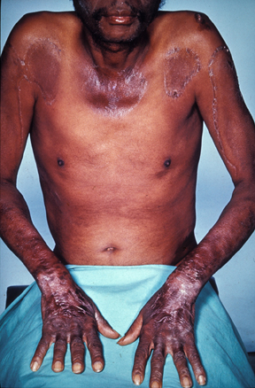
Pellagra is a nutritional disorder caused by a deficiency in niacin (vitamin B3) or its precursor (tryptophan) that is mainly observed in Asia and Africa where it is generally due to poor nutrition. It is characterized by dermatitis (symmetrical photodistributed erythema that may be accompanied by vesicles and bullae, and that develops into hyperkeratotic and hyperpigmented skin), gastrointestinal symptoms (diarrhea), and neuropsychiatric disorders (dementia). It can be life-threatening without a correct management.
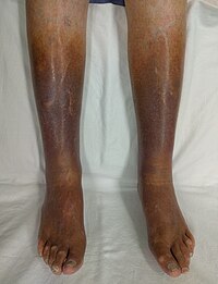
Venous stasis Other names Venostasis Skin changes as a result of long term venous stasis Venostasis , or venous stasis , is a condition of slow blood flow in the veins , usually of the legs. Contents 1 Causes 2 Complications 3 See also 4 References Causes [ edit ] Causes of venous stasis include: Chronic venous insufficiency . [1] Congestive heart failure . [2] Long periods of immobility that can be encountered from driving, [3] flying, bed rest/hospitalization, or having an orthopedic cast . Recommendations by clinicians to reduce venous stasis and DVT/PE often encourage increasing walking, calf exercises, and intermittent pneumatic compression when possible. [4] [5] [6] Complications [ edit ] Potential complications of venous stasis are: Venous ulcers Blood clot formation in veins ( venous thrombosis ), as with the deep veins of the legs ( deep vein thrombosis or DVT), potentially indicating thrombosis prophylaxis . [7] See also [ edit ] Virchow's triad References [ edit ] ^ "Chronic Venous Insufficiency (CVI)" . Cleveland Clinic . Last reviewed by a Cleveland Clinic medical professional on 05/14/2019. ^ Zhu, Ruiqi; Hu, Yu; Tang, Liang (2017). "Reduced cardiac function and risk of venous thromboembolism in Asian countries" .