The proband was a 32-year-old woman who presented at age 26 years with palpitations, at which time electrocardiography (ECG) showed atrial fibrillation with a slow ventricular rate response. During periods of sinus rhythm, extremely low amplitude P-waves and prolonged atrioventricular (AV) conduction (first-degree AV block) were observed. ... In addition, 48-hour ambulatory heart rate monitoring showed profound sinus bradycardia with sinus rates as low as 33 beats per minute.
The patient's AF was refractory to antiarrhythmic medical management, but was eliminated after electrical isolation of the vein of Marshall; she remained in sinus rhythm and symptom free over a 2.5-year follow-up period.
Christophersen et al. (2013) studied a Scandinavian woman who had onset of palpitations at age 16 years and documented persistent atrial fibrillation by age 18. Electrocardiogram (ECG) showed sinus rhythm with severely prolonged PR interval (360 ms), incomplete right bundle branch block, and prolonged QTc (490 ms). ... Event recordings and an exercise test revealed intermittent supraventricular extrasystoles and sinus tachycardia, but atrial fibrillation was not documented, and a 12-lead ECG was normal.
The mother had onset of paroxysmal AF at 32 years of age, and required cardioversion 10 times over the next 14 years to return to normal sinus rhythm. She had a normal QTc, and her older daughter, who developed AF at 16 years of age, also had a normal QTc. ... Guerrier et al. (2013) reported a family in which the proband was a male infant with intermittent bradycardia, in whom ECG showed sinus bradycardia with normal intervals, including a QTc of 439 ms (upper limit of normal).
She was diagnosed at 58 years of age, and electrocardiogram (ECG) showed saddleback-type ST segment elevation in leads V1 to V3 that was present both during AF and during sinus rhythm, with beat-to-beat and day-to-day variability.
Description Atrial fibrillation is the most common sustained cardiac rhythm disturbance, affecting more than 2 million Americans, with an overall prevalence of 0.89%. The prevalence increases rapidly with age, to 2.3% between the ages of 40 and 60 years, and to 5.9% over the age of 65. The most dreaded complication is thromboembolic stroke (Brugada et al., 1997). For a discussion of genetic heterogeneity of atrial fibrillation, see 608583. Mapping Gudbjartsson et al. (2009) expanded the genomewide association study on atrial fibrillation in Iceland, which had identified risk variants on 4q25 (see 611494), and tested the most significant associations in samples from Iceland, Norway, and the United States.
Familial atrial fibrillation is an inherited abnormality of the heart's normal rhythm. Atrial fibrillation is characterized by episodes of uncoordinated electrical activity (fibrillation) in the heart's upper chambers (the atria), which cause a fast and irregular heartbeat. If untreated, this abnormal heart rhythm (arrhythmia) can lead to dizziness, chest pain, a sensation of fluttering or pounding in the chest (palpitations), shortness of breath, or fainting (syncope). Atrial fibrillation also increases the risk of stroke and sudden death. Complications of atrial fibrillation can occur at any age, although some people with this heart condition never experience any health problems associated with the disorder.
Echocardiogram was normal at 15 months of age, and medical treatment appeared to maintain the patient in sinus rhythm with a heart rate of 125 bpm; however, she died suddenly at 19 months of age.
Description Atrial fibrillation (AF) is the most common sustained cardiac rhythm disturbance, affecting more than 2 million Americans, with an overall prevalence of 0.89%. The prevalence increases rapidly with age, to 2.3% between the ages of 40 and 60 years, and to 5.9% over the age of 65. The most dreaded complication is thromboembolic stroke (Brugada et al., 1997). Genetic Heterogeneity of Familial Atrial Fibrillation ATFB1 shows linkage to chromosome 10q22-q24. ATFB2 (608988) maps to chromosome 6q. ATFB3 (607554) is caused by mutation in the KCNQ1 gene (607542) on chromosome 11.
A number sign (#) is used with this entry because of evidence that familial atrial fibrillation-11 (ATFB11) is caused by heterozygous mutation in the GJA5 gene (121013) on chromosome 1q21. Atrial fibrillation has also been associated with somatic mutation in the GJA5 gene. Description Atrial fibrillation is the most common sustained cardiac rhythm disturbance, affecting more than 2 million Americans, with an overall prevalence of 0.89%. The prevalence increases rapidly with age, to 2.3% between the ages of 40 and 60 years, and to 5.9% over the age of 65. The most dreaded complication is thromboembolic stroke (Brugada et al., 1997).
The proband's 75-year-old father presented with syncope at 55 years of age and was found to be in atrial fibrillation; he was diagnosed with sick sinus syndrome and a pacemaker was placed.
Description Atrial fibrillation is the most common sustained cardiac rhythm disturbance, affecting more than 2 million Americans, with an overall prevalence of 0.89%. The prevalence increases rapidly with age, to 2.3% between the ages of 40 and 60 years, and to 5.9% over the age of 65. The most dreaded complication is thromboembolic stroke (Brugada et al., 1997). For a discussion of genetic heterogeneity of atrial fibrillation, see 608583. Mapping Gudbjartsson et al. (2007) performed a genomewide association scan followed by replication studies in 3 populations of European descent and a Chinese population from Hong Kong and found a strong association between 2 sequence variants on chromosome 4q25, rs2200733 and rs10033464, and atrial fibrillation.
Familial atrial fibrillation is an inherited heart condition that disrupts the heart's rhythm. It is characterized by erratic electrical activity in the heart's upper chambers (the atria), causing an irregular response in the heart's lower chambers (the ventricles). This causes a fast and irregular heartbeat ( arrhythmia ). Signs and symptoms may include dizziness, chest pain, palpitations , shortness of breath, or fainting. Affected people also have an increased risk of stroke and sudden death. While complications may occur at any age, some affected people never have associated health problems.
A number sign (#) is used with this entry because of evidence that familial atrial fibrillation-9 (ATFB9) is caused by heterozygous mutation in the KCNJ2 gene (600681) on chromosome 17q24.3. Description Atrial fibrillation is the most common sustained cardiac rhythm disturbance, affecting more than 2 million Americans, with an overall prevalence of 0.89%. The prevalence increases rapidly with age, to 2.3% between the ages of 40 and 60 years, and to 5.9% over the age of 65. The most dreaded complication is thromboembolic stroke (Brugada et al., 1997). For a discussion of genetic heterogeneity of atrial fibrillation, see 608583.
Description Atrial fibrillation is the most common sustained cardiac rhythm disturbance, affecting more than 2 million Americans, with an overall prevalence of 0.89%. The prevalence increases rapidly with age, to 2.3% between the ages of 40 and 60 years, and to 5.9% over the age of 65. The most dreaded complication is thromboembolic stroke (Brugada et al., 1997). For a discussion of genetic heterogeneity of atrial fibrillation, see 608583. Clinical Features Ellinor et al. (2003) reported a large kindred in which atrial fibrillation segregated as a simple autosomal dominant trait.
Branchiootic syndrome is a rare, genetic multiple congenital anomalies syndrome characterized by second branchial arch anomalies (branchial cysts and fistulae), malformations of the outer, middle and inner ear associated with sensorineural, mixed or conductive hearing loss, and the absence of renal abnormalities. Typical ear findings consist of malformed auricles (e.g. lop or cupped ears), preauricular pits and/or tags, and middle and/or inner ear dysplasias (inculding cochlear, vestibular and semicircular channel hypoplasia, malformation of the ossicles and of middle ear space).
Tachycardia and loss of the carotid sinus reflex may indicate involvement of the cardiac vagus. ... Neuro - Progressive neuropathy Inheritance - Autosomal recessive Lab - Low serum protein Cardiac - Tachycardia - Carotid sinus reflex loss GI - Small bowel diverticula - Gastric motility loss - Jejunoileal ulceration - Fat malabsorption Ears - Neural hearing loss ▲ Close
A rare neurologic disease characterized by progressive sensorineural deafness, progressive sensory neuropathy and gastrointestinal abnormalities, including progressive loss of gastric motility and small bowel diverticulosis and ulcerations, resulting in cachexia. Additonal neurological manifestations may include dysarthria and absent tendon reflexes, as well as ptosis and external ophthalmoplegia. There have been no further descriptions in the literature since 1985.
The drains will either drain into a dural venous sinus or into a deep ependymal vein. It appears to look like a palm tree . [1] Location [ edit ] Most common locations for the DVA: [1] Location Percent of DVA Notes Frontoparietal morphoea 36–64% Drains towards the frontal horn of the lateral ventricles . ... Diagnosis [ edit ] DVA can be diagnosed through the cerebral venous sinus thrombosis with collateral drainage.
Inflammation of the paranasal sinuses due to fungal infection Fungal sinusitis Aspergillus is responsible in 90% of cases of fungal sinusitis Specialty Pulmonology Symptoms Facial pain [1] Types Invasive, Non-invasive [1] Diagnostic method CT scan, MRI [1] Treatment Surgical(Management depends on which type) [1] Fungal sinusitis is the inflammation of the lining mucosa of the paranasal sinuses due to fungal infection. [1] It occurs in people with reduced immunity . The maxillary sinus is the most commonly involved. Fungi responsible for fungal sinusitis are Aspergillus fumigatus (90%), Aspergillus flavus , and Aspergillus niger . ... Diabetes mellitus is the most common risk factor involved. [2] Contents 1 Types 2 Signs and symptoms 3 Pathophysiology 4 Diagnosis 5 Treatment 6 Epidemiology 7 See also 8 References 9 Further reading 10 External links Types [ edit ] Granuloma [3] The types of fungal sinusitis are based on invasive and non-invasive as follows: [4] [5] Invasive Acute fulminant Chronic invasive Granulomatous Non Invasive Saprophytic infection Sinus fungal ball Eosinophil related FRS including AFRS Signs and symptoms [ edit ] Individuals with the condition of fungal sinusitis mostly present with features that include facial pain and pain around the eyes, nasal congestion , rhinorrhea (running nose), headache , later there may be ophthalmoplegia (paralysis of ocular muscles). [1] Pathophysiology [ edit ] The mechanism of fungal sinusitis depends on which form, such as: Acute fulminant form – the fungus invades into vessels causing thrombosis, necrosis with minimum inflammation [3] Chronic invasive – fungal hyphae invades tissue leaving necrosis with minimal inflammation [3] Granulomatous form – invasive hyphae invades tissue with inflammation and non-caseating granuloma (with foreign bodies). [3] Saprophytic infection – growth of fungus seen on mucous crusts within sinus cavity. [3] Sinus fungal ball – sequestration of fungal hyphae as densely tangled, and has gritty matted appearance. [3] Eosinophil related Allergic fungal sinusitis – though not completely understood, a possible mechanism sees the protein component of fungus elicits IgE mediated allergic mucosal inflammation. [6] Diagnosis [ edit ] In terms of diagnosis, the clinical examination gives an idea about fungal sinusitis, [5] as well as: MRI Suggestive clinical features include - multiple recurrent episodes, persistent pathology , and absent ability to smell (the Eustachian tube may also be affected). [5] X Ray - can be done if the diagnosis is not certain. [5] CT – can document the presence of sinusitis, in the coronal views [1] MRI – used to find the CNS spread (extent of the disease), to evaluate individuals who demonstrate signs of invasive fungal sinusitis [1] Histology studies [1] Treatment [ edit ] Voriconazole Treatment for fungal sinusitis can include surgical debridement; helps by slowing progression of disease thus allowing time for recovery [7] additionally we see the options below: In cases where the fungus has invaded the sinus tissue, echinocandins , oral voriconazole , and I.V amphoterecin may be used [8] For allergic fungal sinusitis, systemic corticosteroids like prednisolone , methylprednisolone are added for their anti-inflammatory effect, bronchodilators and expectorants help to clear secretions in the sinuses. [ medical citation needed ] Epidemiology [ edit ] Though it is widely held that fungal infections of the nose and paranasal sinuses are not common, most agree that their frequency has been increasing over past decades. [9] See also [ edit ] Granuloma References [ edit ] ^ a b c d e f g h i "Fungal Sinusitis: Background, History of the Procedure, Problem" . eMedicine . 28 June 2016 .
Variation in PR intervals has not been included in the diagnostic criteria because the PR interval varies with the length of the preceding RP interval. [4] Other diagnoses that may present with similar findings on electrocardiogram that should be included in the differential diagnosis include sinus tachycardia with frequent premature atrial contractions (this would have regular PP intervals), atrial flutter with variable AV node conduction (this would have regular PP intervals and flutter waves), atrial fibrillation (this would not have discrete P-wave morphologies), and wandering atrial pacemaker which would have a heart rate less than 100 beats per minute). [4] Additional workup [ edit ] If arrhythmia persists despite the treatment of underlying medical conditions it may be worth checking a complete blood count and serum chemistry for signs of infection, anemia, or electrolyte abnormalities such as hypokalemia and hypomagnesemia. [4] Causes [ edit ] MAT usually arises because of an underlying medical condition. ... Studies have found an average decrease in heart rate of 51 beats per minute and 79% of patients reverted to sinus rhythm. Most patients did not need beta-blocker therapy long term as studies found long-term therapy was needed in only 25% of patients. ... Studies have found an average reduction in the ventricular rate of 31 beats per minute and 43% of patients reverted to sinus rhythm. Caution should be used in patients with preexisting heart failure or hypotension due to negative inotropic effects and peripheral vasodilation. ... External links [ edit ] Classification D ICD - 9-CM : 427.89 DiseasesDB : 31111 External resources MedlinePlus : 000186 eMedicine : article/759135 v t e Cardiovascular disease (heart) Ischaemic Coronary disease Coronary artery disease (CAD) Coronary artery aneurysm Spontaneous coronary artery dissection (SCAD) Coronary thrombosis Coronary vasospasm Myocardial bridge Active ischemia Angina pectoris Prinzmetal's angina Stable angina Acute coronary syndrome Myocardial infarction Unstable angina Sequelae hours Hibernating myocardium Myocardial stunning days Myocardial rupture weeks Aneurysm of heart / Ventricular aneurysm Dressler syndrome Layers Pericardium Pericarditis Acute Chronic / Constrictive Pericardial effusion Cardiac tamponade Hemopericardium Myocardium Myocarditis Chagas disease Cardiomyopathy Dilated Alcoholic Hypertrophic Tachycardia-induced Restrictive Loeffler endocarditis Cardiac amyloidosis Endocardial fibroelastosis Arrhythmogenic right ventricular dysplasia Endocardium / valves Endocarditis infective endocarditis Subacute bacterial endocarditis non-infective endocarditis Libman–Sacks endocarditis Nonbacterial thrombotic endocarditis Valves mitral regurgitation prolapse stenosis aortic stenosis insufficiency tricuspid stenosis insufficiency pulmonary stenosis insufficiency Conduction / arrhythmia Bradycardia Sinus bradycardia Sick sinus syndrome Heart block : Sinoatrial AV 1° 2° 3° Intraventricular Bundle branch block Right Left Left anterior fascicle Left posterior fascicle Bifascicular Trifascicular Adams–Stokes syndrome Tachycardia ( paroxysmal and sinus ) Supraventricular Atrial Multifocal Junctional AV nodal reentrant Junctional ectopic Ventricular Accelerated idioventricular rhythm Catecholaminergic polymorphic Torsades de pointes Premature contraction Atrial Junctional Ventricular Pre-excitation syndrome Lown–Ganong–Levine Wolff–Parkinson–White Flutter / fibrillation Atrial flutter Ventricular flutter Atrial fibrillation Familial Ventricular fibrillation Pacemaker Ectopic pacemaker / Ectopic beat Multifocal atrial tachycardia Pacemaker syndrome Parasystole Wandering atrial pacemaker Long QT syndrome Andersen–Tawil Jervell and Lange-Nielsen Romano–Ward Cardiac arrest Sudden cardiac death Asystole Pulseless electrical activity Sinoatrial arrest Other / ungrouped hexaxial reference system Right axis deviation Left axis deviation QT Short QT syndrome T T wave alternans ST Osborn wave ST elevation ST depression Strain pattern Cardiomegaly Ventricular hypertrophy Left Right / Cor pulmonale Atrial enlargement Left Right Athletic heart syndrome Other Cardiac fibrosis Heart failure Diastolic heart failure Cardiac asthma Rheumatic fever
Multifocal atrial tachycardia is a rare supraventricular arrhythmia in neonates and young infants that is characterized by multiple P waves with varying P wave morphology and is usually asymptomatic. Epidemiology It is a very rare condition occurring in around 1 per 150,000 live births. Clinical description 'The disease mainly affects newborn infants (or those younger than 6 months of age) with a normal heart and no other underlying illness. Most infants are asymptomatic but some may show shortness of breath or respiratory distress. Less often, the disorder may occur in children with heart malformations (such as hypertrophic cardiomyopathy, tetralogy of Fallot, or atrioventricular canal defect; see these terms) or in those having recently undergone an open-heart surgery.
Atrioventricular canal was reported in 2 patients, 1 of whom was also diagnosed with cleft mitral valve, left superior vena cava to coronary sinus, and coarctation of the aorta, whereas the other was diagnosed with left pulmonary artery stenosis, hypoplastic left lung and pulmonary veins, as well as small secundum atrial septal defect (ASD) and trisomy 21. ... All 10 patients were reported to have conotruncal cardiac defects and heterotaxy, with diagnoses that included TOF, TGA, DORV, TAPVR, pulmonary stenosis or atresia, left superior vena cava to coronary sinus, coronary artery anomalies, right aortic arch, left ventricular outflow tract obstruction, hypoplastic left or right ventricle, and single ventricle. ... INHERITANCE - Autosomal dominant CARDIOVASCULAR Heart - Tetralogy of Fallot - Double-outlet right ventricle - Atrioventricular canal - Left ventricular outflow tract obstruction (LVOTO) - Ventricular septal defect - Ostium secundum atrial septal defect - Hypoplastic left ventricle - Hypoplastic right ventricle - Superior-inferior ventricles - Single ventricle - Cleft mitral valve - Left superior vena cava to coronary sinus Vascular - Dextro-looped transposition of the great arteries - Total anomalous pulmonary venous return - Pulmonary stenosis - Pulmonary atresia - Hypoplastic pulmonary veins - Coarctation of the aorta - Right aortic arch - Coronary artery anomalies RESPIRATORY Lung - Hypoplastic left lung (rare) NEUROLOGIC Central Nervous System - Neurodevelopmental disorders (in some patients) MOLECULAR BASIS - Caused by mutation in the growth/differentiation factor-1 gene (GDF1, 602880.0001 ) ▲ Close
This article is about the type of cancer. For 2016 album by Ngaiire , see Blastoma (album) . A blastoma is a type of cancer , more common in children, that is caused by malignancies in precursor cells , often called blasts. Examples are nephroblastoma , medulloblastoma , and retinoblastoma . The suffix -blastoma is used to imply a tumor of primitive, incompletely differentiated (or precursor) cells, e.g., chondroblastoma is composed of cells resembling the precursor of chondrocytes . Contents 1 Molecular biology and treatment 2 Types of blastomas 2.1 Hepatoblastoma 2.2 Medulloblastoma 2.3 Nephroblastoma 2.4 Neuroblastoma 2.5 Pancreatoblastoma 2.6 Pleuropulmonary blastoma 2.7 Retinoblastoma 2.8 Glioblastoma multiforme 3 References Molecular biology and treatment [ edit ] Many types of blastoma have been linked to a mutation in tumor suppressor genes. For example, pleuropulmonary blastomas have been linked to a mutation of the coding for p53 .
Description Intellectual developmental disorder with cardiac arrhythmia is an autosomal recessive multisystem disorder characterized by delayed psychomotor development, severe intellectual disability with poor or absent speech, and bradycardia and/or cardiac sinus arrhythmias. Additional features include visual abnormalities, seizures, hypotonia, and gastric reflux (summary by Lodder et al., 2016). ... All also had cardiac abnormalities, most commonly sick sinus syndrome with bradycardia, escape beats, and other arrhythmias in the absence of structural abnormalities, except for a patent foramen ovale in 1 patient. ... INHERITANCE - Autosomal recessive HEAD & NECK Eyes - Nystagmus - Retinal degeneration - Abnormal electroretinogram CARDIOVASCULAR Heart - Sick sinus syndrome - Bradycardia - Arrhythmias ABDOMEN Gastrointestinal - Gastric reflux MUSCLE, SOFT TISSUES - Hypotonia NEUROLOGIC Central Nervous System - Delayed psychomotor development - Intellectual disability - Speech delay - Seizures (in some patients) MISCELLANEOUS - Onset in early childhood - Some patients are severely affected with no head control, visual contact, or speech MOLECULAR BASIS - Caused by mutation in the guanine nucleotide-binding protein, beta-5 gene (GNB5, 604447.0001 ) ▲ Close
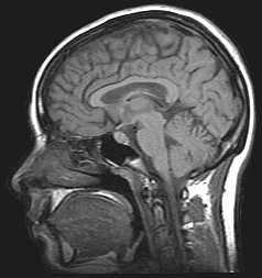
An MR venogram is also performed in most cases to exclude the possibility of venous sinus stenosis/obstruction or cerebral venous sinus thrombosis . [5] [7] [8] A contrast-enhanced MRV (ATECO) scan has a high detection rate for abnormal transverse sinus stenoses. [10] These stenoses can be more adequately identified and assessed with catheter cerebral venography and manometry. [11] Buckling of the bilateral optic nerves with increased perineural fluid is also often noted on MRI imaging. ... They added the requirement that the person is awake and alert, as coma precludes adequate neurological assessment, and require exclusion of venous sinus thrombosis as an underlying cause. ... These may be used in severe papilledema, but otherwise their use is discouraged. [5] Venous sinus stenting [ edit ] Venous sinus stenoses leading to venous hypertension appear to play a significant part in relation to raised ICP , and stenting of a transverse sinus may resolve venous hypertension, leading to improved CSF resorption, decreased ICP, cure of papilledema and other symptoms of IIH. [11] A self-expanding metal stent is permanently deployed within the dominant transverse sinus across the stenosis under general anaesthesia. ... PMID 12743224 . ^ a b c d Ahmed, RM; Wilkinson, M; Parker, GD; Thurtell, MJ; Macdonald, J; McCluskey, PJ; Allan, R; Dunne, V; Hanlon, M; Owler, BK; Halmagyi, GM (Sep 2011). "Transverse sinus stenting for idiopathic intracranial hypertension: a review of 52 patients and of model predictions" . ... A Systematic Analysis of Transverse Sinus Stenting" . Interventional Neurology . 2 (3): 132–143. doi : 10.1159/000357503 .
Idiopathic intracranial hypertension (IIH) , formerly known as pseudotumor cerebri, is a condition that affects the brain. Pseudotumor cerebri literally translates to "false brain tumor." This term was used because symptoms of IIH resemble those of brain tumors depsite no tumor being present. Symptoms of IIH may include severe headache, nausea and vomiting, altered vision, and pulsating sounds within the head. A person with IIH may also have symptoms such as a stiff neck, back or arm pain, eye pain, and memory problems. If the condition remains untreated, permanent visual loss or blindness may develop.
Buchheit et al. (1969) described 2 sisters with idiopathic intracranial hypertension with papilledema (pseudotumor cerebri). Traviesa et al. (1976) described 3 affected sisters. The patients are typically young females who are obese and may be pregnant or suffering from chronic dysfunctional uterine bleeding. Johnston and Morgan (1991) described a mother and 2 of 4 daughters who had a diagnosis of pseudotumor cerebri and 1 son who developed communicating hydrocephalus. Warner et al. (2002) quantified vitamin A in the cerebrospinal fluid of patients with idiopathic intracranial hypertension, elevated intracranial pressure of other causes, and normal intracranial pressure. Vitamin A could be detected by high-pressure liquid chromatography in most of the specimens.
Idiopathic intracranial hypertension is a neurological disorder characterized by isolated increased intracranial pressure manifesting with recurrent and persistent headaches, nausea, vomiting, progressive and transient obstruction of the visual field, papilledema. Visual loss can be irreversible.
Other rare but dangerous complications include osteomyelitis , cavernous sinus thrombosis , and deep neck space infection . [2] Contents 1 Signs and symptoms 1.1 Complications 1.1.1 Osteomyelitis 1.1.2 Cavernous sinus thrombosis 1.1.3 Deep neck space infection 2 Causes 3 Anatomy of mouth 3.1 Oral cavity 3.2 Spread of oral infection 3.2.1 Primary space 3.2.2 Secondary space 4 Diagnosis 5 Treatment 6 References Signs and symptoms [ edit ] Dental pain and swelling are the two hallmark symptoms of a mouth infection. ... The three main, albeit rare, complications of mouth infections are osteomyelitis , cavernous sinus thrombosis , and deep neck space infections . [5] Osteomyelitis [ edit ] Mouth infections that persist for months have the potential to cause a chronic infection of the surrounding bone, also known as osteomyelitis . [ citation needed ] Cavernous sinus thrombosis [ edit ] Although rare, mouth infections may also spread through the nasal and facial veins that drain into a reservoir of deoxygenated blood called the cavernous sinus . Once the infection has spread to the cavernous sinus, it can compress important nerves ( cranial nerves III, IV, V1, V2, and VI ) within this space and obstruct venous drainage from the upper face. ... Rarely, the infection will spread upwards into the maxillary sinus and cause a sinusitis. [2] In the lower jaw (mandible), the primary spaces are the sublingual, submandibular, and submental spaces. ... PMID 27390486 . ^ Plewa, Michael C.; Gupta, Mohit (2018), "Cavernous Sinus, Thrombosis" , StatPearls , StatPearls Publishing, PMID 28846357 , retrieved 2018-12-10 ^ a b Samaranayake, Lakshman; Matsubara, Victor H. (2017-04-01).
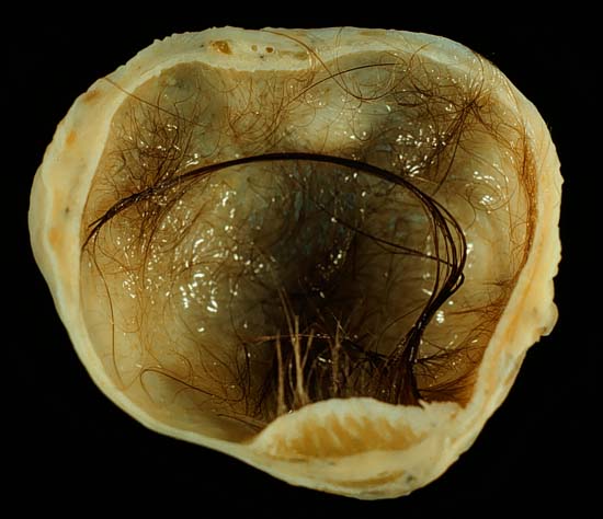
In those rare cases wherein the dermoid cyst is malignant , a squamous cell carcinoma usually develops in adults, while infants and children usually present with an endodermal sinus tumor . [1] : 781 Contents 1 Location 1.1 Vaginal and ovarian dermoid cysts 1.2 Periorbital dermoid cysts 1.3 Spinal dermoid cysts 2 Diagnosis 2.1 Differential diagnosis 3 Treatment 4 See also 5 References 6 External links Location [ edit ] Due to its classification, a dermoid cyst can occur wherever a teratoma can occur. ... This is partly because both can be full of hair. A pilonidal cyst is a pilonidal sinus that is obstructed. Any teratoma near the body surface may develop a sinus or a fistula, or even a cluster of these. ... See also [ edit ] Wikimedia Commons has media related to Dermoid cysts . Dermoid sinus , more commonly known as a pilonidal cyst Proliferating trichilemmal cyst List of cutaneous conditions References [ edit ] ^ Freedberg, et al. (2003). ... External links [ edit ] Classification D ICD - 10 : D27.9 MeSH : D003884 SNOMED CT : 189117002 v t e Germ cell tumors Germinomatous Germinoma Seminoma Dysgerminoma Nongerminomatous Embryonal carcinoma Endodermal sinus tumor/Yolk sac tumor Teratoma : Fetus in fetu Dermoid cyst Struma ovarii Strumal carcinoid Trophoblastic neoplasm : Gestational trophoblastic disease Hydatidiform mole Choriocarcinoma Placental site trophoblastic tumor Polyembryoma Gonadoblastoma
Electrophysiologic testing showed coexisting sinus and AV node dysfunction. The second patient was a 9-year-old girl who was diagnosed with fetal bradycardia (55 bpm) at 24 weeks of gestation. ... The son, who was healthy and athletic, showed sinus bradycardia on ECG with a slightly shortened QTc interval of 356 ms. ... INHERITANCE - Autosomal dominant CARDIOVASCULAR Heart - Bradycardia - Short QT interval - Atrial fibrillation - Slow ventricular response - Coexisting sinus and atrioventricular node dysfunction - Ventricular fibrillation (in some patients) MISCELLANEOUS - Some patients are clinically asymptomatic MOLECULAR BASIS - Caused by mutation in the potassium voltage-gated channel, KQT-like subfamily, member 1 gene (KCNQ1, 607542.0037 ) ▲ Close