• FindZebra
  • About
  • Contact
  • Help
  • Login
Deep Research Advanced
  • Gastrinoma Wikipedia
    Wiener Klinische Wochenschrift . 119 (19–20): 573–578. doi : 10.1007/s00508-007-0884-2 . ISSN 0043-5325 . PMID 17985090 .
    MEN1, GAST, CHGA, SST, SCT, CDKN2A, ACTB, SCTR, GRPR, GRP, ERBB2, POTEF, CASR, MET, NMBR, POMC, PYGM, S100B, BBS2, ATP4A, ATP12A, SSTR5, TCF3, TNFRSF1B, TP53, VIP, KHSRP, PSIP1, SIGLEC7, NMB, CUX1, EGF, SMAD4, EGFR, CDKN2D, CDKN2B, GFAP, GH1, FFAR1, CDH1, CD44, HCLS1, HGF, APC, IGF1, IGF1R, IGFBP1, IL2RB, HRH2
    • Zollinger-Ellison Syndrome Orphanet
      Zollinger-Ellison syndrome (ZES) is characterized by severe peptic disease (ulcers/esophageal disease) caused by hypergastrinemia secondary to a gastrinoma resulting in increased gastric acid secretion. Epidemiology Annual incidence is estimated at 1-2 cases per million. The condition is slightly more common in females than males (sex ratio of 1.3:1). Clinical description ZES is usually diagnosed in the fifth decade of life. Abdominal pain (typically in the upper abdomen) and diarrhea are the most frequent manifestations. Heartburn is often present (44% of cases). Other signs include nausea, vomiting, malabsorption, and weight loss.
    • Zollinger-Ellison Syndrome GARD
      Zollinger-Ellison syndrome (ZES) is a condition in which tumors called gastrinomas in the pancreas and duodenum (part of the small intestine) cause high levels of the hormone gastrin in the blood. High levels of gastrin then cause production of too much stomach acid. Signs and symptoms may include abdominal pain, peptic ulcers , vomiting blood, and diarrhea. The tumors are sometimes cancerous and may spread to other areas of the body. In most cases, the cause of ZES is unknown. However, about 25-30% of gastrinomas are caused by an inherited condition called multiple endocrine neoplasia type 1 (MEN1) .
  • Lewis Lung Carcinoma Wikipedia
    Margaret Lewis of the Wistar Institute and became one of the first transplantable tumors. [1] Contents 1 Models 1.1 Syngeneic 1.2 Orthotopic 2 Characterization 3 Research 4 References Models [ edit ] Syngeneic [ edit ] According to a 2015 review article, Lewis lung carcinoma is the only reproducible syngeneic lung cancer model, meaning that it is the only reproducible lung cancer model that utilizes a transplant that is immunologically compatible. Syngeneic models have proven to be useful in predicting clinical benefit of therapy in preclinical experiments. ... The activity of the mouse product did not translate to the activity of the human counterpart. [2] Orthotopic [ edit ] Lewis lung carcinoma can also be utilized as an orthotopic model. [2] Orthotopic models focus upon correctly modeling the tumor microenvironment by injecting or implanting tumors into the corresponding organ that they originated from (i.e. implanting a Lewis lung carcinoma into the lung of another C57BL mouse). ... However, the creation of such models is a typically more involved and technically challenging process. ... The model is also useful for chemotherapeutic testing in vivo .
    LECT2, TXNRD1, COL18A1, PTGS2, IL6, CHRM3, TSPO, CD274, EGFR, DDX53, SERPINF1, HIF1A, TLR4, LGALS1, CCL2, CSF2, GLB1, IL17A, IFNG, IL2, FLT1, MTCO2P12, BABAM2, KDR, PNPLA2, COX2, PECAM1, PGF, VEGFA, TNF, TGFB1, DLL4, STAT3, MAPK14, TIMM8A, RTN4R, ATN1, DUSP3, MLRL, C20orf85, GPR65, CCN4, SQSTM1, LRG1, ANGPTL1, IL1RL1, AIMP1, GRAP2, ADAMTS1, FBXO32, PRDX6, MVP, AMOT, AIMP2, EML5, POU5F1P3, TCF7L2, LINC01672, ZEB1, TERT, TGFA, POU5F1P4, THBS1, FSD1L, MIR342, MIR126, IL27, IDO2, XBP1, XPA, UCN3, ANO2, NOD1, TLR7, RNF19A, SYF2, POLDIP2, CD209, DESI2, DCTN4, CHST15, AHSA1, ANGPT4, IL17D, NOD2, ROBO4, RHOJ, MYDGF, FSD1, SOAT1, BIRC7, NUP62, SMUG1, SIRT1, UBR2, VASH1, WDHD1, PPARGC1A, FGL2, ELMO3, HPSE, NFAT5, DLL3, KHDRBS1, SPHK2, EML2, ACAT1, SDC2, NDST1, EPAS1, ERBB2, ERN1, EXTL3, F2, FGF2, FGFR1, FGFR2, FLT3, FN1, MTOR, MSTN, GFAP, GJA1, GTF2H1, H1-0, HGF, DPYSL3, DPP4, DNASE1, CASP3, ADM, PARP1, AGTR2, ALCAM, BGLAP, BRAF, VPS51, CD9, DCK, CD40LG, CEBPB, CCR6, COL4A3, COL11A2, CRK, VCAN, HSPD1, IL1B, CCL20, IL4, MSH3, MYD88, PAX7, PCNA, PF4, POMC, PON1, POU5F1, MAPK1, ACTB, PRTN3, PSMD4, PTGER3, PTGIS, RNASE1, RNPEP, RPS10, ABCC1, MMP12, MMP9, IL18, CXCL8, CXCR1, CXCR2, IL10, IL15, IL15RA, TNFRSF9, IDO1, MMP2, ITGA5, CD82, KRT19, LDLR, MDM4, MET, CXCL9, MAPK3
  • Metaphyseal Modeling Abnormality, Skin Lesions, And Spastic Paraplegia OMIM
    Roy et al. (1968) described a 14-year-old girl with defective metaphyseal modeling as in Pyle disease, increased bone density, plaque-like skin lesions, and signs of spastic paraplegia. The parents were not related. Skel - Metaphyseal modeling - Increased bone density Inheritance - Autosomal recessive Neuro - Spastic paraplegia Skin - Plaque-like skin lesions ▲ Close
  • Cadasil Wikipedia
    "A series of Notch3 mutations in CADASIL; insights from 3D molecular modelling and evolutionary analyses". Journal of Molecular Biochemistry . 3 (3): 97–105. ^ Ropper AH, Brown RH, eds. (2005). ... In Pagon RA, Bird TD, Dolan CR, et al., eds. (1993). GeneReviews [Internet] . Seattle WA: University of Washington, Seattle.
    NOTCH3, HTRA1, EGF, COL4A1, COL18A1, NOTCH1, JAG1, APOE, APP, LPA, TREX1, GFAP, PLXNA2, CTSA, PROC, ACTB, SOD1, TGFB1, NOTCH2, LAP, MRS2, RNF213, NOX5, THBD, KDR, NEFL, ACTG2, RBPJ, FN1, FBN1, ELN, DVL1, DCN, CSF3, CSF1R, CLU, CACNA1A, BGN, APCS, LINC01191
    • Cadasil GeneReviews
      Several therapeutic approaches are in pre-clinical development: testing in cells and mouse models including immunotherapy [Machuca-Parra et al 2017, Ghezali et al 2018], antisense mediated NOTCH3 exon skipping [Rutten et al 2016b], and treatment with stem cell factor and granulocyte colony-stimulating factor [Liu et al 2015].
    • Cerebral Autosomal Dominant Arteriopathy-Subcortical Infarcts-Leukoencephalopathy Orphanet
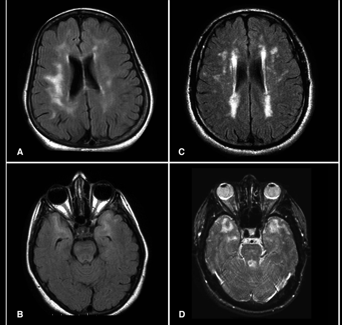
      CADASIL (Cerebral Autosomal Dominant Arteriopathy with Subcortical Infarcts and Leukoencephalopathy) is a hereditary cerebrovascular disorder characterized by mid-adult onset of recurrent subcortical ischemic stroke and cognitive impairment progressing to dementia in addition to migraines with aura and mood disturbances seen in about a third of patients. Epidemiology In Europe, the prevalence of CADASIL has been estimated to range between 1/50 000- 1/25 000. Clinical description The first manifestation of the disease occurs at a mean age of 45-50, usually in the form of ischemic stroke or cognitive decline. The disease onset and course is variable, but more than two thirds of patients suffer from (recurrent) stroke or dementia. Migraine, usually with aura, occurs in about a third of patients and often precedes stroke and dementia symptoms, with a mean age of onset of about 30 years.
    • Cerebral Arteriopathy, Autosomal Dominant, With Subcortical Infarcts And Leukoencephalopathy, Type 1 OMIM
      A number sign (#) is used with this entry because of evidence that autosomal dominant cerebral arteriopathy with subcortical infarcts and leukoencephalopathy type 1 (CADASIL1) is caused by heterozygous mutation in the NOTCH3 gene (600276) on chromosome 19p13. Description Autosomal dominant cerebral arteriopathy with subcortical infarcts and leukoencephalopathy (CADASIL) is a progressive disorder of the small arterial vessels of the brain manifest by migraine, strokes, and white matter lesions, with resultant cognitive impairment in some patients (review by Kalimo et al., 1999). Clinical Features Stevens et al. (1977) reported an English family with onset of recurrent cerebral ischemic strokes between 39 and 57 years resulting in progressive neurologic dysfunction and eventual dementia. Affected individuals did not have hypertension, diabetes, or increased cholesterol, but neuropathologic investigation showed abnormalities of the cerebral vasculature; the authors suggested that it was a form of 'vascular encephalopathy.' Low et al. (2007) provided a follow-up of the family reported by Stevens et al. (1977), including confirmation of the CADASIL diagnosis by identification of a pathogenic mutation in the NOTCH3 gene (see MOLECULAR GENETICS).
    • Cerebral Autosomal Dominant Arteriopathy With Subcortical Infarcts And Leukoencephalopathy MedlinePlus
      Cerebral autosomal dominant arteriopathy with subcortical infarcts and leukoencephalopathy, usually called CADASIL, is an inherited condition that causes stroke and other impairments. This condition affects blood flow in small blood vessels , particularly cerebral vessels within the brain. The muscle cells surrounding these blood vessels (vascular smooth muscle cells ) are abnormal and gradually die. In the brain, the resulting blood vessel damage (arteriopathy) can cause migraines, often with visual sensations or auras, or recurrent seizures (epilepsy). Damaged blood vessels reduce blood flow and can cause areas of tissue death (infarcts) throughout the body.
    • Cadasil GARD
      CADASIL (Cerebral Autosomal Dominant Arteriopathy with Sub-cortical Infarcts and Leukoencephalopathy) is an inherited disease of the blood vessels that occurs when the thickening of blood vessel walls blocks the flow of blood to the brain. The disease primarily affects the small blood vessels in the white matter of the brain. CADASIL is characterized by migraine headaches and multiple strokes , which progresses to dementia. Other symptoms include white matter lesions throughout the brain, cognitive deterioration, seizures, vision problems, and psychiatric problems such as severe depression and changes in behavior and personality. Individuals may also be at higher risk of heart attack. Symptoms and disease onset vary widely, with signs typically appearing in the mid-30s.
  • Environmental Enteropathy Wikipedia
    . ^ a b c d e f g h i j k l m Korpe PS, Petri WA (June 2012). "Environmental enteropathy: critical implications of a poorly understood condition" . ... PMID 26542185 . ^ Gilmartin AA, Petri WA (June 2015). "Exploring the role of environmental enteropathy in malnutrition, infant development and oral vaccine response" .
  • Medullary Cystic Kidney Disease Wikipedia
    Autosomal Dominant Tubulointerstitial Kidney Disease, MUC1-Related (ADTKD-MUC1) . Seattle (WA): University of Washington, Seattle. ... Autosomal Dominant Tubulointerstitial Kidney Disease, UMOD-Related (ADTKD-UMOD) . Seattle (WA): University of Washington, Seattle.
    UMOD, GLI2, KCNJ1, MUC1, HNF1B
    • Autosomal Dominant Tubulointerstitial Kidney Disease Due To Umod Mutations GARD
      Autosomal dominant tubulointerstitial kidney disease due to UMOD mutations (ADTKD–UMOD) is an inherited disorder that causes a gradual loss of kidney function that eventually leads to the need for kidney transplantation or dialysis between the ages of 30 and 70. Patients with ADTKD-UMOD have high blood levels of uric acid before kidney failure develops, and some affected individuals may develop gout. Gout is a form of arthritis (inflammation) that occurs often in the big toe, ankle, knee, or other joints. ADTKD-UMOD is caused by a mistake (mutation) in the UMOD gene, which leads to the build-up of the altered uromodulin protein in the tubules of the kidney, leading to slow loss of kidney function. ADTKD-UMOD is inherited in a dominant pattern in families. It is diagnosed based on the symptoms, laboratory testing, family history and genetic testing.
    • Autosomal Dominant Tubulointerstitial Kidney Disease, Umod-Related GeneReviews
      Summary Clinical characteristics. Autosomal dominant tubulointerstitial kidney disease caused by UMOD pathogenic variants (ADTKD- UMOD ) was previously known as familial juvenile hyperuricemic nephropathy type 1 (FJHN1), medullary cystic kidney disease type 2 (MCKD2), and UMOD -associated kidney disease (or uromodulin-associated kidney disease). Typical clinical findings: Urinalysis revealing minimal protein and no blood Slowly progressive chronic kidney failure, usually first noted in the teen years and progressing to end-stage renal disease (ESRD) between the fourth and seventh decades (Age at ESRD varies among and within families.) Hyperuricemia and gout (resulting from reduced kidney excretion of uric acid) that occurs as early as the teenage years Diagnosis/testing. ADTKD- UMOD is defined by the presence of a heterozygous pathogenic variant in UMOD , the gene encoding uromodulin. The majority of persons with ADTKD- UMOD have the following laboratory test abnormalities: elevated serum creatinine (decreased estimated glomerular filtration rate), bland urinary sediment, elevated serum urate level, and reduced fractional excretion of uric acid.
    • Medullary Cystic Kidney Disease 2 OMIM
      A number sign (#) is used with this entry because medullary cystic kidney disease-2 (MCKD2) is caused by heterozygous mutation in the UMOD gene (191845) on chromosome 16p12. Mutation in the same gene causes familial juvenile hyperuricemic nephropathy-1 (HNFJ1; 162000). Description Medullary cystic kidney disease (MCKD) is an autosomal dominant form of tubulointerstitial nephropathy characterized by formation of renal cysts at the corticomedullary junction. It is characterized by adult onset of impaired renal function and salt wasting resulting in end-stage renal failure by the sixth decade (Wolf et al., 2004). For a general phenotypic description and a discussion of genetic heterogeneity of medullary cystic kidney disease, see MCKD1 (174000).
  • Post-Ebola Virus Syndrome Wikipedia
    Retrieved 5 August 2016 . ^ Fischer WA, Brown J, Wohl DA, Loftis AJ, Tozay S, Reeves E, et al. (2017). ... Retrieved 1 October 2016 . Fischer WA, Brown J, Wohl DA, Loftis AJ, Tozay S, Reeves E, et al. (2017).
  • Mouse Model Of Colorectal And Intestinal Cancer Wikipedia
    There are genes modifying the cancer susceptibility of these mouse models. The most well-established is the modifier of Min locus (Mom1). [10] With combination of Min and Mom1 mutations the lifespan of FAP mouse models of colorectal cancer is increased. ... Thus the K-ras G12D mutant is a valuable mouse model of proximal colon carcinogenesis. ... PMID 26295972 . ^ Oh, BY; Hong, HK; Lee, WY; Cho, YB (28 February 2017). "Animal models of colorectal cancer with liver metastasis". ... "Development of colonic adenocarcinomas in a mouse model of ulcerative colitis". Inflamm. ... "A novel inflammation-related mouse colon carcinogenesis model induced by azoxymethane and dextran sodium sulfate".
  • Thought Insertion Wikipedia
    This model has come under criticism due to its definition of sense of ownership. ... An altered functional connectivity between the supplementary motor area and brain regions involved in language processing and movement implementation was also found. [2] Theory of misattributed inner speech [ edit ] According to the model of misattributed inner speech, during the generation of inner speech, speech production areas fail to inhibit the speech perception area and this leads to a misattribution of one's thoughts to an external source. [8] Comparator-model (forward model) [ edit ] The comparator-model, also known as the forward model, is an elaboration of theory of misattributed inner speech. This theory relies on a model involved in inner speech known as the forward model. ... They argue that this provides evidence that a model for motor agency cannot explain thought agency. Executive control model [ edit ] The executive control model argues that thought insertion may be mediated by altered activity in motor planning regions, specifically the supplementary motor area .
  • Mouse Models Of Breast Cancer Metastasis Wikipedia
    The availability of hundreds of mutations affecting almost every tissue and aspect of development. Mice may not be an ideal model for breast cancer. This is mainly due to the lack of precision in many of the models. ... In vivo imaging of metastatic mouse models [ edit ] Transgenic mouse models can be imaged by various non-invasive techniques. ... PMID 17409287 . ^ Gupta, PB; Kuperwasser, C. (2004). Disease models of breast cancer. Drug Discovery Today: Disease Models 1(1), 9-16. doi: 10.1016/j.ddmod.2004.05.001 ^ Palmiter, R. ... PMID 12466850 . ^ Wagner, KW. (2003). Models of Breast Cancer: quo vadis, animal modeling? ... Oncogenic and tumor-suppressive mouse models for breast cancer engaging HER2/neu.
  • Tetrasomy 18p Wikipedia
    . ^ a b Sebold C, Roeder E, Zimmerman M, Soileau B, Heard P, Carter E, Schatz M, White WA, Perry B, Reinker K, O'Donnell L, Lancaster J, Li J, Hasi M, Hill A, Pankratz L, Hale DE, Cody JD (2010).
    HTC2
    • Tetrasomy 18p MedlinePlus
      Tetrasomy 18p is a chromosomal condition that affects many parts of the body. This condition usually causes feeding difficulties in infancy, delayed development, intellectual disability that is often mild to moderate but can be severe, changes in muscle tone, distinctive facial features, and other birth defects. However, the signs and symptoms vary among affected individuals. Babies with tetrasomy 18p often have trouble feeding and may vomit frequently, which makes it difficult for them to gain weight. Some affected infants also have breathing problems and jaundice, which is a yellowing of the skin and the whites of the eyes. Changes in muscle tone are commonly seen with tetrasomy 18p. Some affected children have weak muscle tone (hypotonia), while others have increased muscle tone (hypertonia) and stiffness (spasticity).
    • Chromosome 18p Tetrasomy GARD
      Chromosome 18p tetrasomy is a chromosomal disorder that affects many parts of the body. It occurs when the short arm of chromosome 18 (18p) appears four times (tetrasomy) rather than the normal two times in cells of the body. The symptoms of chromosomy 18p tetrasomy vary from case to case but may include abnormalities of the head and face (craniofacial) area, malformations of the spine, hands, and/or feet, neuromuscular abnormalities, kidney malformations, intellectual disability, speech delays, and behavioral abnormalities. In most cases, chromosome 18p tetrasomy is the result of a spontaneous (de novo) genetic change (mutation) early in fetal development during pregnancy. Although there is no specific treatment or cure for chromosome 18p tetrasomy, there are ways to manage the symptoms.
    • Tetrasomy 18p OMIM
      A number sign (#) is used with this entry because this dysmorphic condition is caused by tetrasomy of chromosome 18p. Clinical Features Sebold et al. (2010) summarized the phenotype of tetrasomy 18p with a list of findings reported in more than 25% of theretofore published cases: neonatal feeding problems, growth retardation, microcephaly, strabismus, muscle tone abnormalities, scoliosis/kyphosis, and variants on brain MRI. Developmental delay and cognitive impairment are universally present. To more fully describe the molecular features and clinical presentation of tetrasomy 18p, Sebold et al. (2010) performed array CGH on samples from 42 individuals with tetrasomy 18p, and reviewed the medical records of these individuals. Forty-one of these individuals had an isochromosome 18p in all cells examined; the remaining individual had mosaicism.
    • Tetrasomy 18p Orphanet
      Tetrasomy 18p is a very rare structural chromosomal anomaly affecting multiple body systems and characterized clinically by craniofacial abnormalities, delayed development, cognitive impairment, changes in muscle tone, distinctive facial features, and rarely renal malformations.
  • Emanuel Syndrome Wikipedia
    .; Ledbetter, Nikki; Mefford, Heather C. (eds.). GeneReviews . Seattle (WA): University of Washington, Seattle.
    TBX1, DGCR, GRM8, RANBP1, ZNF74, MFRP
    • Emanuel Syndrome GeneReviews
      Summary Clinical characteristics. Emanuel syndrome is characterized by pre- and postnatal growth deficiency, microcephaly, hypotonia, severe developmental delays, ear anomalies, preauricular tags or pits, cleft or high-arched palate, congenital heart defects, kidney abnormalities, and genital abnormalities in males. Diagnosis/testing. The diagnosis of Emanuel syndrome is established in a proband by detection of a duplication of 22q10-22q11 and duplication of 11q23-qter on a supernumerary derivative chromosome 22 [der(22)]. Management. Treatment of manifestations: Care by a multidisciplinary team is usually necessary; standard management of gastroesophageal reflux, nutrition, anal atresia (or stenosis), inguinal hernias, cardiac defects, cleft palate, hip dysplasia, other skeletal complications, hearing loss, cryptorchidism and/or micropenis, refractive errors, and strabismus or other ophthalmologic issues; ongoing physical, occupational, and speech therapies; alternative communication methods to facilitate communication. Prevention of secondary complications: Attention to the airway during sedation and/or operative procedures in an institution with pediatric anesthesiologists. Surveillance: Follow up as needed based on the extent of systemic involvement in each individual; regular developmental assessments; periodic reevaluation by a clinical geneticist.
    • Emanuel Syndrome MedlinePlus
      Emanuel syndrome is a chromosomal disorder that disrupts normal development and affects many parts of the body. Infants with Emanuel syndrome have weak muscle tone (hypotonia) and fail to gain weight and grow at the expected rate (failure to thrive). Their development is significantly delayed, and most affected individuals have severe to profound intellectual disability. Other features of Emanuel syndrome include an unusually small head (microcephaly), distinctive facial features, and a small lower jaw (micrognathia). Ear abnormalities are common, including small holes in the skin just in front of the ears (preauricular pits or sinuses).
    • Emanuel Syndrome OMIM
      A number sign (#) is used with this entry because Emanuel syndrome is caused by malsegregation of the t(11;22)(q23;q11.2) translocation, one of only a few recurrent non-Robertsonian constitutional translocations in humans (Fraccaro et al., 1980; Zackai and Emanuel, 1980). See also supernumerary der(22)t(8;22) syndrome (613700). Description Emanuel syndrome is characterized by multiple congenital anomalies, craniofacial dysmorphism, and significant developmental delay and mental retardation. Features include ear anomalies, preauricular tag or sinus, cleft or high-arched palate, micrognathia, microcephaly, kidney abnormalities, heart defects, and genital abnormalities in males (summary by Carter et al., 2009). Carriers of the balanced constitutional t(11;22) translocation are phenotypically normal but have a 10% risk of having progeny with supernumerary der(22)t(11;22) syndrome as a result of malsegregation of the der(22). The affected progeny are genotypically unbalanced because they carry the der(22) as a supernumerary chromosome--either 47,XX,+der(22)t(11;22) or 47,XY,+der(22)t(11;22) (Zackai and Emanuel, 1980; Lin et al., 1986).
    • Emanuel Syndrome Orphanet
      Emanuel syndrome is a constitutional genomic disorder due to the presence of a supernumerary derivative 22 chromosome and characterized by severe intellectual disability, characteristic facial dysmorphism (micrognathia, hooded eyelids, upslanting downslanting parebral fissures, deep set eyes, low hanging columnella and long philtrum), congenital heart defects and kidney abnormalities.
    • Emanuel Syndrome GARD
      Emanuel syndrome is a chromosomal disorder that is characterized by learning problems and stunted growth and development. The signs and symptoms are varied and may include decreased muscle tone (hypotonia) and developmental delay in childhood, intellectual disability severe, extremely small head ( microcephaly ), distinctive facial features, small jaw, ear anomalies, arched palate (roof of the mouth), cleft palate , heart defects , kidney malformations , and genital abnormalities (in males). Emanuel syndrome is caused by the presence of additional genetic material of the chromosomes 11 and 22 in each cell . This condition is usually inherited from a parent who has a balanced translocation between chromosomes 11 and 22. Treatment depends on the signs and symptoms present in the individual.
  • Carnitine Palmitoyltransferase I Deficiency Wikipedia
    .; Ledbetter, Nikki; Mefford, Heather C. (eds.). GeneReviews . Seattle (WA): University of Washington, Seattle.
    CPT1A, CPT2, CHPT1, DHDDS
    • Carnitine Palmitoyltransferase I Deficiency OMIM
      A number sign (#) is used with this entry because carnitine palmitoyltransferase deficiency I is caused by homozygous or compound heterozygous mutation in the gene encoding carnitine palmitoyltransferase IA (CPT1A; 600528) on chromosome 11q13. Description CPT I deficiency is an autosomal recessive metabolic disorder of long-chain fatty acid oxidation characterized by severe episodes of hypoketotic hypoglycemia usually occurring after fasting or illness. Onset is in infancy or early childhood (Bougneres et al., 1981) Clinical Features Bougneres et al. (1981) reported 2 sisters who developed severe hypoketotic hypoglycemia at age 8 months, resulting in death in 1 of them. Other features included hepatomegaly, nonketotic hypoglycemia, and coma. Liver CPT activity was absent in the patient who was tested. Demaugre et al. (1988) reported 2 patients with carnitine palmitoyltransferase deficiency and hepatic symptoms.
    • Carnitine Palmitoyltransferase 1a Deficiency GeneReviews
      Summary Clinical characteristics. Carnitine palmitoyltransferase 1A (CPT1A) deficiency is a disorder of long-chain fatty acid oxidation. Clinical manifestations usually occur in an individual with a concurrent febrile or gastrointestinal illness when energy demands are increased; onset of symptoms is usually rapid. The recognized phenotypes are: acute fatty liver of pregnancy, in which the fetus has biallelic pathogenic variants in CPT1A that causes CPT1A deficiency; and hepatic encephalopathy, in which individuals (typically children) present with hypoketotic hypoglycemia and sudden onset of liver failure. Individuals with hepatic encephalopathy typically present with hypoglycemia, absent or low levels of ketones, and elevated serum concentrations of liver transaminases, ammonia, and total carnitine. Between episodes of hepatic encephalopathy, individuals appear developmentally and cognitively normal unless previous metabolic decompensation has resulted in neurologic damage.
    • Carnitine Palmitoyl Transferase 1a Deficiency Orphanet
      Carnitine palmitoyltransferase 1A (CPT-1A) deficiency is an inborn error of metabolism that affects mitochondrial oxidation of long chain fatty acids (LCFA) in the liver and kidneys, and is characterized by recurrent attacks of fasting-induced hypoketotic hypoglycemia and risk of liver failure. Epidemiology Since the description of the disease in 1981, less than 50 cases have been reported. Clinical description CPT-1A deficiency manifests between birth and 18 months of age with recurrent attacks of hypoketotic hypoglycemia of varying severity, triggered by fasting or intercurrent illness, that can lead to severe neurological sequelae. CPT-1A-deficient patients can also present with hepatic encephalopathy with loss of consciousness, seizures, coma, or even sudden death. There may be a risk of progression to liver failure. Patients with severe CPT-1A deficiency may also have renal tubular acidosis.
    • Carnitine Palmitoyltransferase I Deficiency MedlinePlus
      Carnitine palmitoyltransferase I (CPT I) deficiency is a condition that prevents the body from using certain fats for energy, particularly during periods without food (fasting). The severity of this condition varies among affected individuals. Signs and symptoms of CPT I deficiency often appear during early childhood. Affected individuals usually have low blood sugar (hypoglycemia) and a low level of ketones, which are produced during the breakdown of fats and used for energy. Together these signs are called hypoketotic hypoglycemia. People with CPT I deficiency can also have an enlarged liver (hepatomegaly), liver malfunction, and elevated levels of carnitine in the blood. Carnitine, a natural substance acquired mostly through the diet, is used by cells to process fats and produce energy.
    • Carnitine Palmitoyl Transferase 1a Deficiency GARD
      Carnitine palmitoyltransferase I deficiency (CPT1A deficiency) is an inherited metabolic condition that prevents the body from converting certain fats (long-chain fatty acids) into energy, particularly during periods without food. Carnitine, a natural substance acquired mostly through the diet, is required by cells to process fats and produce energy. Symptoms of this condition often appear early in life and include low blood sugar (hypoglycemia) and low levels of ketones, which are produced when the body breaks down fat for energy (hypoketotic hypoglycemia). This can lead to a greater risk for loss of consciousness or seizures. People with this disorder typically also have an enlarged liver (hepatomegaly), muscle weakness, nervous system damage, and elevated levels of carnitine in the blood.
  • Phobia, Specific OMIM
    Parametric analysis yielded a maximum lod score of 3.17 (dominant model) and 2.86 (recessive model) at marker D14S75. Using a simple parametric model, the lod scores at D14S75 increased to 3.70 (dominant model) and 3.30 (recessive model). ... They also noted that the homologous genomic region has been implicated in a mouse model for fear.
    OPN1SW, DDOST, FMR1, MCF2L
    • Specific Phobia Wikipedia
      Phobic disorder that is characterized by an unreasonable or irrational fear related to exposure to specific objects or situations This article needs additional citations for verification . Please help improve this article by adding citations to reliable sources . Unsourced material may be challenged and removed. Find sources: "Specific phobia" – news · newspapers · books · scholar · JSTOR ( August 2009 ) ( Learn how and when to remove this template message ) Specific phobia is an anxiety disorder , characterized by an unreasonable fear associated with a specific object or situation. Specific phobia can lead to avoidance of the object or situation, persistence of the fear, and significant distress or problems functioning associated with the fear. [1] The fear or anxiety may be triggered both by the presence and the anticipation of the specific object or situation. In most adults, the person may logically know the fear is unreasonable but still find it difficult to control the anxiety.
    • Specific Phobias Mayo Clinic
      Over time, urge your child to keep closing the distance. Model positive behavior. Because children learn by watching, you can show how to respond when faced with something your child fears or that you fear.
  • Pulmonary Hypertension, Neonatal, Susceptibility To OMIM
    Multiple dimensionality reduction (MDR) was used to both internally validate observations and develop optimal 2-variable through 5-variable models that were tested prospectively in a validation cohort of 41 children. Unconditional logistic regression analysis of the modeling chohort revealed that age (OR = 0.92, p = 0.01), CPS1 T1405N (608307.0006) genotype (AC vs AA: OR = 4.08, p = 0.04; CC vs AA: OR = 5.96, p = 0.01), and Down syndrome (OR = 5.25, p = 0.04) were independent predictors of this complex phenotype. MDR predicted that the best 2-variable model consisted of age and CPS1 T1405N genotype (p less than 0.001). This 2-variable model correctly predicted 73% of the outcomes from the validation cohort. A 5-variable model that added race, gender, and Down syndrome was not significantly better than the 2-variable model.
    CPS1
  • Kifafa Seizure Disorder OMIM
    Among the mendelian single-locus models, an additive model was favored over either a dominant, recessive, or codominant model. The single-locus model could be rejected when compared with the mixed mendelian model (inclusion of a polygenic background), although the major-gene component tends to be recessive.
  • Culture-Bound Syndrome Wikipedia
    In a 130-page report on the condition commissioned by the government and published in 2006, a team of psychologists, political scientists, and sociologists hypothesized that it was a culture-bound syndrome. [28] A startle disorder similar to latah, called imu (sometimes spelled imu: ), is found among Ainu people , both Sakhalin Ainu and Hokkaido Ainu. [29] [30] A condition similar to piblokto, called menerik [ ru ] (sometimes meryachenie ), is found among Yakuts , Yukaghirs , and Evenks living in Siberia. [31] See also [ edit ] Psychology portal Cross-cultural psychiatry Cross-cultural psychology Cultural competence in healthcare Mass psychogenic illness Hikikomori Hi-wa itck Medical anthropology Neurasthenia Zen sickness References [ edit ] ^ a b Diagnostic criteria for research , p. 213–225 ( WHO 1993) ^ Porta, Miquel, ed. (2008). ... "EPA-1025 - Integrative psychotherapy model of anxiety disorders" . European Psychiatry . 29 (1): 1–10. doi : 10.1016/S0924-9338(14)78319-1 . ... Further reading [ edit ] Kleinman, Arthur (1991), Rethinking psychiatry: from cultural category to personal experience , New York: Free Press, ISBN 978-0-02-917441-8 , retrieved 8 January 2011 Landy, David, ed. (1977), Culture, Disease, and Healing: Studies in Medical Anthropology , New York: Macmillan, ISBN 978-0-02-367390-0 Launer, John (November 2003). "Folk illness and medical models". QJM . 96 (11): 875–876. doi : 10.1093/qjmed/hcg136 .
  • Congenital Mirror Movement Disorder Wikipedia
    Therefore, quality of life can be severely hampered. [3] CMM disorder’s prevalence in the world is thought to be less than 1 in 1 million people. [1] [5] Because of its rarity, researchers suggest that some mildly affected individuals may never be diagnosed. [2] [6] It is important not to confuse congenital mirror movement disorders, a rare genetically based neurologic disease, with acquired mirror movement disorders that present themselves during one’s lifetime due to other reasons (stroke for example). [2] Contents 1 Causes 2 Pathophysiology 2.1 Interhemispheric connections 2.2 Motor cortex 2.3 Corticospinal tract 3 Diagnosis 4 Treatment and Management 5 Related Diseases 6 References 7 External links Causes [ edit ] The specific molecular mechanism that underpins this movement disorder is not well known. [2] However, most researchers suggest that it follows an autosomal dominant genetic inheritance pattern in which mutations in certain genes give rise to structural abnormalities in nervous system networks responsible for voluntary skeletal muscle movement , which, in turn, result in the functional movement abnormalities seen in patients. [1] [2] [7] [8] [9] Despite being autosomal dominant, it is important to note that the disease has variable expressivity . [3] That is, patients who have inherited a mutated dominant allele, along with their genetically affected parent, can be symptomatic or asymptomatic for CMM disorder. [4] The genes that currently have evidence to be associated with CMM disorder include DCC (deleted in colorectal carcinoma), DNAL4 (dynein axonemal light chain 4), and RAD51 (recombination protein A) . [6] [10] DCC encodes a receptor for NTN1 (netrin-1), a protein thought to be responsible for axon guidance and neuronal cell migration during development . [11] [12] A mutation of this gene (including nonsense , splice site mutation , insertions , frameshift ) has been identified as a possible cause for CMM disorder. [2] [13] [14] Experiments in mice also support the claim that CMM disorder is associated with genetic mutations in DCC . [9] Kanga mice, lacking the P3 intracellular domain of the DCC receptor, show a hopping gait, moving their hind legs in a strictly paired fashion, as do kangaroos . [3] [15] DNAL4 encodes a component of dynein motor complex in commissural neurons of the corpus callosum . [1] [6] [3] In contrast to DCC , DNAL4 is thought to have a recessive inheritance pattern for the CMM disorder. [8] In CMM disorder patients, researchers found splice site mutations on DNAL4 , which caused skipping of exon 3, and thereby omission of 28 amino acids from DNAL4 protein. [8] This mutant DNAL4 protein, in turn, could lead to faulty cross-hemisphere wiring, resulting in CMM. [8] [16] RAD51 maintains genome integrity by repairing DNA double-strand breaks through homologous recombination . [7] RAD51 heterozygous mutations, specifically premature termination codons , have been found in many CMM disorder patients through genome-wide linkage analysis and exome sequencing . [1] [2] [4] [6] In a mouse model, researchers also found RAD51 products in corticospinal tract axons at the pyramidal decussation . [7] They therefore suggest that RAD51 might be a gene that, when haploinsufficient, causes CMM disorder in humans. [7] Despite identification of three prospective genes, no genotype - phenotype correlations have yet been found. [1] [2] That is, the severity of clinical signs and symptoms does not correlate with the type of genetic variant. [3] [17] Mutations in the above genes account for a total of about 35 percent of cases. [1] Mutations in other genes that have not been identified likely account for the other cases of this disorder. [1] Pathophysiology [ edit ] There are three main pathophysiological hypotheses for congenital mirror movement disorder that exist. ... This might provide an alternate explanation for the presence of mild mirror movements in normally developing young children that typically disappear before the age of 7. [24] Some researchers propose that DCC mutations cause a reduction in gene expression and less robust midline guidance , which may lead to a partial failure of axonal fiber crossing and encourage development of an abnormal ipsilateral connection. [7] This is confirmed by other researchers who demonstrate that patients with DDC mutants show an increased proportion of ipsilateral axonal projections, and show that even a very small number of aberrant ipsilateral descending axons is sufficient to induce incorrect movement patterns. [11] [14] [25] These findings are corroborated by evidence from mice models, Kanga mice with a deletion of DCC , whose CST has been shown not to be altered , but rather partially rerouted ipsilaterally. [16] Diagnosis [ edit ] Currently, clinical diagnosis of CMM disorder has been based on clinical findings or molecular genetic testing . [2] Clinical Findings (Signs and Symptoms) [1] [2] [10] [26] [14] : onset of mirror movements in infancy or early childhood persistence of mirror movements into and throughout adulthood with the absence of other neurologic disorders little improvement nor deterioration of mirror movements over the course of one’s life intensity of mirrored movements increasing with the complexity of the voluntary movement involuntary mirror movements that are generally of lesser amplitude compared with voluntary movements predominant mirror movement in upper limbs, with increasing severity in more distal appendages (fingers) inability to perform tasks requiring skilled bimanual coordination occasional pain in the upper limbs during prolonged manual activities occasional observed subclinical mirroring movement, but detectable with accelerometer gloves Molecular genetic testing [1] : identification of a heterozygous mutant DCC, DNAL4, or RAD51 gene ( single gene test or multi-gene panel) Treatment and Management [ edit ] CMM has clear severe impacts on a patient’s ability to carry out daily manual tasks . [27] [17] It is recommended that children be placed under more forgiving school environments, allowing more time for written evaluations and limiting handwritten assignments, to ease the burden of the movement disability. [1] [3] Furthermore, because of patients’ inability to perform pure unilateral movements and their difficulty with tasks requiring skilled bimanual coordination, young and new members to the workforce are encouraged to consider professions that do not require complex bimanual movements, repetitive or sustained hand movements, or extensive handwriting, to reduce overuse, pain, and discomfort in upper limbs. [2] [5] Because of its pronounced and obviously noticeable signs and symptoms, CMM patients can suffer social stigma ; however, physicians need to make it clear to parents, family, and friends that the disorder bears no relation to intellectual abilities . [5] [28] However, the rarity of this neurologic disease, found in one in a million people, makes its societal and cultural significance quite limited. [6] Related Diseases [ edit ] Movement disorders Chiari malformation Klippel-Feil Syndrome Dystonia Cerebral palsy Parkinson's disease Epilepsies Amyotrophic lateral sclerosis Kallman's syndrome Alien hand syndrome Obsessive compulsive disorder Schizophrenia Congenital hemiplegia Moebius syndrome Seckel syndrome Wildervanck syndrome Polymicrogyria References [ edit ] ^ a b c d e f g h i j k l m n o Reference, Genetics Home. ... In Adam, Margaret P.; Ardinger, Holly H.; Pagon, Roberta A.; Wallace, Stephanie E.; Bean, Lora J.H.; Mefford, Heather C.; Stephens, Karen; Amemiya, Anne; Ledbetter, Nikki (eds.). GeneReviews® . Seattle (WA): University of Washington, Seattle.
    CDH2, DCC, RAD51, ANOS1, DNAL4, NTN1, FGFR1, FGF8, FEZF1, CCDC141, PROKR2, KISS1R, SPRY4, PROK2, KNL1, WDR11, CHD7, IL17RD, NSMF, FLRT3, CEP152, LINC01917, SEMA3A, CREBBP, HS6ST1, FGF17, HESX1, TACR3, SOX10, DUSP6, EP300, RAD51B, GDF6, MEF2C, SMN2, SCN8A, POMK, SMN1, NTNG1
  • Specific Language Impairment 3 OMIM
    Bartlett et al. (2002) conducted a genomewide categorical linkage analysis, using model-based lod score techniques, in 5 Canadian families of Celtic ancestry that segregated SLI. Analysis was conducted under both dominant and recessive models by use of 3 phenotypic classifications: clinical diagnosis, language impairment (spoken language quotient less than 85) and reading discrepancy (nonverbal IQ minus nonword reading greater than 15). Chromosome 13 yielded a maximum multipoint lod score of 3.92 under the recessive reading discrepancy model. Stimulation to correct for multiple models and multiple phenotypes indicated that the genomewide empirical P value was less than 0.01. ... Under the recessive reading impairment model, a maximum lod score of 2.616 was obtained at D13S1317.
  • Congenital Muscular Dystrophy Wikipedia
    Congenital Muscular Dystrophy Overview . Seattle (WA): University of Washington, Seattle. ... LAMA2-Related Muscular Dystrophy . Seattle (WA): University of Washington, Seattle.
    COL6A3
  • ←
  • 1
  • 2
  • 3
  • 4
  • 5
  • 6
  • 7
  • 8
  • 9
  • ...
  • 142
  • 143
  • →

FindZebra

contact@findzebra.com