-
Onychomycosis
Wikipedia
Causes [ edit ] The causative pathogens of onychomycosis are all in the fungus kingdom and include dermatophytes , Candida ( yeasts ), and nondermatophytic molds . [2] Dermatophytes are the fungi most commonly responsible for onychomycosis in the temperate western countries; while Candida and nondermatophytic molds are more frequently involved in the tropics and subtropics with a hot and humid climate. [12] Dermatophytes [ edit ] When onychomycosis is due to a dermatophyte infection, it is termed tinea unguium. ... Other [ edit ] Other causative pathogens include Candida and nondermatophytic molds , in particular members of the mold genus Scytalidium (name recently changed to Neoscytalidium ), Scopulariopsis , and Aspergillus . ... Scytalidium mainly affects people in the tropics , though it persists if they later move to areas of temperate climate . Other molds more commonly affect people older than 60 years, and their presence in the nail reflects a slight weakening in the nail's ability to defend itself against fungal invasion. ... Other risk factors include perspiring heavily, being in a humid or moist environment, psoriasis , wearing socks and shoes that hinder ventilation and do not absorb perspiration, going barefoot in damp public places such as swimming pools, gyms and shower rooms, having athlete's foot (tinea pedis), minor skin or nail injury, damaged nail, or other infection, and having diabetes, circulation problems, which may also lead to lower peripheral temperatures on hands and feet, or a weakened immune system. [13] Diagnosis [ edit ] The diagnosis is generally suspected based on the appearance and confirmed by laboratory testing. [2] The four main tests are a potassium hydroxide smear , culture , histology examination, and polymerase chain reaction . [2] [3] The sample examined is generally nail scrapings or clippings. [2] These being from as far up the nail as possible. [3] Nail plate biopsy with periodic acid-Schiff stain appear more useful than culture or direct KOH examination. [14] To reliably identify nondermatophyte molds, several samples may be necessary. [15] Classification [ edit ] There are five classic types of onychomycosis: [16] [17] Distal subungual onychomycosis is the most common form of tinea unguium [2] and is usually caused by Trichophyton rubrum , which invades the nail bed and the underside of the nail plate . ... External links [ edit ] Classification D ICD - 10 : B35.1 ICD - 9-CM : 110.1 MeSH : D014009 DiseasesDB : 13125 External resources MedlinePlus : 001330 eMedicine : derm/300 Patient UK : Onychomycosis v t e Diseases of the skin and appendages by morphology Growths Epidermal Wart Callus Seborrheic keratosis Acrochordon Molluscum contagiosum Actinic keratosis Squamous-cell carcinoma Basal-cell carcinoma Merkel-cell carcinoma Nevus sebaceous Trichoepithelioma Pigmented Freckles Lentigo Melasma Nevus Melanoma Dermal and subcutaneous Epidermal inclusion cyst Hemangioma Dermatofibroma (benign fibrous histiocytoma) Keloid Lipoma Neurofibroma Xanthoma Kaposi's sarcoma Infantile digital fibromatosis Granular cell tumor Leiomyoma Lymphangioma circumscriptum Myxoid cyst Rashes With epidermal involvement Eczematous Contact dermatitis Atopic dermatitis Seborrheic dermatitis Stasis dermatitis Lichen simplex chronicus Darier's disease Glucagonoma syndrome Langerhans cell histiocytosis Lichen sclerosus Pemphigus foliaceus Wiskott–Aldrich syndrome Zinc deficiency Scaling Psoriasis Tinea ( Corporis Cruris Pedis Manuum Faciei ) Pityriasis rosea Secondary syphilis Mycosis fungoides Systemic lupus erythematosus Pityriasis rubra pilaris Parapsoriasis Ichthyosis Blistering Herpes simplex Herpes zoster Varicella Bullous impetigo Acute contact dermatitis Pemphigus vulgaris Bullous pemphigoid Dermatitis herpetiformis Porphyria cutanea tarda Epidermolysis bullosa simplex Papular Scabies Insect bite reactions Lichen planus Miliaria Keratosis pilaris Lichen spinulosus Transient acantholytic dermatosis Lichen nitidus Pityriasis lichenoides et varioliformis acuta Pustular Acne vulgaris Acne rosacea Folliculitis Impetigo Candidiasis Gonococcemia Dermatophyte Coccidioidomycosis Subcorneal pustular dermatosis Hypopigmented Tinea versicolor Vitiligo Pityriasis alba Postinflammatory hyperpigmentation Tuberous sclerosis Idiopathic guttate hypomelanosis Leprosy Hypopigmented mycosis fungoides Without epidermal involvement Red Blanchable Erythema Generalized Drug eruptions Viral exanthems Toxic erythema Systemic lupus erythematosus Localized Cellulitis Abscess Boil Erythema nodosum Carcinoid syndrome Fixed drug eruption Specialized Urticaria Erythema ( Multiforme Migrans Gyratum repens Annulare centrifugum Ab igne ) Nonblanchable Purpura Macular Thrombocytopenic purpura Actinic/solar purpura Papular Disseminated intravascular coagulation Vasculitis Indurated Scleroderma / morphea Granuloma annulare Lichen sclerosis et atrophicus Necrobiosis lipoidica Miscellaneous disorders Ulcers Hair Telogen effluvium Androgenic alopecia Alopecia areata Systemic lupus erythematosus Tinea capitis Loose anagen syndrome Lichen planopilaris Folliculitis decalvans Acne keloidalis nuchae Nail Onychomycosis Psoriasis Paronychia Ingrown nail Mucous membrane Aphthous stomatitis Oral candidiasis Lichen planus Leukoplakia Pemphigus vulgaris Mucous membrane pemphigoid Cicatricial pemphigoid Herpesvirus Coxsackievirus Syphilis Systemic histoplasmosis Squamous-cell carcinoma v t e Fungal infection and mesomycetozoea Superficial and cutaneous ( dermatomycosis ): Tinea = skin ; Piedra ( exothrix / endothrix ) = hair Ascomycota Dermatophyte ( Dermatophytosis ) By location Tinea barbae / tinea capitis Kerion Tinea corporis Ringworm Dermatophytids Tinea cruris Tinea manuum Tinea pedis (athlete's foot) Tinea unguium/onychomycosis White superficial onychomycosis Distal subungual onychomycosis Proximal subungual onychomycosis Tinea corporis gladiatorum Tinea faciei Tinea imbricata Tinea incognito Favus By organism Epidermophyton floccosum Microsporum canis Microsporum audouinii Trichophyton interdigitale/mentagrophytes Trichophyton tonsurans Trichophyton schoenleini Trichophyton rubrum Trichophyton verrucosum Other Hortaea werneckii Tinea nigra Piedraia hortae Black piedra Basidiomycota Malassezia furfur Tinea versicolor Pityrosporum folliculitis Trichosporon White piedra Subcutaneous , systemic , and opportunistic Ascomycota Dimorphic (yeast+mold) Onygenales Coccidioides immitis / Coccidioides posadasii Coccidioidomycosis Disseminated coccidioidomycosis Primary cutaneous coccidioidomycosis .CLEC7A, MMEL1, CARD9, DOCK8, CDH23, TNPO3, TRAF3IP2, USP8, STAT1, TNFSF15, IL17RC, POU2AF1, SDR9C7, IRF5, IL12RB1, IL12A, SPIB, LYST, VPS13B, RNA28SN5, ATR, TNMD, SLN, POLR2A, CYLD, COX8A, MFT2

-
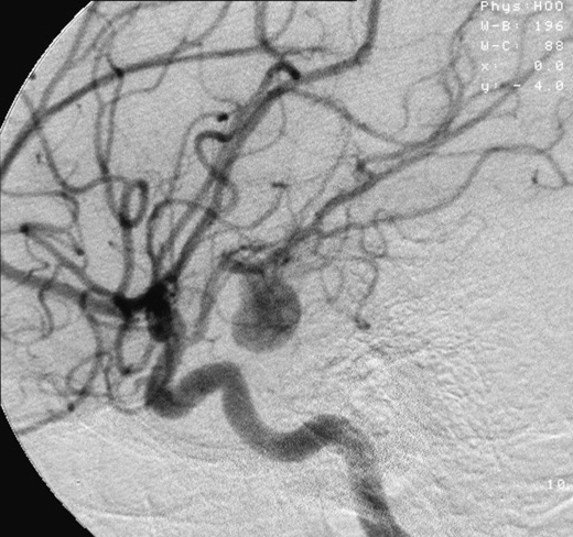
Aneurysm
Wikipedia
Bulge in the wall of a blood vessel For other uses, see Aneurysm (disambiguation) . ... At a certain point, the stiffness of the arterial wall starts to decrease with increasing load. ... Analyzing the velocity and pressure profiles of the blood flow leads to obtaining the resulting wall shear stress on the vessel and aneurysm wall. The neck of the aneurysm is the most at risk due to the combination of a small wall thickness and high wall shear stress. ... Similarly, sometimes it is difficult to model the varying wall thickness in small vessels, so researchers treat wall thickness as constant.MMP1, NOX4, TIMP1, COL3A1, SLC2A10, TGFBR2, COL5A1, COL5A2, FBN1, FARSB, SMAD3, COL1A1, MYH11, MYLK, NF1, MMP9, PRF1, CXCL8, TNF, IL6, GZMB, CHI3L1
-
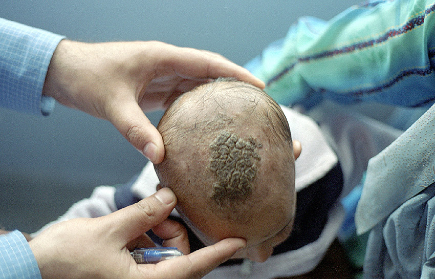
Kerion
Wikipedia
External links [ edit ] Classification D ICD - 10 : B35.0 ( ILDS B35.020) ICD - 9-CM : 110.0 MeSH : C536165 DiseasesDB : 29142 DermAtlas 850027489 v t e Fungal infection and mesomycetozoea Superficial and cutaneous ( dermatomycosis ): Tinea = skin ; Piedra ( exothrix / endothrix ) = hair Ascomycota Dermatophyte ( Dermatophytosis ) By location Tinea barbae / tinea capitis Kerion Tinea corporis Ringworm Dermatophytids Tinea cruris Tinea manuum Tinea pedis (athlete's foot) Tinea unguium/onychomycosis White superficial onychomycosis Distal subungual onychomycosis Proximal subungual onychomycosis Tinea corporis gladiatorum Tinea faciei Tinea imbricata Tinea incognito Favus By organism Epidermophyton floccosum Microsporum canis Microsporum audouinii Trichophyton interdigitale/mentagrophytes Trichophyton tonsurans Trichophyton schoenleini Trichophyton rubrum Trichophyton verrucosum Other Hortaea werneckii Tinea nigra Piedraia hortae Black piedra Basidiomycota Malassezia furfur Tinea versicolor Pityrosporum folliculitis Trichosporon White piedra Subcutaneous , systemic , and opportunistic Ascomycota Dimorphic (yeast+mold) Onygenales Coccidioides immitis / Coccidioides posadasii Coccidioidomycosis Disseminated coccidioidomycosis Primary cutaneous coccidioidomycosis . Primary pulmonary coccidioidomycosis Histoplasma capsulatum Histoplasmosis Primary cutaneous histoplasmosis Primary pulmonary histoplasmosis Progressive disseminated histoplasmosis Histoplasma duboisii African histoplasmosis Lacazia loboi Lobomycosis Paracoccidioides brasiliensis Paracoccidioidomycosis Other Blastomyces dermatitidis Blastomycosis North American blastomycosis South American blastomycosis Sporothrix schenckii Sporotrichosis Talaromyces marneffei Talaromycosis Yeast -like Candida albicans Candidiasis Oral Esophageal Vulvovaginal Chronic mucocutaneous Antibiotic candidiasis Candidal intertrigo Candidal onychomycosis Candidal paronychia Candidid Diaper candidiasis Congenital cutaneous candidiasis Perianal candidiasis Systemic candidiasis Erosio interdigitalis blastomycetica C. auris C. glabrata C. lusitaniae C. tropicalis Pneumocystis jirovecii Pneumocystosis Pneumocystis pneumonia Mold -like Aspergillus Aspergillosis Aspergilloma Allergic bronchopulmonary aspergillosis Primary cutaneous aspergillosis Exophiala jeanselmei Eumycetoma Fonsecaea pedrosoi / Fonsecaea compacta / Phialophora verrucosa Chromoblastomycosis Geotrichum candidum Geotrichosis Pseudallescheria boydii Allescheriasis Basidiomycota Cryptococcus neoformans Cryptococcosis Trichosporon spp Trichosporonosis Zygomycota ( Zygomycosis ) Mucorales ( Mucormycosis ) Rhizopus oryzae Mucor indicus Lichtheimia corymbifera Syncephalastrum racemosum Apophysomyces variabilis Entomophthorales ( Entomophthoramycosis ) Basidiobolus ranarum Basidiobolomycosis Conidiobolus coronatus / Conidiobolus incongruus Conidiobolomycosis Microsporidia ( Microsporidiosis ) Enterocytozoon bieneusi / Encephalitozoon intestinalis Mesomycetozoea Rhinosporidium seeberi Rhinosporidiosis Ungrouped Alternariosis Fungal folliculitis Fusarium Fusariosis Granuloma gluteale infantum Hyalohyphomycosis Otomycosis Phaeohyphomycosis

-
Damping Off
Wikipedia
Leaf spotting sometimes accompanies other symptoms, as does a grey mold growth on stems and leaves. Roots sometimes rot completely or back to just discolored stumps. [2] Causal agents [ edit ] A number of different fungi and fungi-like organisms cause the symptoms of damping off, including: Alternaria species. ... Cause leaf spotting. [2] Phytophthora – a genus of plant-damaging oomycetes ( water molds ), whose member species are capable of causing enormous economic losses on crops worldwide, as well as environmental damage in natural ecosystems . [3] Pseudomonas species.

-
Tinea Barbae
Wikipedia
. ^ a b c d Rutecki; Wurtz & Thomson (2000), "From Animal to Man: Tinea Barbae", Current Infectious Disease Reports , 2 (5): 433–437, doi : 10.1007/s11908-000-0073-1 External links [ edit ] Classification D ICD - 10 : B35.0 ( ILDS B35.040) ICD - 9-CM : 110.0 External resources eMedicine : derm/419 v t e Fungal infection and mesomycetozoea Superficial and cutaneous ( dermatomycosis ): Tinea = skin ; Piedra ( exothrix / endothrix ) = hair Ascomycota Dermatophyte ( Dermatophytosis ) By location Tinea barbae / tinea capitis Kerion Tinea corporis Ringworm Dermatophytids Tinea cruris Tinea manuum Tinea pedis (athlete's foot) Tinea unguium/onychomycosis White superficial onychomycosis Distal subungual onychomycosis Proximal subungual onychomycosis Tinea corporis gladiatorum Tinea faciei Tinea imbricata Tinea incognito Favus By organism Epidermophyton floccosum Microsporum canis Microsporum audouinii Trichophyton interdigitale/mentagrophytes Trichophyton tonsurans Trichophyton schoenleini Trichophyton rubrum Trichophyton verrucosum Other Hortaea werneckii Tinea nigra Piedraia hortae Black piedra Basidiomycota Malassezia furfur Tinea versicolor Pityrosporum folliculitis Trichosporon White piedra Subcutaneous , systemic , and opportunistic Ascomycota Dimorphic (yeast+mold) Onygenales Coccidioides immitis / Coccidioides posadasii Coccidioidomycosis Disseminated coccidioidomycosis Primary cutaneous coccidioidomycosis . Primary pulmonary coccidioidomycosis Histoplasma capsulatum Histoplasmosis Primary cutaneous histoplasmosis Primary pulmonary histoplasmosis Progressive disseminated histoplasmosis Histoplasma duboisii African histoplasmosis Lacazia loboi Lobomycosis Paracoccidioides brasiliensis Paracoccidioidomycosis Other Blastomyces dermatitidis Blastomycosis North American blastomycosis South American blastomycosis Sporothrix schenckii Sporotrichosis Talaromyces marneffei Talaromycosis Yeast -like Candida albicans Candidiasis Oral Esophageal Vulvovaginal Chronic mucocutaneous Antibiotic candidiasis Candidal intertrigo Candidal onychomycosis Candidal paronychia Candidid Diaper candidiasis Congenital cutaneous candidiasis Perianal candidiasis Systemic candidiasis Erosio interdigitalis blastomycetica C. auris C. glabrata C. lusitaniae C. tropicalis Pneumocystis jirovecii Pneumocystosis Pneumocystis pneumonia Mold -like Aspergillus Aspergillosis Aspergilloma Allergic bronchopulmonary aspergillosis Primary cutaneous aspergillosis Exophiala jeanselmei Eumycetoma Fonsecaea pedrosoi / Fonsecaea compacta / Phialophora verrucosa Chromoblastomycosis Geotrichum candidum Geotrichosis Pseudallescheria boydii Allescheriasis Basidiomycota Cryptococcus neoformans Cryptococcosis Trichosporon spp Trichosporonosis Zygomycota ( Zygomycosis ) Mucorales ( Mucormycosis ) Rhizopus oryzae Mucor indicus Lichtheimia corymbifera Syncephalastrum racemosum Apophysomyces variabilis Entomophthorales ( Entomophthoramycosis ) Basidiobolus ranarum Basidiobolomycosis Conidiobolus coronatus / Conidiobolus incongruus Conidiobolomycosis Microsporidia ( Microsporidiosis ) Enterocytozoon bieneusi / Encephalitozoon intestinalis Mesomycetozoea Rhinosporidium seeberi Rhinosporidiosis Ungrouped Alternariosis Fungal folliculitis Fusarium Fusariosis Granuloma gluteale infantum Hyalohyphomycosis Otomycosis Phaeohyphomycosis

-
Entomophthoramycosis
Wikipedia
External links [ edit ] Classification D ICD - 10 : B46.8 ( ILDS B46.810) ICD - 9-CM : 117.7 MeSH : D020096 DiseasesDB : 35031 v t e Fungal infection and mesomycetozoea Superficial and cutaneous ( dermatomycosis ): Tinea = skin ; Piedra ( exothrix / endothrix ) = hair Ascomycota Dermatophyte ( Dermatophytosis ) By location Tinea barbae / tinea capitis Kerion Tinea corporis Ringworm Dermatophytids Tinea cruris Tinea manuum Tinea pedis (athlete's foot) Tinea unguium/onychomycosis White superficial onychomycosis Distal subungual onychomycosis Proximal subungual onychomycosis Tinea corporis gladiatorum Tinea faciei Tinea imbricata Tinea incognito Favus By organism Epidermophyton floccosum Microsporum canis Microsporum audouinii Trichophyton interdigitale/mentagrophytes Trichophyton tonsurans Trichophyton schoenleini Trichophyton rubrum Trichophyton verrucosum Other Hortaea werneckii Tinea nigra Piedraia hortae Black piedra Basidiomycota Malassezia furfur Tinea versicolor Pityrosporum folliculitis Trichosporon White piedra Subcutaneous , systemic , and opportunistic Ascomycota Dimorphic (yeast+mold) Onygenales Coccidioides immitis / Coccidioides posadasii Coccidioidomycosis Disseminated coccidioidomycosis Primary cutaneous coccidioidomycosis . Primary pulmonary coccidioidomycosis Histoplasma capsulatum Histoplasmosis Primary cutaneous histoplasmosis Primary pulmonary histoplasmosis Progressive disseminated histoplasmosis Histoplasma duboisii African histoplasmosis Lacazia loboi Lobomycosis Paracoccidioides brasiliensis Paracoccidioidomycosis Other Blastomyces dermatitidis Blastomycosis North American blastomycosis South American blastomycosis Sporothrix schenckii Sporotrichosis Talaromyces marneffei Talaromycosis Yeast -like Candida albicans Candidiasis Oral Esophageal Vulvovaginal Chronic mucocutaneous Antibiotic candidiasis Candidal intertrigo Candidal onychomycosis Candidal paronychia Candidid Diaper candidiasis Congenital cutaneous candidiasis Perianal candidiasis Systemic candidiasis Erosio interdigitalis blastomycetica C. auris C. glabrata C. lusitaniae C. tropicalis Pneumocystis jirovecii Pneumocystosis Pneumocystis pneumonia Mold -like Aspergillus Aspergillosis Aspergilloma Allergic bronchopulmonary aspergillosis Primary cutaneous aspergillosis Exophiala jeanselmei Eumycetoma Fonsecaea pedrosoi / Fonsecaea compacta / Phialophora verrucosa Chromoblastomycosis Geotrichum candidum Geotrichosis Pseudallescheria boydii Allescheriasis Basidiomycota Cryptococcus neoformans Cryptococcosis Trichosporon spp Trichosporonosis Zygomycota ( Zygomycosis ) Mucorales ( Mucormycosis ) Rhizopus oryzae Mucor indicus Lichtheimia corymbifera Syncephalastrum racemosum Apophysomyces variabilis Entomophthorales ( Entomophthoramycosis ) Basidiobolus ranarum Basidiobolomycosis Conidiobolus coronatus / Conidiobolus incongruus Conidiobolomycosis Microsporidia ( Microsporidiosis ) Enterocytozoon bieneusi / Encephalitozoon intestinalis Mesomycetozoea Rhinosporidium seeberi Rhinosporidiosis Ungrouped Alternariosis Fungal folliculitis Fusarium Fusariosis Granuloma gluteale infantum Hyalohyphomycosis Otomycosis Phaeohyphomycosis This mycology -related article is a stub .
-
Phycomycosis
Wikipedia
The condition is caused by a variety of molds and fungi , and individual forms include pythiosis , zygomycosis , and lagenidiosis . ... External links [ edit ] Oomycosis: Pythiosis in the Dog, Horse and Cat and Lagenidiosis in the Dog v t e Fungal infection and mesomycetozoea Superficial and cutaneous ( dermatomycosis ): Tinea = skin ; Piedra ( exothrix / endothrix ) = hair Ascomycota Dermatophyte ( Dermatophytosis ) By location Tinea barbae / tinea capitis Kerion Tinea corporis Ringworm Dermatophytids Tinea cruris Tinea manuum Tinea pedis (athlete's foot) Tinea unguium/onychomycosis White superficial onychomycosis Distal subungual onychomycosis Proximal subungual onychomycosis Tinea corporis gladiatorum Tinea faciei Tinea imbricata Tinea incognito Favus By organism Epidermophyton floccosum Microsporum canis Microsporum audouinii Trichophyton interdigitale/mentagrophytes Trichophyton tonsurans Trichophyton schoenleini Trichophyton rubrum Trichophyton verrucosum Other Hortaea werneckii Tinea nigra Piedraia hortae Black piedra Basidiomycota Malassezia furfur Tinea versicolor Pityrosporum folliculitis Trichosporon White piedra Subcutaneous , systemic , and opportunistic Ascomycota Dimorphic (yeast+mold) Onygenales Coccidioides immitis / Coccidioides posadasii Coccidioidomycosis Disseminated coccidioidomycosis Primary cutaneous coccidioidomycosis . Primary pulmonary coccidioidomycosis Histoplasma capsulatum Histoplasmosis Primary cutaneous histoplasmosis Primary pulmonary histoplasmosis Progressive disseminated histoplasmosis Histoplasma duboisii African histoplasmosis Lacazia loboi Lobomycosis Paracoccidioides brasiliensis Paracoccidioidomycosis Other Blastomyces dermatitidis Blastomycosis North American blastomycosis South American blastomycosis Sporothrix schenckii Sporotrichosis Talaromyces marneffei Talaromycosis Yeast -like Candida albicans Candidiasis Oral Esophageal Vulvovaginal Chronic mucocutaneous Antibiotic candidiasis Candidal intertrigo Candidal onychomycosis Candidal paronychia Candidid Diaper candidiasis Congenital cutaneous candidiasis Perianal candidiasis Systemic candidiasis Erosio interdigitalis blastomycetica C. auris C. glabrata C. lusitaniae C. tropicalis Pneumocystis jirovecii Pneumocystosis Pneumocystis pneumonia Mold -like Aspergillus Aspergillosis Aspergilloma Allergic bronchopulmonary aspergillosis Primary cutaneous aspergillosis Exophiala jeanselmei Eumycetoma Fonsecaea pedrosoi / Fonsecaea compacta / Phialophora verrucosa Chromoblastomycosis Geotrichum candidum Geotrichosis Pseudallescheria boydii Allescheriasis Basidiomycota Cryptococcus neoformans Cryptococcosis Trichosporon spp Trichosporonosis Zygomycota ( Zygomycosis ) Mucorales ( Mucormycosis ) Rhizopus oryzae Mucor indicus Lichtheimia corymbifera Syncephalastrum racemosum Apophysomyces variabilis Entomophthorales ( Entomophthoramycosis ) Basidiobolus ranarum Basidiobolomycosis Conidiobolus coronatus / Conidiobolus incongruus Conidiobolomycosis Microsporidia ( Microsporidiosis ) Enterocytozoon bieneusi / Encephalitozoon intestinalis Mesomycetozoea Rhinosporidium seeberi Rhinosporidiosis Ungrouped Alternariosis Fungal folliculitis Fusarium Fusariosis Granuloma gluteale infantum Hyalohyphomycosis Otomycosis Phaeohyphomycosis Classification D ICD - 10 : B46 ICD - 9-CM : 117.7 MeSH : D020096 DiseasesDB : 31329
-
Phaeohyphomycosis
Wikipedia
Phaeohyphomycosis is a diverse group of mycotic infections [1] caused by dematiaceous fungi whose morphologic characteristics in tissue include hyphae , yeast-like cells, or a combination of these. [2] : 324 It can be associated an array of melanistic filamentous fungi including Alternaria species , [3] Exophiala jeanselmei , [4] and Rhinocladiella mackenziei . [5] The term “phaeohyphomycosis” was introduced to determine infections caused by dematiaceous (pigmented) filamentous fungi which contain melanin in their cell walls. [6] Phaeohyphomycosis is an uncommon infection, however the number of case reported has been increasing in recent years. ... This high resilience may be contributed to the presence of melanin in their cell walls, as well as the greater similarity to host cells which are both eukaryotes than other pathogens such as bacteria or viruses. ... See also [ edit ] Skin lesion References [ edit ] ^ Naggie S, Perfect JR (June 2009). "Molds: hyalohyphomycosis, phaeohyphomycosis, and zygomycosis" .
-
Otomycosis
Wikipedia
External links [ edit ] Classification D ICD - 10 : H62.2, B36.9 ( ILDS B36.920) MeSH : D059249 External resources Patient UK : Otomycosis v t e Fungal infection and mesomycetozoea Superficial and cutaneous ( dermatomycosis ): Tinea = skin ; Piedra ( exothrix / endothrix ) = hair Ascomycota Dermatophyte ( Dermatophytosis ) By location Tinea barbae / tinea capitis Kerion Tinea corporis Ringworm Dermatophytids Tinea cruris Tinea manuum Tinea pedis (athlete's foot) Tinea unguium/onychomycosis White superficial onychomycosis Distal subungual onychomycosis Proximal subungual onychomycosis Tinea corporis gladiatorum Tinea faciei Tinea imbricata Tinea incognito Favus By organism Epidermophyton floccosum Microsporum canis Microsporum audouinii Trichophyton interdigitale/mentagrophytes Trichophyton tonsurans Trichophyton schoenleini Trichophyton rubrum Trichophyton verrucosum Other Hortaea werneckii Tinea nigra Piedraia hortae Black piedra Basidiomycota Malassezia furfur Tinea versicolor Pityrosporum folliculitis Trichosporon White piedra Subcutaneous , systemic , and opportunistic Ascomycota Dimorphic (yeast+mold) Onygenales Coccidioides immitis / Coccidioides posadasii Coccidioidomycosis Disseminated coccidioidomycosis Primary cutaneous coccidioidomycosis . Primary pulmonary coccidioidomycosis Histoplasma capsulatum Histoplasmosis Primary cutaneous histoplasmosis Primary pulmonary histoplasmosis Progressive disseminated histoplasmosis Histoplasma duboisii African histoplasmosis Lacazia loboi Lobomycosis Paracoccidioides brasiliensis Paracoccidioidomycosis Other Blastomyces dermatitidis Blastomycosis North American blastomycosis South American blastomycosis Sporothrix schenckii Sporotrichosis Talaromyces marneffei Talaromycosis Yeast -like Candida albicans Candidiasis Oral Esophageal Vulvovaginal Chronic mucocutaneous Antibiotic candidiasis Candidal intertrigo Candidal onychomycosis Candidal paronychia Candidid Diaper candidiasis Congenital cutaneous candidiasis Perianal candidiasis Systemic candidiasis Erosio interdigitalis blastomycetica C. auris C. glabrata C. lusitaniae C. tropicalis Pneumocystis jirovecii Pneumocystosis Pneumocystis pneumonia Mold -like Aspergillus Aspergillosis Aspergilloma Allergic bronchopulmonary aspergillosis Primary cutaneous aspergillosis Exophiala jeanselmei Eumycetoma Fonsecaea pedrosoi / Fonsecaea compacta / Phialophora verrucosa Chromoblastomycosis Geotrichum candidum Geotrichosis Pseudallescheria boydii Allescheriasis Basidiomycota Cryptococcus neoformans Cryptococcosis Trichosporon spp Trichosporonosis Zygomycota ( Zygomycosis ) Mucorales ( Mucormycosis ) Rhizopus oryzae Mucor indicus Lichtheimia corymbifera Syncephalastrum racemosum Apophysomyces variabilis Entomophthorales ( Entomophthoramycosis ) Basidiobolus ranarum Basidiobolomycosis Conidiobolus coronatus / Conidiobolus incongruus Conidiobolomycosis Microsporidia ( Microsporidiosis ) Enterocytozoon bieneusi / Encephalitozoon intestinalis Mesomycetozoea Rhinosporidium seeberi Rhinosporidiosis Ungrouped Alternariosis Fungal folliculitis Fusarium Fusariosis Granuloma gluteale infantum Hyalohyphomycosis Otomycosis Phaeohyphomycosis v t e Diseases of the outer and middle ear Outer ear Otitis externa Otomycosis Middle ear and mastoid Otitis media Mastoiditis Bezold's abscess Gradenigo's syndrome Tympanosclerosis Cholesteatoma Perforated eardrum Symptoms Ear pain Hearing loss Tests Otoscope pneumatic tympanometry
-
Hip Dysplasia
Mayo Clinic
The ball and socket need to fit together well because they act as molds for each other. If the ball isn't seated firmly into the socket, the socket will not fully form around the ball and will become too shallow. ... Infants are usually treated with a soft brace, such as a Pavlik harness, that holds the ball portion of the joint firmly in its socket for several months. This helps the socket mold to the shape of the ball. The brace doesn't work as well for babies older than 6 months.COL2A1, UFSP2, BICD2, ATAD3A, SETD5, INPP5K, NIN, GMNN, ANKRD11, PGAP2, VPS33B, POLR1A, NIPBL, SHPK, SLC35A3, AP4E1, ARSB, CHD7, PUF60, RNF13, AP4S1, POLR3A, PRDM5, PLK4, AP4B1, EBP, FARS2, TRAIP, IRX5, RERE, CEP152, PIGV, PIGL, NGLY1, SH3PXD2B, PIGW, KANSL1, SPATA5, PGAP3, PIGY, PIGO, MYO18B, ZNF469, ATRIP, CCDC8, SLC2A10, FA2H, VPS33A, SIL1, PRDM16, PIEZO2, VIPAS39, TRAPPC11, TRPV4, WDR19, SELENON, THOC2, CCDC47, HDAC8, CENPJ, MBD5, TBX4, ADAMTS2, RAB11B, SLC26A2, HOXA11, HNRNPK, GUSB, GLI3, GLE1, GABRD, FZD2, BPTF, EXT1, MECOM, DVL3, DVL1, ATN1, LMNA, COMP, COL9A3, COL9A2, COL9A1, COL6A1, COL1A2, COL1A1, CENPE, CACNA1C, ATR, ATP7A, ATP6V1E1, AP4M1, HSPG2, GDF5, PCNT, TRPS1, SLC6A3, USP9X, SKI, RBBP8, RAD21, SMC1A, WNT5A, TONSL, TTN, KCNAB2, MYH7, MMP13, UMPS, KMT2A, MATN3, AP3B1, SMC3, LZTR1, ACTD, SEMA4D, GSC, IFNA13, VDR, PPP1R2C, IFNA1, MAPK8, FOXC1, GH1, MMP9, HOXC@, ASPN, DICER1
-
Sleepwalking
Wikipedia
Sleepwalkers should aim to have their bedrooms on the ground floor of a home, apartment, dorm, hotel, etc. Sleepwalkers should not have easily accessible weapons (loaded guns, knives) in the bedroom or any room of the house for that matter. ... Ugeskr Laeger. 2010;172(50):3489–90 ^ "Sleepwalker dies after falling from hotel window following night out with work colleagues" . 2014-01-03. ^ "Tragedy as sleepwalker plunges to death from hotel window" . 2014-01-02. ^ "Naked sleepwalker stumbles out of city hotel then makes an odd request to police" . 2016-05-22. ^ "Sleep walker mows lawn naked" . 2005-03-21. ^ Pressman.

-
Recurrent Airway Obstruction
Wikipedia
Typically, a BAL recovers an abnormally high percentage of neutrophils from an RAO horse - up to 50 or 70% in severe cases. [1] [2] Causes [ edit ] An allergic reaction to certain otherwise innocuous substances - allergens , typically dust and mold spores (e.g. Aspergillus spp.). It is therefore most common in horses fed on hay and bedded on straw. [3] Endotoxins from organisms in the bedding and feedstuffs may also play a role. [4] [5] The condition is most common in the Northern Hemisphere - it is rare in the South. [4] This is probably because northern horses are more likely to be overwintered in stables or barn, and therefore become sensitised more readily. In contrast to human asthma (which it otherwise resembles), RAO is associated primarily with neutrophil mediated inflammation , and IgE and mast cells are much less important in the pathophysiology . [6] In animals maintained in stable during several hours of the day, other causes, like inhalation of ammonia (mainly from urine), a gas that is potentially damaging to the lungs, should also be considered. [7] [8] Another point to think, is about liver dysfunctions, which could lead and/ or be associated with the occurrence of allergies. [9] [10] Remembering that liver dysfunctions are commonly present in mold intoxications. [11] [12] [13] [14] NOTE: A similar condition, Summer Pasture Associated RAO also exists. ... Stored hay contains dust and fungal spores that can cause irritation and inflammation, that can lead to COPD, airway and digestive conditions. 5) Liver Function Monitoring: because mold may potentially cause liver damage, it is advisable to evaluate parameters related to liver health and, if possible, to adopt hepatoprotective foods or medicines, as well as other measures favorable to the reestablishment of liver functions Prognosis [ edit ] RAO often limits the horse's ability to work, and it may find strenuous activity difficult.
-
Favus
Wikipedia
External links [ edit ] Classification D ICD - 10 : B35.0 ( ILDS B35.030) ICD - 9-CM : 110.9 MeSH : D014007 DiseasesDB : 32462 External resources eMedicine : article/1090828 v t e Fungal infection and mesomycetozoea Superficial and cutaneous ( dermatomycosis ): Tinea = skin ; Piedra ( exothrix / endothrix ) = hair Ascomycota Dermatophyte ( Dermatophytosis ) By location Tinea barbae / tinea capitis Kerion Tinea corporis Ringworm Dermatophytids Tinea cruris Tinea manuum Tinea pedis (athlete's foot) Tinea unguium/onychomycosis White superficial onychomycosis Distal subungual onychomycosis Proximal subungual onychomycosis Tinea corporis gladiatorum Tinea faciei Tinea imbricata Tinea incognito Favus By organism Epidermophyton floccosum Microsporum canis Microsporum audouinii Trichophyton interdigitale/mentagrophytes Trichophyton tonsurans Trichophyton schoenleini Trichophyton rubrum Trichophyton verrucosum Other Hortaea werneckii Tinea nigra Piedraia hortae Black piedra Basidiomycota Malassezia furfur Tinea versicolor Pityrosporum folliculitis Trichosporon White piedra Subcutaneous , systemic , and opportunistic Ascomycota Dimorphic (yeast+mold) Onygenales Coccidioides immitis / Coccidioides posadasii Coccidioidomycosis Disseminated coccidioidomycosis Primary cutaneous coccidioidomycosis . Primary pulmonary coccidioidomycosis Histoplasma capsulatum Histoplasmosis Primary cutaneous histoplasmosis Primary pulmonary histoplasmosis Progressive disseminated histoplasmosis Histoplasma duboisii African histoplasmosis Lacazia loboi Lobomycosis Paracoccidioides brasiliensis Paracoccidioidomycosis Other Blastomyces dermatitidis Blastomycosis North American blastomycosis South American blastomycosis Sporothrix schenckii Sporotrichosis Talaromyces marneffei Talaromycosis Yeast -like Candida albicans Candidiasis Oral Esophageal Vulvovaginal Chronic mucocutaneous Antibiotic candidiasis Candidal intertrigo Candidal onychomycosis Candidal paronychia Candidid Diaper candidiasis Congenital cutaneous candidiasis Perianal candidiasis Systemic candidiasis Erosio interdigitalis blastomycetica C. auris C. glabrata C. lusitaniae C. tropicalis Pneumocystis jirovecii Pneumocystosis Pneumocystis pneumonia Mold -like Aspergillus Aspergillosis Aspergilloma Allergic bronchopulmonary aspergillosis Primary cutaneous aspergillosis Exophiala jeanselmei Eumycetoma Fonsecaea pedrosoi / Fonsecaea compacta / Phialophora verrucosa Chromoblastomycosis Geotrichum candidum Geotrichosis Pseudallescheria boydii Allescheriasis Basidiomycota Cryptococcus neoformans Cryptococcosis Trichosporon spp Trichosporonosis Zygomycota ( Zygomycosis ) Mucorales ( Mucormycosis ) Rhizopus oryzae Mucor indicus Lichtheimia corymbifera Syncephalastrum racemosum Apophysomyces variabilis Entomophthorales ( Entomophthoramycosis ) Basidiobolus ranarum Basidiobolomycosis Conidiobolus coronatus / Conidiobolus incongruus Conidiobolomycosis Microsporidia ( Microsporidiosis ) Enterocytozoon bieneusi / Encephalitozoon intestinalis Mesomycetozoea Rhinosporidium seeberi Rhinosporidiosis Ungrouped Alternariosis Fungal folliculitis Fusarium Fusariosis Granuloma gluteale infantum Hyalohyphomycosis Otomycosis Phaeohyphomycosis
-
Abortion In Brunei
Wikipedia
. ^ Cohen, Sandy (6 May 2014). "Beverly Hills Hotel Boycotted Over Brunei's Sharia Penal Code" .
-
Diverticulitis
Wikipedia
Specialty General surgery Symptoms Abdominal pain , fever , nausea , diarrhea , constipation , blood in the stool [1] Complications Abscess , fistula , bowel perforation [1] Usual onset Sudden, age > 50 [1] Causes Uncertain [1] Risk factors Obesity , lack of exercise, smoking , family history, nonsteroidal anti-inflammatory drugs [1] [2] Diagnostic method Blood tests, CT scan , colonoscopy , lower gastrointestinal series [1] Differential diagnosis Irritable bowel syndrome [2] Prevention Mesalazine , rifaximin [2] Treatment Antibiotics , liquid diet, hospital admission [1] Frequency 3.3% (developed world) [1] [3] Diverticulitis , specifically colonic diverticulitis , is a gastrointestinal disease characterized by inflammation of abnormal pouches— diverticula —which can develop in the wall of the large intestine . [1] Symptoms typically include lower abdominal pain of sudden onset, but the onset may also occur over a few days. [1] There may also be nausea ; and diarrhea or constipation . [1] Fever or blood in the stool suggests a complication. [1] Repeated attacks may occur. [2] The causes of diverticulitis are uncertain. [1] Risk factors may include obesity , lack of exercise, smoking , a family history of the disease, and use of nonsteroidal anti-inflammatory drugs (NSAIDs). [1] [2] The role of a low fiber diet as a risk factor is unclear. [2] Having pouches in the large intestine that are not inflamed is known as diverticulosis . [1] Inflammation occurs in between 10% and 25% at some point in time, and is due to a bacterial infection . [2] [4] Diagnosis is typically by CT scan , though blood tests, colonoscopy , or a lower gastrointestinal series may also be supportive. [1] The differential diagnoses include irritable bowel syndrome . [2] Preventive measures include altering risk factors such as obesity, inactivity, and smoking. [2] Mesalazine and rifaximin appear useful for preventing attacks in those with diverticulosis. [2] Avoiding nuts and seeds as a preventive measure is no longer recommended since there is no evidence these play a role in initiating inflammation in diverticula. [1] [5] For mild diverticulitis, antibiotics by mouth and a liquid diet are recommended. [1] For severe cases, intravenous antibiotics, hospital admission, and complete bowel rest may be recommended. [1] Probiotics are of unclear value. [2] Complications such as abscess formation, fistula formation, and perforation of the colon may require surgery. [1] The disease is common in the Western world and uncommon in Africa and Asia. [1] In the Western world about 35% of people have diverticulosis while it affects less than 1% of those in rural Africa, [4] and 4 to 15% of those may go on to develop diverticulitis. [3] In North America and Europe the abdominal pain is usually on the left lower side (sigmoid colon), while in Asia it is usually on the right (ascending colon). [2] [6] The disease becomes more frequent with age, being particularly common in those over the age of 50. [1] It has also become more common in all parts of the world. [2] In 2003 in Europe, it resulted in approximately 13,000 deaths. [2] It is the most frequent anatomic disease of the colon. [2] Costs associated with diverticular disease were around US $2.4 billion a year in the United States in 2013. [2] Contents 1 Signs and symptoms 1.1 Complications 2 Causes 2.1 Diet 3 Pathology 4 Diagnosis 4.1 Classification by severity 4.2 Differential diagnoses 5 Treatment 5.1 Diet 5.2 Antibiotics 5.3 Surgery 5.3.1 Technique 5.3.2 Approach 5.3.3 Maneuvers 5.3.4 Bowel resection with colostomy 6 Epidemiology 7 References 8 External links Signs and symptoms [ edit ] Diverticulitis typically presents with lower quadrant abdominal pain of a sudden onset. [1] In North America and Europe the abdominal pain is usually on the left lower side (sigmoid colon), while in Asia it is usually on the right (ascending colon). [2] [6] There may also be fever , nausea , diarrhea or constipation , and blood in the stool. [1] Complications [ edit ] This section does not cite any sources . ... In order to extract the most information possible about the person's condition, thin section (5 mm) transverse images are obtained through the entire abdomen and pelvis after oral and intravascular contrast have been administered. Images reveal localized colon wall thickening, with inflammation extending into the fat surrounding the colon. [13] The diagnosis of acute diverticulitis is made confidently when the involved segment contains diverticula. [14] CT may also identify people with more complicated diverticulitis, such as those with an associated abscess. ... A colostomy implies creating a temporary opening of the colon on the skin surface, and the end of the colon is passed through the abdominal wall with a removable bag attached to it. ... The surgeon makes an opening in the abdominal wall (a colostomy) which helps clear the infection and inflammation. ... PMID 21928041 . ^ Rottier, SJ; van Dijk, ST; van Geloven, AAW; Schreurs, WH; Draaisma, WA; van Enst, WA; Puylaert, JBCM; de Boer, MGJ; Klarenbeek, BR; Otte, JA; Felt, RJF; Boermeester, MA (July 2019).FAM155A, COLQ, ARHGAP15, CALCB, PDE7B, CRP, ARSA, ACAD8, MMP13, MMP1, TNFSF15, ADIPOQ, VEGFA, PHOX2B, LAMB4, IL18BP, TH, CHPT1, CLEC7A, DHDDS, HSP90B1, ACR, TRBV20OR9-2, SLC6A4, SAG, RET, SERPINA1, MMP9, MMP2, LEP, LCN2, IL18, IL6, IBD2, FOLR2, CD40LG, CD40, CCL11

-
Triskaidekaphobia
Wikipedia
Instead, the decks are numbered up to 12 and skip straight to number 14. Hotels, buildings and elevator manufacturers have also avoided using the number 13 for rooms and floors based on triskaidekaphobia. [27] Famous people with triskaidekaphobia [ edit ] This section contains information of unclear or questionable importance or relevance to the article's subject matter . ... In China , Taiwan , Singapore , Japan , Korea and Vietnam , as well as in some other East Asian and South East Asian countries, it is not uncommon for buildings (including offices , apartments , hotels ) to omit floors with numbers that include the digit 4, and Finnish mobile phone manufacturer Nokia's 1xxx-9xxx series of mobile phones does not include any model numbers beginning with a 4 (except Series 40 , Nokia 3410 and Nokia 4.2 ). ... In Italy, some planes have no row 17 and some hotels have no room 17. [33] Number 39 ( Triakontenneaphobia ).

-
Loeys–dietz Syndrome
Wikipedia
It has features similar to Marfan syndrome and Ehlers–Danlos syndrome . [3] [4] [5] The disorder is marked by aneurysms in the aorta , often in children, and the aorta may also undergo sudden dissection in the weakened layers of the wall of the aorta. Aneurysms and dissections also can occur in arteries other than the aorta. ... Both Marfan syndrome and Loeys–Dietz syndrome are associated with increased TGF-beta signaling in the vessel wall. Therefore, losartan also holds promise for the treatment of Loeys–Dietz syndrome. ... In Adam, MP; Ardinger, HH; Pagon, RA (eds.). GeneReviews® . Seattle (WA): University of Washington, Seattle.

-
Latah
Wikipedia
., Giving Away the Basic American Root[ed]ness ^ a b Tanner, C M; Chamberland, J (2001). "Latah in Jakarta, Indonesia". Movement Disorders . 16 (1): 526–529. doi : 10.1002/mds.1088 .
-
Interstitial Lung Disease
Mayo Clinic
Certain bloodwork can detect proteins, antibodies and other markers of autoimmune diseases or inflammatory responses to environmental exposures, such as those caused by molds or bird protein. Imaging tests Computerized tomography (CT) scan. ... Have you recently had new contact with the following: air conditioners, humidifiers, pools, hot tubs, or water-damaged walls or carpet? Are you exposed to mold or dust in your home or other homes where you spend a lot of time?SFTPC, COPA, FARSB, SOD2, IL18, IL1A, CCL2, MUC5B, SFTPB, HLA-DRB1, ITGA3, STING1, MUC2, NKX2-1, MARS1, TRIM21, ZKSCAN1, WNT3, AP3D1, NSF, SMPD1, SFTPD, AKAP13, MUC1, MAPT, IVD, FAM13A, CTNNA3, ATP11A, LINC02210, LINC02210-CRHR1, TSBP1-AS1, MAPT-AS1, ADAMTS7P4, KRTAP5-4, KANSL1, COPD, RNF168, SPPL2C, LRRC34, DPP9, ERF, DISP2, ACTRT3, STN1, DEPTOR, CSMD1, IFIH1, INPP5E, BTNL2, MYNN, TOLLIP, GBA, ABCA3, LINC02689, SCARB2, DSP, EGFR, CRP, TNF, ALK, PLF, RGS6, CHI3L1, RASA1, SLC27A5, DCPS, CEL, SLURP1, RIEG2, PRTN3, PARP9, RBM45, CCL18, TGFB1, SMUG1, TRIM33, PAH, MORC3, IFNG, MMP7, PADI4, PROS2P, IL17A, SFTPA1, CXCL8, IL2, CD274, IL1B, ADAM17, TERT, CSF2, TNFSF13B, ACR, SFTPA2, CYCS, KRT20, FLI1, PADI3, WNT5A, BLK, CABIN1, ABCA4, TP53, CXCL13, CAV1, FLNA, HARS1, FEV, GPT, TFF1, CRYGD, IL33, MS4A1, CX3CL1, HARS2, ROS1, CXCL10, CDC42EP1, PLEKHM3, HSPA4, ALB, IFNA1, IFNA13, IGF1, BTG3, IL4, IL6, IL10, IRF5, CNTRL, COX8A, AP3B1, LOXL2, TRBV28, TRBV20-1, CXCR6, PSS, MPC1, CD226, ACTB, SCLY, CD40LG, PADI2, SEC14L2, LILRB1, FOXP3, CD53, ERVK-12, IL17RA, PRPF31, ADH5, ERVK-11, CNMD, ERVK-2, BRD4, TRBV3-1, HPGDS, WDHD1, ERVK-22, PSIP1, BICD2, MIR7-1, TLR9, AQP4, HPS4, BCL2, BPIFB1, PIK3IP1, FOXP4, PGA3, C11orf40, IL34, ATF3, AKT1, CD22, SPAAR, FAS, AIRE, MIR7-3, GLIS3, ALOX5AP, ALOX5, MIR7-2, MIR200C, TSLP, BDNF, NT5C1A, SLA2, ADM, BANK1, RNPC3, CD19, CD14, MIR214, NCF1, CXCL16, ACE2, NOD2, CANX, CALR, PINK1, FSD1, KLF5, SLC2A10, MAP1LC3B, PGA4, FSD1L, POSTN, ETS1, YAP1, DNASE1L3, IL1RN, IL15, EGF, ITGAE, ITGAM, SLC26A3, KIT, KRT7, KRT14, LGALS1, LYZ, CYP2B6, NTF3, MATN1, MMP1, MMP9, MPO, MRC1, MSN, MST1, CX3CR1, CTNNB1, MUC5AC, NARS1, IDUA, ICAM2, ICAM1, HSPG2, FCER2, FCGR2B, FCN2, FGF2, FOXF1, FOLR2, FOSL2, ELN, GH1, GLRX, GPR15, GSTM1, HCRT, HDAC2, HGF, HLA-B, HLA-DPB1, EIF2B1, HLA-DRB5, HPS1, HSPA1A, HSPA1B, HSP90AA1, CCN2, PDGFA, TRAIP, EIF2B4, VCAM1, VEGFA, EZR, VTN, VWF, CCR3, FBN1, CXCR4, HMGA2, SEMA7A, FCN3, EIF2B2, PGA5, EIF2S2, LRRFIP1, CD163, PPIG, COX5A, NTN1, ITM2B, CXCL14, CCS, CENPB, PLXNC1, TTF1, TNFRSF1B, TNFRSF1A, COL4A5, PLG, SERPINF2, PSMB2, PTPRC, BRD2, S100A9, SAFB, CSF2RA, CCL3, CCL5, CCL17, CCL21, SELPLG, CPOX, SNRNP70, SPP1, SSTR2, STAT3, STAT4, TAL1, TRB, TGFA, TGFBR1, CHIT1

-
Subacromial Bursitis
Wikipedia
At US , an abnormal bursa may show fluid distension, synovial proliferation, and/or thickening of the bursal walls. [11] In any case, the magnitude of pathological findings does not correlate with the magnitude of the symptoms. [11] Special considerations [ edit ] In patients with bursitis who have rheumatoid arthritis , short term improvements are not taken as a sign of resolution and may require long term treatment to ensure recurrence is minimized. ... Active assisted range of motion - creeping the hand up the wall in abduction, scaption and flexion and door pulley manoeuvre Help to improve active range of motion and gravity assists with shoulder depression Active internal and external rotator exercises with the use of a bar or a theraband Improves strength of rotator cuff and improves mobility in internal and external rotation Electrophysical modalities Heat Improves muscle extensibility Low intensity pulsed ultrasound (3 megaHz) Facilitates healing External physical aids May use head of humerus repositioning tape if necessary To maintain the head of humerus in its optimal position for optimal muscle recruitment Late / return to function [ edit ] Return to function phase of physiotherapy rehabilitation Goals of treatment Return the patient to their previous level of function Achieve full active and passive range of motion Treatment Justification Education and advice Education about the importance of a home based exercise program in the late stage of rehabilitation Ensures patient compliance Correction of techniques performed Ensures that the correct target muscles are being used Education to ensure that the patient performs activities and exercises within pain free limits This reduces the chance that the patient may work too hard and cause reinjury Manual therapy PNF functional patterns with increasing resistance Continues to strengthen muscles, improves motor control and scapulohumeral rhythm Therapeutic exercise Exercises specific for the patient’s functional needs e.g. functional reaching To improve the patients functional ability Proprioception exercises e.g. Wall push ups with the hands resting on medicine balls or dura disks Improves proprioception important to reduce reinjury as return to function/sport Strengthen the shoulder elevators – deltoid, flexors and also latissimus dorsi. ... The poorer outcome for patients over 60 years old was thought to be potentially related to "undiagnosed full-thickness tears of the rotator cuff". [13] References [ edit ] ^ a b Salzman KL, Lillegard WA, Butcher JD (1997). "Upper extremity bursitis". ... PMID 10226960 . Butcher JD, Salzman KL, Lillegard WA (1996). "Lower extremity bursitis".


