• FindZebra
  • About
  • Contact
  • Help
  • Login
Deep Research Advanced
  • Acephalgic Migraine Wikipedia
    ISBN 1-55009-180-8 . ^ Al-Twaijri, WA; Shevell, MI (May 2002). "Pediatric migraine equivalents: occurrence and clinical features in practice".
  • Uterine Incarceration Wikipedia
    The bladder is decompressed by a Foley catheter and the obstetrician may attempt to manipulate the uterus if necessary using general or spinal anesthesia. [3] Rarely will a woman with an incarcerated uterus reach term, - if so, a cesarean delivery is called for. [8] References [ edit ] ^ a b Lettieri L, Rodis JF, McLean DA, Campbell WA, Vintzileos AM (September 1994). "Incarceration of the gravid uterus".
  • Ovine Pulmonary Adenocarcinoma Wikipedia
    ISBN 9783642628979 . ^ Youssef, G; Wallace, WA; Dagleish, MP; Cousens, C; Griffiths, DJ (2015).
  • Noma (Disease) Wikipedia
    PMID 12655218 . ^ a b c Enwonwu CO, Falkler WA, Phillips RS (July 8, 2006). "Noma (cancrum oris)". ... Saunders Book Company, 062008. 5.11.2 ^ Barmes DE, Enwonwu CO, Leclercq MH, Bourgeois D, Falkler WA (1997). "The need for action against oro-facial gangrene (noma)".
    • Noma Orphanet
      Noma is a gangrenous disease that causes severe destruction of the soft and osseous tissues of the face. Epidemiology Its exact prevalence is unknown. The disease was present in the Western world up until the start of the 20th century, but it now mainly affects children between 2 and 6 years of age living in the poorest regions of the world. Rare cases of noma have been described in adults with severe immunodeficiency (individuals with AIDS or myelopathy, or those being treated with immunosuppressants) in Africa and in the Western world. Clinical description In addition to the severe facial destruction, children with noma often present with rhinolalia aperta, uncontrollable drooling, and socially handicapping halitosis. The most debilitating sequela is permanent jaw constriction. Spontaneous resolution of the disease is associated with the formation of extremely dense and fibrous scars that may lead to osseous ankylosis between the mandible and maxilla or the mandible and the malar bone.
  • Ngly1 Deficiency Wikipedia
    In Adam MP, Ardinger HH, Pagon RA, Wallace SE, Bean LJ, Stephens K, Amemiya A, Lam C, Wolfe L, Need A, Shashi V, Enns G (eds.). GeneReviews . Seattle (WA): University of Washington, Seattle.
    NGLY1, PGM1, ENGASE
    • Congenital Disorder Of Deglycosylation OMIM
      A number sign (#) is used with this entry because congenital disorder of deglycosylation (CDDG) is caused by homozygous or compound heterozygous mutation in the NGLY1 gene (610661) on chromosome 3p24. Description Congenital disorder of deglycosylation is an autosomal recessive multisystem disorder characterized by global developmental delay, hypotonia, abnormal involuntary movements, and alacrima or poor tear production. Other common features include microcephaly, intractable seizures, abnormal eye movements, and evidence of liver dysfunction. Liver biopsy shows cytoplasmic accumulation of storage material in vacuoles (summary by Enns et al., 2014). For a discussion of the classification of congenital disorders of glycosylation, see CDG1A (212065).
    • Deficiency Of N-Glycanase 1 GARD
      Deficiency of N-glycanase 1 (NGLY1 deficiency) is a complex neurological syndrome in which there is a deficiency of an enzyme known as N-glycanase 1 (NGLY1). This enzyme normally helps the body remove proteins that are not functioning properly. The typical features of NGLY1 deficiency include abnormal tear production, a movement disorder (choreoathetosis), and liver disease. Additional features may include developmental delay, hypotonia (weak muscle tone), peripheral neuropathy , EEG abnormalities, and a small head size (microcephaly). The condition is caused by mutations in the N-glycanase 1 gene ( NGLY1 gene ) and is inherited in an autosomal recessive manner.
    • Ngly1-Congenital Disorder Of Deglycosylation MedlinePlus
      NGLY1 -congenital disorder of deglycosylation ( NGLY1 -CDDG) is an inherited condition that affects many parts of the body. The severity of the signs and symptoms varies widely among people with the condition. Individuals with NGLY1 -CDDG typically develop features of the condition during infancy. They often have delayed development of speech and motor skills, such as sitting and walking, and weak muscle tone (hypotonia). Many affected individuals have movement abnormalities, such as uncontrolled movements of the limbs (choreoathetosis), and some develop seizures that are difficult to treat.
    • Alacrimia-Choreoathetosis-Liver Dysfunction Syndrome Orphanet
      A rare, genetic, inborn error of metabolism disorder characterized by global developmental delay, hypotonia, choreoathetosis, hypo-/alacrimia, and liver dysfunction which manifests with elevated liver transanimases and hepatocyte cytoplasmic storage material or vacuolization on liver biposy. Additional features reported include acquired microcephaly, hypo-/areflexia, seizures, peripheral neuropathy, intellectual and language/speech disability, additional ocular anomalies and EEG and brain imaging abnomalities.
    • Ngly1-Related Congenital Disorder Of Deglycosylation GeneReviews
      Summary Clinical characteristics. Individuals with NGLY1 -related congenital disorder of deglycosylation (NGLY1-CDDG) typically display a clinical tetrad of developmental delay / intellectual disability in the mild to profound range, hypo- or alacrima, elevated liver transaminases that may spontaneously resolve in childhood, and a complex hyperkinetic movement disorder that can include choreiform, athetoid, dystonic, myoclonic, action tremor, and dysmetric movements. About half of affected individuals will develop clinical seizures. Other findings may include obstructive and/or central sleep apnea, oral motor defects that affect feeding ability, auditory neuropathy, constipation, scoliosis, and peripheral neuropathy. Diagnosis/testing. The diagnosis of NGLY1-CDDG is established in a proband by the identification of biallelic pathogenic variants in NGLY1 on molecular genetic testing. Typical serum screening tests for congenital disorders of glycosylation (i.e., analysis of serum transferrin glycoforms, N and O glycan profiling) will NOT reliably detect NGLY1-CDDG. Management. Treatment of manifestations: Lubricating eye drops and/or bland ointments for hypolacrima; feeding therapy and/or supplemental tube feeding for those with oromotor deficits and feeding difficulties; adequate access to water and a cool environment (including a cooling vest for those who live in hot climates) for hypohydrosis; vitamin D supplementation for those with vitamin D deficiency; evaluation by a developmental pediatrician and supportive therapies for developmental and cognitive issues; standard treatment for hearing loss, sleep apnea, constipation, scoliosis, and seizure disorder; consideration of referral to a hematologist for abnormal hematologic studies; consideration of referral to a gastroenterologist for elevated liver transaminases.
  • Salla Disease Wikipedia
    . ^ Online Mendelian Inheritance in Man (OMIM): 604322 ^ Kleta R, Morse RP, Orvisky E, Krasnewich D, Alroy J, Ucci AA, Bernardini I, Wenger DA, Gahl WA (2004). "Clinical, biochemical, and molecular diagnosis of a free sialic acid storage disease patient of moderate severity" .
    SLC17A5, GNE, CEACAM5, MATN1, VEGFA, RENBP, CD274, HPGDS, SGCB, TAF2, TGFB1, ZFPM2-AS1, TNF, EFL1, PCGF2, ZBTB16, PER2, ADIPOQ, KDM5A, DLL3, BACE1, RETN, LAMP3, APP, PIK3CD, PIK3CG, ATP7A, CALB2, CASP3, CD28, CDKN2B, CHIT1, DBH, DCC, DCX, EPO, ERBB2, GH1, HSP90AA1, IFNG, IDO1, LEP, LSAMP, NPY, PIK3CA, PIK3CB, LOC108281177
  • Andermann Syndrome Wikipedia
    .; Ledbetter, Nikki; Mefford, Heather C. (eds.). GeneReviews . Seattle (WA): University of Washington, Seattle.
    SLC12A6, ACACA, BMS1
    • Agenesis Of The Corpus Callosum With Peripheral Neuropathy OMIM
      A number sign (#) is used with this entry because autosomal recessive agenesis of the corpus callosum with peripheral neuropathy (ACCPN), also known as Andermann syndrome, is caused by homozygous or compound heterozygous mutation in the SLC12A6 gene (604878) on chromosome 15q14. Description Andermann syndrome is an autosomal recessive motor and sensory neuropathy with agenesis of the corpus callosum associated with developmental and neurodegenerative defects and dysmorphic features. It has a high prevalence in the French Canadian population in the Charlevoix and Saguenay-Lac-Saint-Jean region of Quebec (Uyanik et al., 2006). Dupre et al. (2003) provided a comprehensive review of the disorder. Dobyns (1996) reviewed the many genetic causes of agenesis of the corpus callosum. Clinical Features Naiman and Fraser (1955) described 2 sisters, and Ziegler (1958) described 2 brothers with agenesis of the corpus callosum associated with mental and physical retardation.
    • Hereditary Motor And Sensory Neuropathy With Agenesis Of The Corpus Callosum GeneReviews
      Summary Clinical characteristics. Hereditary motor and sensory neuropathy with agenesis of the corpus callosum (HMSN/ACC), a neurodevelopmental and neurodegenerative disorder, is characterized by severe progressive sensorimotor neuropathy with resulting hypotonia, areflexia, and amyotrophy, and by variable degrees of dysgenesis of the corpus callosum. Mild-to-severe intellectual disability and "psychotic episodes" during adolescence are observed. Sensory modalities are moderately to severely affected beginning in infancy. The average age of onset of walking is 3.8 years; the average age of loss of walking is 13.8 years; the average age of death is 33 years. Diagnosis/testing. The diagnosis of HMSN/ACC is established in a proband with suggestive findings and biallelic pathogenic variants in SLC12A6 identified by molecular genetic testing.
    • Andermann Syndrome GARD
      Andermann syndrome (AS) is a disorder that damages the nerves used for muscle movement and sensation (motor and sensory neuropathy). Agenesis or malformation of the corpus callosum also occurs in most people with this disorder. Signs and symptoms of the disorder include areflexia ; hypotonia; amyotrophy ; severe progressive weakness and loss of sensation in the limbs; and tremors . Affected individuals typically begin walking late and lose this ability by their teenage years. Other features may include intellectual disability, seizures, contractures, scoliosis, various psychiatric symptoms, various atypical physical features, and cranial nerve problems that cause facial muscle weakness, ptosis, and difficulty following movements with the eyes (gaze palsy).
    • Corpus Callosum Agenesis-Neuronopathy Syndrome Orphanet
      Corpus callosum agenesis-neuronopathy syndrome is a neurodegenerative disorder characterized by severe progressive sensorimotor neuropathy beginning in infancy with resulting hypotonia, areflexia, amyotrophy and variable degrees of dysgenesis of the corpus callosum. Additional features include mild-to-severe intellectual and developmental delays, and psychiatric manifestations that include paranoid delusions, depression, hallucinations, and 'autistic-like' features. Affected individuals are usually wheelchair restricted in the second decade of life and die in the third decade of life. The disease is inherited as an autosomal recessive trait.
    • Andermann Syndrome MedlinePlus
      Andermann syndrome is a disorder that damages the nerves used for muscle movement and sensation (motor and sensory neuropathy). Absence (agenesis) or malformation of the tissue connecting the left and right halves of the brain (corpus callosum ) also occurs in most people with this disorder. People affected by Andermann syndrome have abnormal or absent reflexes (areflexia) and weak muscle tone (hypotonia). They experience muscle wasting (amyotrophy), severe progressive weakness and loss of sensation in the limbs, and rhythmic shaking (tremors). They typically begin walking between ages 3 and 4 and lose this ability by their teenage years.
  • Erythropoietic Protoporphyria Wikipedia
    Annals of Hematology . 89 (7): 743–4. doi : 10.1007/s00277-009-0859-7 . PMID 19902211 . S2CID 36170513 . ^ Kong, Xiao-Fei; Ye, Jing; Gao, De-Yong; Gong, Qi-Ming; Zhang, Dong-Hua; Lu, Zhi-Meng; Lu, Yi-Ming; Zhang, Xin-Xin (2008-02-01).
    FECH, AREG, BTC, EREG, ABCG2, ALAS2, ALAS1, SLC25A37, POMC, UROD, CLPX, GDF15, SUCLA2, TNFSF14, UROS, TFRC, TXNRD1, TXN, SMARCA4, PPOX, HMOX1, HMBS, CD38, HAMP
    • Autosomal Erythropoietic Protoporphyria GARD
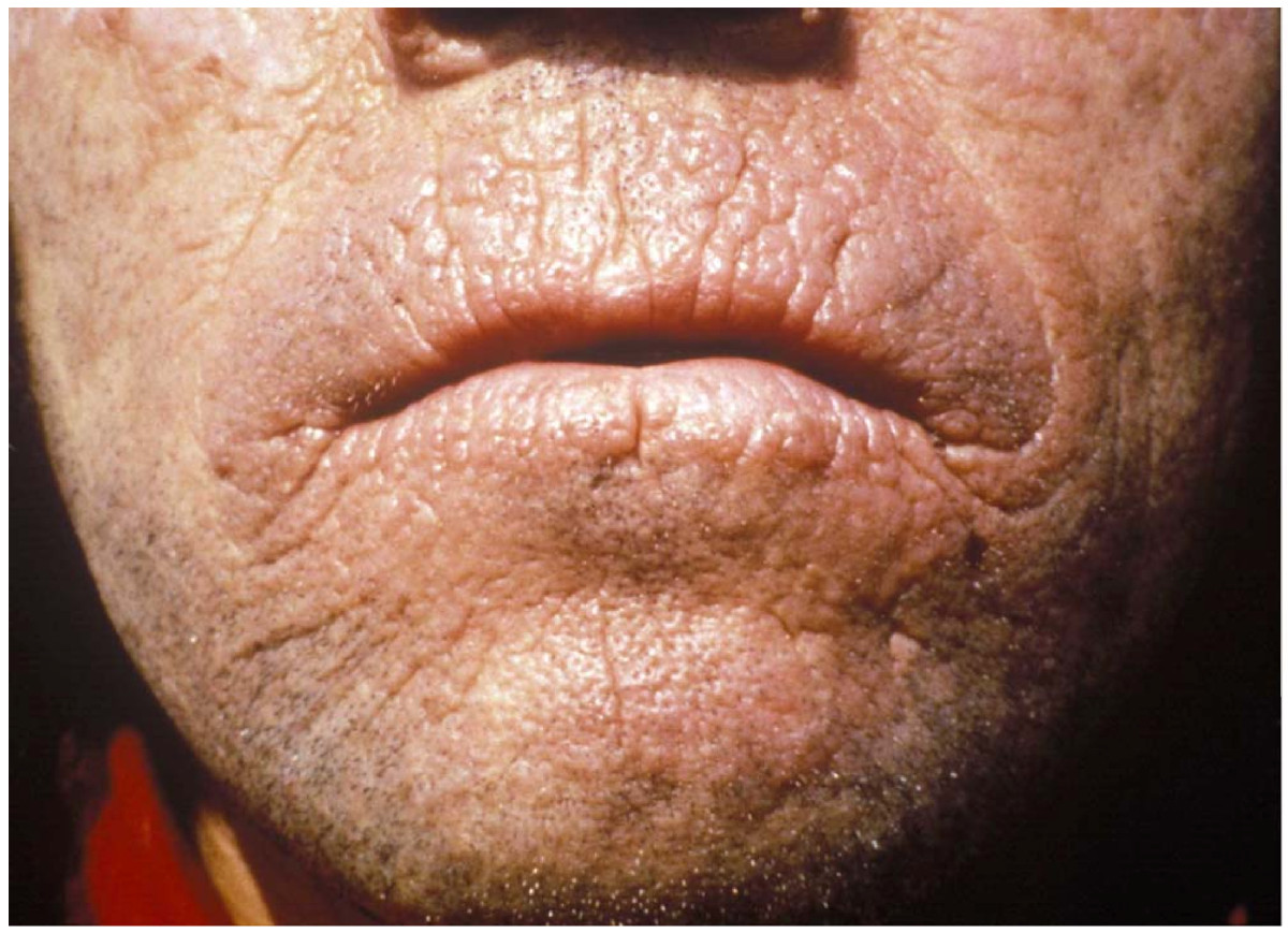
      Erythropoietic protoporphyria (EPP) is a type of porphyria . Porphyrias are caused by an abnormality in the heme production process. Heme is essential in enabling our blood cells to carry oxygen and in breaking down chemical compounds in the liver. Erythropoietic protoporphyria is caused by pathogenic variants (mutations) in the FECH gene which lead to an impaired activity of ferrocheletase (FECH), an important enzyme in heme production. This results in the build-up of protoporphyrin in the bone marrow, red blood cells, blood plasma, skin, and eventually liver. Build up of protoporphyrin can cause extreme sensitivity to sunlight, liver damage, abdominal pain, gallstones, and enlargement of the spleen.
    • Protoporphyria, Erythropoietic, 1 OMIM
      A number sign (#) is used with this entry because erythropoietic protoporphyria-1 (EPP1) is caused by compound heterozygous or homozygous mutation in the gene encoding ferrochelatase (FECH; 612386) on chromosome 18q21. The disorder most often results from inheritance of a null FECH allele in trans with a low-expression FECH mutation (612386.0015) prevalent in some populations, resembling autosomal dominant inheritance with incomplete penetrance. Description Erythropoietic protoporphyria-1 is an inborn error of porphyrin metabolism caused by decreased activity of the enzyme ferrochelatase, the terminal enzyme of the heme biosynthetic pathway, which catalyzes the insertion of iron into protoporphyrin to form heme. EPP is characterized clinically by photosensitivity to visible light commencing in childhood, and biochemically by elevated red cell protoporphyrin levels (Todd, 1994). Genetic Heterogeneity of Erythropoietic Protoporphyria Also see X-linked erythropoietic protoporphyria (XLEPP; 300752), caused by mutation in the ALAS2 gene (301300) on chromosome Xp11, and EPP2 (618015), caused by mutation in the CLPX gene (615611) on chromosome 15q22.
    • Autosomal Erythropoietic Protoporphyria Orphanet
      Erythropoietic protoporphyria (EPP) is an inherited disorder of the heme metabolic pathway characterized by accumulation of protoporphyrin in blood, erythrocytes and tissues, and cutaneous manifestations of photosensitivity. Epidemiology EPP has been reported worldwide, with prevalence ranging between 1/75,000 and 1/200,000. Clinical description EPP usually manifests in early infancy upon the first exposure to sun. EPP is characterized by cutaneous manifestations of acute painful photosensitivity with erythema and edema, sometimes with petechiae, together with stinging and burning sensations without blistering, upon exposure to sunlight or artificial light (400-700 nm). These episodes have a variable severity depending on the exposure duration and may result in chronic permanent lesions on exposed skin.
  • Hiv/aids In Namibia Wikipedia
    Namibia: State of the Nation’s Health: Findings from the Global Burden of Disease. Seattle, WA: IHME, 2016. ^ a b c d e f Ministry of Health and Social Services. (2015). ... Namibia: State of the Nation’s Health: Findings from the Global Burden of Disease. Seattle, WA: IHME, 2016. ^ Ministry of Health and Social Services, Directorate of Special Programmes, Results of the 2008 HIV Sentinel Survey, 2008 ^ http://www.pepfar.gov/countries namibia/index.htm "2008 Country Profile: Namibia".
  • Merrf Syndrome Wikipedia
    .; Stephens, Karen; Amemiya, Anne; Ledbetter, Nikki (eds.). GeneReviews . Seattle (WA): University of Washington, Seattle. ... In Adam, Margaret P.; Ardinger, Holly H.; Pagon, Roberta A.; Wallace, Stephanie E.; Bean, Lora J.H.; Mefford, Heather C.; Stephens, Karen; Amemiya, Anne; Ledbetter, Nikki (eds.). GeneReviews . Seattle (WA): University of Washington, Seattle.
    TRNL1, TRNP, TRNF, TRNK, ND5, TRNI, IL1A, TRNS2, TRNS1, TRNH, RNR1, IL1B, TRNQ, TRNT, TK2, POLG, HSPB2, HSPB3, HSPB1, MMP1, NAE1, EPM2A, TNFSF10, CARS2, RARS2, COQ8A, COQ9, TRMU, FGF21, PRPF6, AFG3L2, SOD2, COX5A, SPG7, ACTB, RFX3, PRKCD, PLXNA2, OPA1, AHSG, COX2, ATP6, CSTB, CPOX, COX8A, CLN5, CLN3, TPP1, CA8, SLC25A4, MTCO2P12
    • Myoclonic Epilepsy Associated With Ragged-Red Fibers OMIM
      A number sign (#) is used with this entry because this syndrome represents a phenotype that can be produced by mutation in more than 1 mitochondrial gene, e.g., MTTK (590060), MTTL1 (590050), MTTH (590040), MTTS1 (590080), MTTS2 (590085), MTTF (590070). Features of the MERRF syndrome have also been associated with mutation in the MTND5 gene (516005). Clinical Features Fukuhara et al. (1980) provided an early report of myoclonic epilepsy associated with ragged-red fibers (MERRF). For detailed clinical features, see MOLECULAR GENETICS Inheritance Rosing et al. (1985) described an extensive family in which many members showed this combination of abnormalities which goes by the acronymic designation MERRF syndrome. Autosomal dominant, autosomal recessive, and X-linked inheritance could be excluded.
    • Myoclonic Epilepsy With Ragged-Red Fibers MedlinePlus
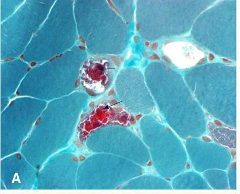
      Myoclonic epilepsy with ragged-red fibers (MERRF) is a disorder that affects many parts of the body, particularly the muscles and nervous system. In most cases, the signs and symptoms of this disorder appear during childhood or adolescence. The features of MERRF vary widely among affected individuals, even among members of the same family. MERRF is characterized by muscle twitches (myoclonus), weakness (myopathy), and progressive stiffness (spasticity). When the muscle cells of affected individuals are stained and viewed under a microscope, these cells usually appear abnormal.
    • Merrf GeneReviews
      Summary Clinical characteristics. MERRF ( m yoclonic e pilepsy with r agged r ed f ibers) is a multisystem disorder characterized by myoclonus (often the first symptom) followed by generalized epilepsy, ataxia, weakness, exercise intolerance, and dementia. Onset can occur from childhood to adulthood, occurring after normal early development. Common findings are ptosis, hearing loss, short stature, optic atrophy, cardiomyopathy, cardiac dysrhythmias such as Wolff-Parkinson-White syndrome, and peripheral neuropathy. Pigmentary retinopathy, optic neuropathy, diabetes mellitus, and lipomatosis have been observed. Diagnosis/testing. A clinical diagnosis of MERRF can be established in a proband with the following four "canonic" features: myoclonus, generalized epilepsy, ataxia, and ragged red fibers (RRF) in the muscle biopsy.
    • Myoclonic Epilepsy With Ragged Red Fibers GARD
      Myoclonic epilepsy with ragged red fibers (MERRF) is a multisystem disorder characterized by myoclonus , which is often the first symptom, followed by generalized epilepsy , ataxia, weakness, and dementia. Symptoms usually first appear in childhood or adolescence after normal early development. The features of MERRF vary widely from individual to individual, even within families. Other common findings include hearing loss, short stature, optic atrophy , and cardiomyopathy with Wolff-Parkinson-White (WPW) syndrome . The diagnosis is based on clinical features and a muscle biopsy finding of ragged red fibers (RRF).
    • Merrf Orphanet
      MERRF (Myoclonic Epilepsy with Ragged Red Fibers) syndrome is a mitochondrial encephalomyopathy characterized by myoclonic seizures. Epidemiology The prevalence in the general population of Europe has been estimated at 0.9 in 100 000, but the disease seems to be more common in the USA. Clinical description Patients usually present during adolescence or early adulthood with myoclonic epilepsy, sometimes with neurosensory deafness, optic atrophy, short stature or peripheral neuropathy. A few cases have been associated with lipomatosis, cardiomyopathy, pigmentary retinopathy, ophthalmoparesis and/or pyramidal signs. The disease is progressive with worsening of the epilepsy and onset of additional symptoms including ataxia, deafness, muscle weakness, and dementia.
    • Cardiomyopathy And Deafness Due To Trna Lysine Gene Mutation GARD
      A specific change in the MTTK gene causes a condition characterized by weakened heart muscle (cardiomyopathy) and hearing loss. Affected individuals may also have myopathy and ataxia. This mutation replaces the DNA building block (nucleotide) guanine with the nucleotide adenine at position 8363 (written as G8363A) within the gene. It is unclear how this alteration in the MTTK gene results in cardiomyopathy, hearing loss, and other symptoms.
  • Autosomal Dominant Cerebellar Ataxia Wikipedia
    Hereditary Ataxia Overview . Seattle (WA): University of Washington, Seattle. ... Spinocerebellar Ataxia Type 2 . Seattle (WA): University of Washington, Seattle.
    PDYN, ATXN3, ATXN2, ATXN1, LY6E, ATXN7, PLEKHG4, PPP2R2B, CACNA1A, PRKCG, TBP, DNMT1, FXN, TWNK, ATXN8OS, TSHZ1, CACNA1G, PMPCA, AFG3L2, ARHGEF28, SCA25, ATOX1, RHO, SPTBN2, PRPH, KCND3, ITPR1, FGF14, ATN1, CACNA1C, SCA30
    • Autosomal Dominant Cerebellar Ataxia Orphanet
      A clinically and genetically heterogeneous group of neurodegenerative diseases characterized by a slowly progressive ataxia of gait, stance and limbs, dysarthria and/or oculomotor disorder, due to cerebellar degeneration in the absence of coexisting diseases. The degenerative process can be limited to the cerebellum (ADCA type 3) or may additionally involve the retina (ADCA type 2), optic nerve, ponto-medullary systems, basal ganglia, cerebral cortex, spinal tracts or peripheral nerves (ADCA type 1). In ACDA type 4, a cerebellar syndrome is associated with epilepsy.
    • Autosomal Dominant Cerebellar Ataxia GARD
      Autosomal dominant cerebellar ataxia (ADCA) is one of the genetic subtypes of hereditary ataxia . Although the signs and symptoms vary depending on the specific type, the most common symptom of ADCA is poor movement coordination ( ataxia ) especially a jerky, unsteady walking style (gait). Coordination of hands and clearness of speech (dysarthria) are also affected. The area of the brain controlling balance and movement decreases in size ( cerebellar atrophy ). This can be seen on brain imaging . The ataxia usually slowly worsens over time.
  • Factor Xii Deficiency Wikipedia
    . ^ Zeerleder S, Schloesser M, Redondo M, Wuillemin WA, Engel W, Furlan M, Laemmle B (1999).
    F12, BRAF, GRK6, MGAT2, MAP2K1, PTPN11, COX8A, F5, F11, CFH, KNG1, PLG, VWF, PGAP1
    • Factor Xii Deficiency OMIM
      A number sign (#) is used with this entry because of evidence that factor XII deficiency is caused by mutation in the F12 gene (610619) on chromosome 5q35. Clinical Features Factor XII deficiency was usually discovered because of the practice in some hospitals of routinely performing whole blood clotting times before surgical operations (McCain et al., 1959). Ratnoff and Steinberg (1962) analyzed data on 55 cases in 37 families. Parental consanguinity was present in at least 2 instances. Some heterozygotes show partial deficiency of Hageman factor. The Japanese case reported by Miwa et al. (1968) had first-cousin parents.
    • Factor Xii Deficiency GARD
      Factor XII deficiency is an inherited disorder that affects a protein (factor XII) involved in blood clotting. While a lack of factor XII does not cause affected individuals to bleed abnormally, the blood takes longer than normal to clot in a test tube. The condition is usually discovered when prolonged clotting is noticed in the process of running other laboratory tests. Factor XII deficiency is caused by mutations in the F12 gene. It is inherited in an autosomal recessive manner.
    • Congenital Factor Xii Deficiency Orphanet
      A rare, autosomal recessive systemic dysfunction of the hemostatic pathway, that is due to a defect in the coagulation factor XII (FXII or Hageman factor), and is either asymptomatic or characterized by a prolonged activated partial thromboplastin time and an increased risk for thromboembolism. FXII deficiency is strongly associated with primary recurrent abortions.
  • Pmm2 Deficiency Wikipedia
    In Adam MP, Ardinger HH, Pagon RA, Wallace SE, Bean LJ, Stephens K, Amemiya A (eds.). GeneReviews® . Seattle (WA): University of Washington, Seattle.
    PMM2, PMM1, MPI, LEMD3, AHCY, GYPA, GYPB, GYPE, OGA, ALG6
    • Pmm2-Cdg (Cdg-Ia) GeneReviews
      Summary Clinical characteristics. PMM2-CDG ( CDG-Ia ) (previously known as congenital disorder of glycosylation type 1a), the most common of a group of disorders of abnormal glycosylation of N-linked oligosaccharides, is divided into three types: infantile multisystem, late-infantile and childhood ataxia-intellectual disability, and adult stable disability. The three types notwithstanding, clinical presentation and course are highly variable, ranging from infants who die in the first year of life to mildly involved adults. Clinical presentations tend to be similar in sibs. In the infantile multisystem type, infants show axial hypotonia, hyporeflexia, esotropia, and developmental delay. Feeding problems, vomiting, failure to thrive, and impaired growth are frequently seen. Subcutaneous fat may be excessive over the buttocks and suprapubic region.
    • Congenital Disorder Of Glycosylation, Type Ia OMIM
      A number sign (#) is used with this entry because congenital disorder of glycosylation type Ia (CDG Ia, CDG1A) is caused by homozygous or compound heterozygous mutation in the gene encoding phosphomannomutase-2 (PMM2; 601785) on chromosome 16p13. Description Congenital disorders of glycosylation (CDGs) are a genetically heterogeneous group of autosomal recessive disorders caused by enzymatic defects in the synthesis and processing of asparagine (N)-linked glycans or oligosaccharides on glycoproteins. These glycoconjugates play critical roles in metabolism, cell recognition and adhesion, cell migration, protease resistance, host defense, and antigenicity, among others. CDGs are divided into 2 main groups: type I CDGs comprise defects in the assembly of the dolichol lipid-linked oligosaccharide (LLO) chain and its transfer to the nascent protein, whereas type II CDGs (see, e.g., CDG2A, 212066) refer to defects in the trimming and processing of the protein-bound glycans either late in the endoplasmic reticulum or the Golgi compartments. CDG1A is the most common form of CDG and was the first to be characterized at the molecular level (reviews by Marquardt and Denecke, 2003; Grunewald et al., 2002).
    • Pmm2-Cdg (Cdg-Ia) GARD
      Congenital disorder of glycosylation type Ia (CDG-Ia) is an inherited condition that affects many parts of the body. The type and severity of problems associated with CDG-Ia vary widely among affected individuals, sometimes even among members of the same family. Signs and symptoms are typically evident in infancy and can include hypotonia, inverted nipples, an abnormal distribution of fat, strabismus, developmental delay, failure to thrive , seizures, and distinctive facial features. About 20 percent of affected infants do not survive the first year of life due to multiple organ failure. The most severe cases of CDG-Ia are characterized by hydrops fetalis .
    • Pmm2-Cdg Orphanet
      PMM2-CDG is the most frequent form of congenital disorder of N-glycosylation and is characterized by cerebellar dysfunction, abnormal fat distribution, inverted nipples, strabismus and hypotonia. 3 forms of PMM2-CDG can be distinguished: the infantile multisystem type, late-infantile and childhood ataxia-intellectual disability type (3-10 yrs old), and the adult stable disability type. Infants usually develop ataxia, psychomotor delay and extraneurological manifestations including failure to thrive, enteropathy, hepatic dysfunction, coagulation abnormalities and cardiac and renal involvement. The phenotype is however highly variable and ranges from infants who die in the first year of life to mildly involved adults.
    • Pmm2-Congenital Disorder Of Glycosylation MedlinePlus
      PMM2 -congenital disorder of glycosylation ( PMM2 -CDG, also known as congenital disorder of glycosylation type Ia) is an inherited condition that affects many parts of the body. The type and severity of problems associated with PMM2 -CDG vary widely among affected individuals, sometimes even among members of the same family. Individuals with PMM2 -CDG typically develop signs and symptoms of the condition during infancy. Affected infants may have weak muscle tone (hypotonia), retracted (inverted) nipples, an abnormal distribution of fat, eyes that do not look in the same direction (strabismus), developmental delay, and a failure to gain weight and grow at the expected rate (failure to thrive). Infants with PMM2 -CDG also frequently have an underdeveloped cerebellum , which is the part of the brain that coordinates movement.
  • Gastrinoma Wikipedia
    Wiener Klinische Wochenschrift . 119 (19–20): 573–578. doi : 10.1007/s00508-007-0884-2 . ISSN 0043-5325 . PMID 17985090 .
    MEN1, GAST, CHGA, SST, SCT, CDKN2A, ACTB, SCTR, GRPR, GRP, ERBB2, POTEF, CASR, MET, NMBR, POMC, PYGM, S100B, BBS2, ATP4A, ATP12A, SSTR5, TCF3, TNFRSF1B, TP53, VIP, KHSRP, PSIP1, SIGLEC7, NMB, CUX1, EGF, SMAD4, EGFR, CDKN2D, CDKN2B, GFAP, GH1, FFAR1, CDH1, CD44, HCLS1, HGF, APC, IGF1, IGF1R, IGFBP1, IL2RB, HRH2
    • Zollinger-Ellison Syndrome Orphanet
      Zollinger-Ellison syndrome (ZES) is characterized by severe peptic disease (ulcers/esophageal disease) caused by hypergastrinemia secondary to a gastrinoma resulting in increased gastric acid secretion. Epidemiology Annual incidence is estimated at 1-2 cases per million. The condition is slightly more common in females than males (sex ratio of 1.3:1). Clinical description ZES is usually diagnosed in the fifth decade of life. Abdominal pain (typically in the upper abdomen) and diarrhea are the most frequent manifestations. Heartburn is often present (44% of cases). Other signs include nausea, vomiting, malabsorption, and weight loss.
    • Zollinger-Ellison Syndrome GARD
      Zollinger-Ellison syndrome (ZES) is a condition in which tumors called gastrinomas in the pancreas and duodenum (part of the small intestine) cause high levels of the hormone gastrin in the blood. High levels of gastrin then cause production of too much stomach acid. Signs and symptoms may include abdominal pain, peptic ulcers , vomiting blood, and diarrhea. The tumors are sometimes cancerous and may spread to other areas of the body. In most cases, the cause of ZES is unknown. However, about 25-30% of gastrinomas are caused by an inherited condition called multiple endocrine neoplasia type 1 (MEN1) .
  • Environmental Enteropathy Wikipedia
    . ^ a b c d e f g h i j k l m Korpe PS, Petri WA (June 2012). "Environmental enteropathy: critical implications of a poorly understood condition" . ... PMID 26542185 . ^ Gilmartin AA, Petri WA (June 2015). "Exploring the role of environmental enteropathy in malnutrition, infant development and oral vaccine response" .
  • Medullary Cystic Kidney Disease Wikipedia
    Autosomal Dominant Tubulointerstitial Kidney Disease, MUC1-Related (ADTKD-MUC1) . Seattle (WA): University of Washington, Seattle. ... Autosomal Dominant Tubulointerstitial Kidney Disease, UMOD-Related (ADTKD-UMOD) . Seattle (WA): University of Washington, Seattle.
    UMOD, GLI2, KCNJ1, MUC1, HNF1B
    • Autosomal Dominant Tubulointerstitial Kidney Disease Due To Umod Mutations GARD
      Autosomal dominant tubulointerstitial kidney disease due to UMOD mutations (ADTKD–UMOD) is an inherited disorder that causes a gradual loss of kidney function that eventually leads to the need for kidney transplantation or dialysis between the ages of 30 and 70. Patients with ADTKD-UMOD have high blood levels of uric acid before kidney failure develops, and some affected individuals may develop gout. Gout is a form of arthritis (inflammation) that occurs often in the big toe, ankle, knee, or other joints. ADTKD-UMOD is caused by a mistake (mutation) in the UMOD gene, which leads to the build-up of the altered uromodulin protein in the tubules of the kidney, leading to slow loss of kidney function. ADTKD-UMOD is inherited in a dominant pattern in families. It is diagnosed based on the symptoms, laboratory testing, family history and genetic testing.
    • Autosomal Dominant Tubulointerstitial Kidney Disease, Umod-Related GeneReviews
      Summary Clinical characteristics. Autosomal dominant tubulointerstitial kidney disease caused by UMOD pathogenic variants (ADTKD- UMOD ) was previously known as familial juvenile hyperuricemic nephropathy type 1 (FJHN1), medullary cystic kidney disease type 2 (MCKD2), and UMOD -associated kidney disease (or uromodulin-associated kidney disease). Typical clinical findings: Urinalysis revealing minimal protein and no blood Slowly progressive chronic kidney failure, usually first noted in the teen years and progressing to end-stage renal disease (ESRD) between the fourth and seventh decades (Age at ESRD varies among and within families.) Hyperuricemia and gout (resulting from reduced kidney excretion of uric acid) that occurs as early as the teenage years Diagnosis/testing. ADTKD- UMOD is defined by the presence of a heterozygous pathogenic variant in UMOD , the gene encoding uromodulin. The majority of persons with ADTKD- UMOD have the following laboratory test abnormalities: elevated serum creatinine (decreased estimated glomerular filtration rate), bland urinary sediment, elevated serum urate level, and reduced fractional excretion of uric acid.
    • Medullary Cystic Kidney Disease 2 OMIM
      A number sign (#) is used with this entry because medullary cystic kidney disease-2 (MCKD2) is caused by heterozygous mutation in the UMOD gene (191845) on chromosome 16p12. Mutation in the same gene causes familial juvenile hyperuricemic nephropathy-1 (HNFJ1; 162000). Description Medullary cystic kidney disease (MCKD) is an autosomal dominant form of tubulointerstitial nephropathy characterized by formation of renal cysts at the corticomedullary junction. It is characterized by adult onset of impaired renal function and salt wasting resulting in end-stage renal failure by the sixth decade (Wolf et al., 2004). For a general phenotypic description and a discussion of genetic heterogeneity of medullary cystic kidney disease, see MCKD1 (174000).
  • Post-Ebola Virus Syndrome Wikipedia
    Retrieved 5 August 2016 . ^ Fischer WA, Brown J, Wohl DA, Loftis AJ, Tozay S, Reeves E, et al. (2017). ... Retrieved 1 October 2016 . Fischer WA, Brown J, Wohl DA, Loftis AJ, Tozay S, Reeves E, et al. (2017).
  • Tetrasomy 18p Wikipedia
    . ^ a b Sebold C, Roeder E, Zimmerman M, Soileau B, Heard P, Carter E, Schatz M, White WA, Perry B, Reinker K, O'Donnell L, Lancaster J, Li J, Hasi M, Hill A, Pankratz L, Hale DE, Cody JD (2010).
    HTC2
    • Tetrasomy 18p MedlinePlus
      Tetrasomy 18p is a chromosomal condition that affects many parts of the body. This condition usually causes feeding difficulties in infancy, delayed development, intellectual disability that is often mild to moderate but can be severe, changes in muscle tone, distinctive facial features, and other birth defects. However, the signs and symptoms vary among affected individuals. Babies with tetrasomy 18p often have trouble feeding and may vomit frequently, which makes it difficult for them to gain weight. Some affected infants also have breathing problems and jaundice, which is a yellowing of the skin and the whites of the eyes. Changes in muscle tone are commonly seen with tetrasomy 18p. Some affected children have weak muscle tone (hypotonia), while others have increased muscle tone (hypertonia) and stiffness (spasticity).
    • Chromosome 18p Tetrasomy GARD
      Chromosome 18p tetrasomy is a chromosomal disorder that affects many parts of the body. It occurs when the short arm of chromosome 18 (18p) appears four times (tetrasomy) rather than the normal two times in cells of the body. The symptoms of chromosomy 18p tetrasomy vary from case to case but may include abnormalities of the head and face (craniofacial) area, malformations of the spine, hands, and/or feet, neuromuscular abnormalities, kidney malformations, intellectual disability, speech delays, and behavioral abnormalities. In most cases, chromosome 18p tetrasomy is the result of a spontaneous (de novo) genetic change (mutation) early in fetal development during pregnancy. Although there is no specific treatment or cure for chromosome 18p tetrasomy, there are ways to manage the symptoms.
    • Tetrasomy 18p OMIM
      A number sign (#) is used with this entry because this dysmorphic condition is caused by tetrasomy of chromosome 18p. Clinical Features Sebold et al. (2010) summarized the phenotype of tetrasomy 18p with a list of findings reported in more than 25% of theretofore published cases: neonatal feeding problems, growth retardation, microcephaly, strabismus, muscle tone abnormalities, scoliosis/kyphosis, and variants on brain MRI. Developmental delay and cognitive impairment are universally present. To more fully describe the molecular features and clinical presentation of tetrasomy 18p, Sebold et al. (2010) performed array CGH on samples from 42 individuals with tetrasomy 18p, and reviewed the medical records of these individuals. Forty-one of these individuals had an isochromosome 18p in all cells examined; the remaining individual had mosaicism.
    • Tetrasomy 18p Orphanet
      Tetrasomy 18p is a very rare structural chromosomal anomaly affecting multiple body systems and characterized clinically by craniofacial abnormalities, delayed development, cognitive impairment, changes in muscle tone, distinctive facial features, and rarely renal malformations.
  • Emanuel Syndrome Wikipedia
    .; Ledbetter, Nikki; Mefford, Heather C. (eds.). GeneReviews . Seattle (WA): University of Washington, Seattle.
    TBX1, DGCR, GRM8, RANBP1, ZNF74, MFRP
    • Emanuel Syndrome GeneReviews
      Summary Clinical characteristics. Emanuel syndrome is characterized by pre- and postnatal growth deficiency, microcephaly, hypotonia, severe developmental delays, ear anomalies, preauricular tags or pits, cleft or high-arched palate, congenital heart defects, kidney abnormalities, and genital abnormalities in males. Diagnosis/testing. The diagnosis of Emanuel syndrome is established in a proband by detection of a duplication of 22q10-22q11 and duplication of 11q23-qter on a supernumerary derivative chromosome 22 [der(22)]. Management. Treatment of manifestations: Care by a multidisciplinary team is usually necessary; standard management of gastroesophageal reflux, nutrition, anal atresia (or stenosis), inguinal hernias, cardiac defects, cleft palate, hip dysplasia, other skeletal complications, hearing loss, cryptorchidism and/or micropenis, refractive errors, and strabismus or other ophthalmologic issues; ongoing physical, occupational, and speech therapies; alternative communication methods to facilitate communication. Prevention of secondary complications: Attention to the airway during sedation and/or operative procedures in an institution with pediatric anesthesiologists. Surveillance: Follow up as needed based on the extent of systemic involvement in each individual; regular developmental assessments; periodic reevaluation by a clinical geneticist.
    • Emanuel Syndrome MedlinePlus
      Emanuel syndrome is a chromosomal disorder that disrupts normal development and affects many parts of the body. Infants with Emanuel syndrome have weak muscle tone (hypotonia) and fail to gain weight and grow at the expected rate (failure to thrive). Their development is significantly delayed, and most affected individuals have severe to profound intellectual disability. Other features of Emanuel syndrome include an unusually small head (microcephaly), distinctive facial features, and a small lower jaw (micrognathia). Ear abnormalities are common, including small holes in the skin just in front of the ears (preauricular pits or sinuses).
    • Emanuel Syndrome OMIM
      A number sign (#) is used with this entry because Emanuel syndrome is caused by malsegregation of the t(11;22)(q23;q11.2) translocation, one of only a few recurrent non-Robertsonian constitutional translocations in humans (Fraccaro et al., 1980; Zackai and Emanuel, 1980). See also supernumerary der(22)t(8;22) syndrome (613700). Description Emanuel syndrome is characterized by multiple congenital anomalies, craniofacial dysmorphism, and significant developmental delay and mental retardation. Features include ear anomalies, preauricular tag or sinus, cleft or high-arched palate, micrognathia, microcephaly, kidney abnormalities, heart defects, and genital abnormalities in males (summary by Carter et al., 2009). Carriers of the balanced constitutional t(11;22) translocation are phenotypically normal but have a 10% risk of having progeny with supernumerary der(22)t(11;22) syndrome as a result of malsegregation of the der(22). The affected progeny are genotypically unbalanced because they carry the der(22) as a supernumerary chromosome--either 47,XX,+der(22)t(11;22) or 47,XY,+der(22)t(11;22) (Zackai and Emanuel, 1980; Lin et al., 1986).
    • Emanuel Syndrome Orphanet
      Emanuel syndrome is a constitutional genomic disorder due to the presence of a supernumerary derivative 22 chromosome and characterized by severe intellectual disability, characteristic facial dysmorphism (micrognathia, hooded eyelids, upslanting downslanting parebral fissures, deep set eyes, low hanging columnella and long philtrum), congenital heart defects and kidney abnormalities.
    • Emanuel Syndrome GARD
      Emanuel syndrome is a chromosomal disorder that is characterized by learning problems and stunted growth and development. The signs and symptoms are varied and may include decreased muscle tone (hypotonia) and developmental delay in childhood, intellectual disability severe, extremely small head ( microcephaly ), distinctive facial features, small jaw, ear anomalies, arched palate (roof of the mouth), cleft palate , heart defects , kidney malformations , and genital abnormalities (in males). Emanuel syndrome is caused by the presence of additional genetic material of the chromosomes 11 and 22 in each cell . This condition is usually inherited from a parent who has a balanced translocation between chromosomes 11 and 22. Treatment depends on the signs and symptoms present in the individual.
  • Carnitine Palmitoyltransferase I Deficiency Wikipedia
    .; Ledbetter, Nikki; Mefford, Heather C. (eds.). GeneReviews . Seattle (WA): University of Washington, Seattle.
    CPT1A, CPT2, CHPT1, DHDDS
    • Carnitine Palmitoyltransferase I Deficiency OMIM
      A number sign (#) is used with this entry because carnitine palmitoyltransferase deficiency I is caused by homozygous or compound heterozygous mutation in the gene encoding carnitine palmitoyltransferase IA (CPT1A; 600528) on chromosome 11q13. Description CPT I deficiency is an autosomal recessive metabolic disorder of long-chain fatty acid oxidation characterized by severe episodes of hypoketotic hypoglycemia usually occurring after fasting or illness. Onset is in infancy or early childhood (Bougneres et al., 1981) Clinical Features Bougneres et al. (1981) reported 2 sisters who developed severe hypoketotic hypoglycemia at age 8 months, resulting in death in 1 of them. Other features included hepatomegaly, nonketotic hypoglycemia, and coma. Liver CPT activity was absent in the patient who was tested. Demaugre et al. (1988) reported 2 patients with carnitine palmitoyltransferase deficiency and hepatic symptoms.
    • Carnitine Palmitoyltransferase 1a Deficiency GeneReviews
      Summary Clinical characteristics. Carnitine palmitoyltransferase 1A (CPT1A) deficiency is a disorder of long-chain fatty acid oxidation. Clinical manifestations usually occur in an individual with a concurrent febrile or gastrointestinal illness when energy demands are increased; onset of symptoms is usually rapid. The recognized phenotypes are: acute fatty liver of pregnancy, in which the fetus has biallelic pathogenic variants in CPT1A that causes CPT1A deficiency; and hepatic encephalopathy, in which individuals (typically children) present with hypoketotic hypoglycemia and sudden onset of liver failure. Individuals with hepatic encephalopathy typically present with hypoglycemia, absent or low levels of ketones, and elevated serum concentrations of liver transaminases, ammonia, and total carnitine. Between episodes of hepatic encephalopathy, individuals appear developmentally and cognitively normal unless previous metabolic decompensation has resulted in neurologic damage.
    • Carnitine Palmitoyl Transferase 1a Deficiency Orphanet
      Carnitine palmitoyltransferase 1A (CPT-1A) deficiency is an inborn error of metabolism that affects mitochondrial oxidation of long chain fatty acids (LCFA) in the liver and kidneys, and is characterized by recurrent attacks of fasting-induced hypoketotic hypoglycemia and risk of liver failure. Epidemiology Since the description of the disease in 1981, less than 50 cases have been reported. Clinical description CPT-1A deficiency manifests between birth and 18 months of age with recurrent attacks of hypoketotic hypoglycemia of varying severity, triggered by fasting or intercurrent illness, that can lead to severe neurological sequelae. CPT-1A-deficient patients can also present with hepatic encephalopathy with loss of consciousness, seizures, coma, or even sudden death. There may be a risk of progression to liver failure. Patients with severe CPT-1A deficiency may also have renal tubular acidosis.
    • Carnitine Palmitoyltransferase I Deficiency MedlinePlus
      Carnitine palmitoyltransferase I (CPT I) deficiency is a condition that prevents the body from using certain fats for energy, particularly during periods without food (fasting). The severity of this condition varies among affected individuals. Signs and symptoms of CPT I deficiency often appear during early childhood. Affected individuals usually have low blood sugar (hypoglycemia) and a low level of ketones, which are produced during the breakdown of fats and used for energy. Together these signs are called hypoketotic hypoglycemia. People with CPT I deficiency can also have an enlarged liver (hepatomegaly), liver malfunction, and elevated levels of carnitine in the blood. Carnitine, a natural substance acquired mostly through the diet, is used by cells to process fats and produce energy.
    • Carnitine Palmitoyl Transferase 1a Deficiency GARD
      Carnitine palmitoyltransferase I deficiency (CPT1A deficiency) is an inherited metabolic condition that prevents the body from converting certain fats (long-chain fatty acids) into energy, particularly during periods without food. Carnitine, a natural substance acquired mostly through the diet, is required by cells to process fats and produce energy. Symptoms of this condition often appear early in life and include low blood sugar (hypoglycemia) and low levels of ketones, which are produced when the body breaks down fat for energy (hypoketotic hypoglycemia). This can lead to a greater risk for loss of consciousness or seizures. People with this disorder typically also have an enlarged liver (hepatomegaly), muscle weakness, nervous system damage, and elevated levels of carnitine in the blood.
  • ←
  • 1
  • 2
  • 3
  • 4
  • 5
  • 6
  • 7
  • ...
  • 13
  • 14
  • →

FindZebra

contact@findzebra.com