• FindZebra
  • About
  • Contact
  • Help
  • Login
Deep Research Advanced
  • Enamel-Renal Syndrome Wikipedia
    History [ edit ] This condition was first described in 1972. [2] References [ edit ] ^ Jaureguiberry G, De la Dure-Molla M, Parry D, Quentric M, Himmerkus N, Koike T, Poulter J, Klootwijk E, Robinette SL, Howie AJ, Patel V, Figueres ML, Stanescu HC, Issler N, Nicholson JK, Bockenhauer D, Laing C, Walsh SB, McCredie DA, Povey S, Asselin A, Picard A, Coulomb A, Medlar AJ, Bailleul-Forestier I, Verloes A, Le Caignec C, Roussey G, Guiol J, Isidor B, Logan C, Shore R, Johnson C, Inglehearn C, Al-Bahlani S, Schmittbuhl M, Clauss F, Huckert M, Laugel V, Ginglinger E, Pajarola S, Spartà G, Bartholdi D, Rauch A, Addor MC, Yamaguti PM, Safatle HP, Acevedo AC, Martelli-Júnior H, dos Santos Netos PE, Coletta RD, Gruessel S, Sandmann C, Ruehmann D, Langman CB, Scheinman SJ, Ozdemir-Ozenen D, Hart TC, Hart PS, Neugebauer U, Schlatter E, Houillier P, Gahl WA, Vikkula M, Bloch-Zupan A, Bleich M, Kitagawa H, Unwin RJ, Mighell A, Berdal A, Kleta R (2013) Nephrocalcinosis (Enamel Renal Syndrome) caused by autosomal recessive FAM20A Mutations.
    FAM20A, LINC01482, PRKAR1A, SCN5A, KCNJ8, DSP, EGFR, KCND3, KCNE1, CACNA2D1, MYD88, NMT1, DPP6, PRKCE, SCN1B, DDIT3, TLR4, CACNB2, CACNA1C
    • Amelogenesis Imperfecta, Type Ig OMIM
      A number sign (#) is used with this entry because amelogenesis imperfecta type IG (AI1G), also known as enamel-renal syndrome (ERS), is caused by homozygous or compound heterozygous mutation in the FAM20A gene (611062) on chromosome 17q24. Description Amelogenesis imperfecta type IG, also known as enamel-renal syndrome, is characterized by hypoplastic enamel on primary and secondary dentition, pulp stones, delayed or failed eruption of secondary dentition, gingival overgrowth, and nephrocalcinosis. Blood chemistry analyses are typically normal, and nephrocalcinosis, which is found on renal ultrasound, may not appear until later in life (summary by Wang et al., 2013). Clinical Features MacGibbon (1972) reported a brother and sister with absent enamel, nephrocalcinosis, and apparently normal calcium metabolism. Lubinsky et al. (1985) also described an affected brother and sister, aged 11 and 9 years, respectively.
    • Enamel-Renal Syndrome Orphanet
      A extremely rare, genetic malformation syndrome characterized by hypoplastic amelogenesis imperfecta (hypoplastic dental enamel) and nephrocalcinosis (precipitation of calcium salts in renal tissue). Oral manifestations include yellow and misshaped teeth, delayed tooth eruption, and intrapulpal calcifications. Nephrocalcinosis is often asymptomatic but can progress during late childhood or early adulthood to impaired renal function, recurrent urinary infections, renal tubular acidosis, and rarely to end-stage renal failure.
  • Citrullinemia Type I Wikipedia
    (eds.). Citrullinemia Type I . Seattle (WA): University of Washington, Seattle.
    ASS1
    • Citrullinemia, Classic OMIM
      Population Genetics The prevalence of citrullinemia is estimated to be 1 in 100,000 (Testai and Gorelick, 2010). Animal Model In Friesian cattle in Australia, Harper et al. (1986, 1989) reported that citrullinemia-affected calves had a clinical disease similar to the acute neonatal form of citrullinemia in humans.
    • Citrullinemia Type I Orphanet
      Citrullinemia type I is a rare autosomal recessive urea cycle defect characterized biologically by hyperammonemia and clinically by progressive lethargy, poor feeding and vomiting in the neonatal form (Acute neonatal citrullinemia type I, see this term) and by variable hyperammonemia in the later-onset form (Adult-onset citrullinemia type I, see this term).
  • Intestinal Neuronal Dysplasia Wikipedia
    European Journal of Pediatric Surgery . 18 (1): 59–60. doi : 10.1055/s-2008-1038324 . PMID 18302074 . ^ Fadda B, Maier WA, Meier-Ruge W, Schärli A, Daum R (October 1983).
    FLNA, TLX2, SPRY2
    • Intestinal Pseudoobstruction With Patent Ductus Arteriosus And Natal Teeth OMIM
      Harris et al. (1976) described 2 male sibs with mandibular teeth present at birth, patent ductus arteriosus (see 607411), and intestinal pseudoobstruction evident from birth. Vomiting of bile stain material began soon after birth, and there was no passage of meconium. The older brother died at 5 months of age despite 2 gastrointestinal operations and ligation of the patent ductus arteriosus which had led to cardiac failure. The younger brother died at the age of 6 weeks. Either autosomal or X-linked recessive inheritance is possible.
    • Neuronal Intestinal Pseudoobstruction Orphanet
      Neuronal intestinal pseudoobstruction is a form of chronic intestinal pseudoobstruction caused by a developmental failure of the enteric neurons to differentiate or migrate properly and manifests as a bowel obstruction.
  • Craniodiaphyseal Dysplasia Wikipedia
    .), "Craniometaphyseal Dysplasia, Autosomal Dominant" , GeneReviews® , Seattle (WA): University of Washington, Seattle, PMID 20301634 , retrieved 2021-01-18 External links [ edit ] Craniodiaphyseal dysplasia at orpha.net Classification D ICD - 10 : M85.2 OMIM : 218300 MeSH : C562940 External resources Orphanet : 1513 v t e Congenital malformations and deformations of musculoskeletal system / musculoskeletal abnormality Appendicular limb / dysmelia Arms clavicle / shoulder Cleidocranial dysostosis Sprengel's deformity Wallis–Zieff–Goldblatt syndrome hand deformity Madelung's deformity Clinodactyly Oligodactyly Polydactyly Leg hip Hip dislocation / Hip dysplasia Upington disease Coxa valga Coxa vara knee Genu valgum Genu varum Genu recurvatum Discoid meniscus Congenital patellar dislocation Congenital knee dislocation foot deformity varus Club foot Pigeon toe valgus Flat feet Pes cavus Rocker bottom foot Hammer toe Either / both fingers and toes Polydactyly / Syndactyly Webbed toes Arachnodactyly Cenani–Lenz syndactylism Ectrodactyly Brachydactyly Stub thumb reduction deficits / limb Acheiropodia Ectromelia Phocomelia Amelia Hemimelia multiple joints Arthrogryposis Larsen syndrome RAPADILINO syndrome Axial Skull and face Craniosynostosis Scaphocephaly Oxycephaly Trigonocephaly Craniofacial dysostosis Crouzon syndrome Hypertelorism Hallermann–Streiff syndrome Treacher Collins syndrome other Macrocephaly Platybasia Craniodiaphyseal dysplasia Dolichocephaly Greig cephalopolysyndactyly syndrome Plagiocephaly Saddle nose Vertebral column Spinal curvature Scoliosis Klippel–Feil syndrome Spondylolisthesis Spina bifida occulta Sacralization Thoracic skeleton ribs : Cervical Bifid sternum : Pectus excavatum Pectus carinatum
    SOST, CDKL5, CDA, GRIA2, CXCL8, NPPA, CLIP1
    • Craniodiaphyseal Dysplasia, Autosomal Dominant OMIM
      The long bones showed an extreme asymmetric hyperostosis and sclerosis of the diaphyses and evidence of a modeling defect in the metaphyses. The spine, ribs, clavicles, and pelvis all showed some degree of sclerosis and defective modeling but were less severely involved.
    • Craniodiaphyseal Dysplasia Orphanet
      Craniodiaphyseal dysplasia is a rare sclerotic bone disorder with a variable phenotypic expression with massive generalized hyperostosis and sclerosis, particularly of the skull and facial bones, that may lead to severe deformity.
    • Craniodiaphyseal Dysplasia OMIM
      Clinical Features Cranial and facial hyperostosis results in a characteristic clinical and radiographic appearance. The diaphyses of the bones are generally expanded. Halliday (1949) and Stransky et al. (1962) reported isolated cases with similar findings. Facial and cranial thickening and distortion are particularly striking in this form. Most cases have been mentally retarded. Unlike the situation in the craniometaphyseal dysplasias (e.g., 218400), the long bones do not show metaphyseal flaring but show diaphyseal endostosis and are shaped like a policeman's nightstick. Joseph et al. (1958), who first suggested the designation of progressive craniodiaphyseal dysplasia, described a patient with a picture they considered identical to that described by Halliday (1949).
  • Aneurysm Wikipedia
    Velocity at center is near zero. 3- Blood flow exit Modeling of aneurysms consists of creating a 3D model that mimics a particular aneurysm. ... Researchers are able to CT scan a patient's body to create a 3D computer model that possesses the correct geometry. Aneurysms can now be modeled with their distinctive "balloon" shape. ... Current modeling is not able to take into account all variables though. ... "Three-dimensional finite volume modelling of blood flow in simulated angular neck abdominal aortic aneurysm" .
    MMP1, NOX4, TIMP1, COL3A1, SLC2A10, TGFBR2, COL5A1, COL5A2, FBN1, FARSB, SMAD3, COL1A1, MYH11, MYLK, NF1, MMP9, PRF1, CXCL8, TNF, IL6, GZMB, CHI3L1
    • Aneurysms Mayo Clinic
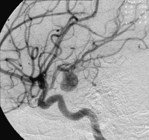
      Overview An aneurysm is an abnormal bulge or ballooning in the wall of a blood vessel. An aneurysm can burst. This is called a rupture. A ruptured aneurysm causes bleeding inside the body and often leads to death. Some aneurysms may not cause symptoms. You might not know you have an aneurysm even if it is large. Aneurysms can develop in several parts of the body, including: The body's main artery, called the aorta ( aortic aneurysm ). The part of the aorta that passes through the belly area ( abdominal aortic aneurysm ).
  • Adenoid Cystic Carcinoma Wikipedia
    Cancer Discov . 622 (2): 176–87. doi : 10.1158/2159-8290.CD-15-0859 . PMC 4744535 . PMID 26631070 . ^ Mitani Y, Liu B, Rao PH, Borra VJ, Zafereo M, Weber RS, Kies M, Lozano G, Futreal PA, Caulin C, El-Naggar AK (2016).
    TP53, NOTCH1, PIK3CA, MYBL1, FBXW7, BCOR, HRAS, MYB, NFIB, CREBBP, CDH1, PTEN, CCND1, SOX4, DAPK1, BRCA1, AQP1, IGFBP2, ATM, ESPL1, SRCAP, HOMER3, TLK1, MAGI2, SMARCA2, MORF4L1, KDM6B, MLC1, FGF16, DTX4, H1-4, GUCY1A1, MGA, RBFOX2, GAS6, GAS2, MAGI1, KAT6A, H2AC16, INSRR, SERPINF1, PRKDC, MAP2K2, MYCN, PYGB, ST3GAL4, SMARCE1, SON, SOX11, MARCKS, TOP2A, KDM6A, KRT15, SMC1A, KRT5, ARID1A, ITGB4, ZIM2, BRD1, MYCBP, NSD1, STAG3L1, CNTN6, MIER2, IL17RD, MAML3, CMTR2, ERBIN, SLC24A3, KMT2C, BCORL1, EFHD1, BCL11A, WNT5B, NETO2, ARID5B, ATRX, FOXP2, JMJD1C, JAG1, ASPM, KANSL1, XAGE1A, VCAN, PDZK1, FAT1, PCSK1N, ARID4B, GINS2, SETD2, FOXO3, DTL, UHRF1, FGFR4, IRX4, ISYNA1, FANCA, MARK2, EN1, EP300, KRAS, KIT, ARID2, SF3B1, CYLD, IDH1, EGFR, CTNNB1, CDKN2A, SOX10, CXCR4, ERBB2, HIF1A, VEGFA, ACCS, SOX2, SMUG1, H3P10, NOS2, BCL2, NOS1, RUNX3, FN1, BCL2A1, MIR21, ACACB, SNAI1, MMP2, MIF, MDM2, TBX1, KRT14, ILK, TP63, BECN1, BMS1, ID1, MMP9, CD274, CTSD, ANO1, MYB-AS1, CD44, BTBD7, SLC2A1, ACACA, PCNA, NOTCH4, MAPK1, PSMD7, CTSB, CTAG1B, CCR5, CENPF, CDK2, CDH5, PECAM1, CDH4, PAX3, DNMT1, KRIT1, PA2G4, CCL28, TMPRSS4, ACKR3, CALM3, NDRG2, MTUS1, CALM2, NNMT, EPHA2, BUB1, BBC3, USP22, GPC3, GJA1, GATA1, FZD2, MTOR, SULT4A1, FOLH1, DKK2, LEF1, PIK3CB, FGF2, FABP7, NGF, EWSR1, OBP2A, ERBB3, TMED7, CALM1, CAMKMT, HOXB7, MIR17HG, MIR125A, MIR140, MIR150, MIR181A2, NTF3, MIR222, MIR320A, MIR93, MIR338, SBSN, MIR375, MIR455, ASIC1, MIR1234, TMED7-TICAM2, ADAMTS9-AS2, H3P23, H3P28, MIRLET7B, TICAM2, BSG, ATF1, PDCD1LG2, CD276, BNIP3, NTRK3, REG4, NTRK1, MINDY4, MAML2, AQP5, MACC1, WDR66, AQP3, SKA1, ALCAM, CTAG1A, ADK, ADAM10, ARMH1, SFN, HES1, PSMD9, HSPB1, SRY, STAT3, PPP2R2B, TFE3, TGFB1, ICAM5, TNF, EPCAM, LYZ, TRAF6, TXN, TYMS, LMNA, UVRAG, LGALS3, VEGFC, VIM, NSD2, RPSA, SPN, SMAD4, MCAM, SDC1, PTCH1, MYC, PTGS2, MMP15, RAC1, RPE65, S100A1, S100B, SGTA, PTPA, PROX1, SKP2, NCAM1, SMARCA1, MAPK3, MMP7, NFKB1, PRKD1, PRRX1, KRT7, KIF22, AGR2, IFI27, PIM1, MFN2, AKT3, RABEPK, NOTCH2, HOXB13, ZNRD2, DCTN6, PIK3CD, HSPG2, SMR3B, HSPB2, MLLT11, EBNA1BP2, CKAP4, PTP4A3, RASSF1, ZEB2, IGF1R, PLAG1, NRP2, RECK, GEMIN2, PPM1D, PIN1, PIK3CG, TNFSF10, ADAM9, IL9, PROM1, ATG5, HSPB3, SCAF11, ARHGEF2, IL2, SLC9A3R2, LHX2, DDX23, CCN1, LANCL1
    • Adenoid Cystic Carcinoma GARD
      Adenoid cystic carcinoma (ACC) is a rare form of adenocarcinoma , a type of cancer that begins in glandular tissues. It most commonly arises in the major and minor salivary glands of the head and neck. It can also occur in the breast, uterus, or other locations in the body. Symptoms depend on the tumor's location. Salivary gland tumors may cause painless masses in the mouth or face. Tumors of the lacrimal gland may cause a bulging eye or changes in vision.
  • Lafora Disease Wikipedia
    Recent research is looking into how inhibition of glycogen synthesis, since increased glucose uptake causes increased glycogen, could potentially stop the formation of the Lafora Bodies in neurons in laforin-deficient mice models while also reducing the chances of seizures . [28] The adipocyte hormone Leptin is what this research targeted by blocking the leptin signaling to reduce glucose uptake and stop Lafora bodies from forming. [28] Other researchers are looking into the ways in which Lafora bodies are being regulated at the level of gene expression. ... Since researchers have found the two genes that cause LD, they are currently aiming to interrupt the process of how these mutations in those genes interfere with normal carbohydrate metabolism in mice models. They predict they will have one or more drugs ready for human clinical trials within the next few years. [30] References [ edit ] ^ http://www.rightdiagnosis.com/medical/melf.htm ^ a b "Progressive Myoclonus Epilepsy, Lafora Type" . ^ a b "Lafora Overview" . ^ a b Ianzano L, Zhang J, Chan EM, Zhao XC, Lohi H, Scherer SW, Minassian BA (2005). ... In Adam, Margaret P.; Ardinger, Holly H.; Pagon, Roberta A.; Wallace, Stephanie E.; Bean, Lora J.H.; Mefford, Heather C.; Stephens, Karen; Amemiya, Anne; Ledbetter, Nikki (eds.). GeneReviews® . Seattle (WA): University of Washington, Seattle. ... "Glycogen metabolism in tissues from a mouse model of Lafora disease" . Archives of Biochemistry and Biophysics . 457 (2): 264–269. doi : 10.1016/j.abb.2006.10.017 . ... "Suppression of leptin signaling reduces polyglucosan inclusions and seizure susceptibility in a mouse model for Lafora disease" . Human Molecular Genetics . 26 (24): 4778–4785. doi : 10.1093/hmg/ddx357 .
    NHLRC1, EPM2A
  • Fg Syndrome Wikipedia
    .), "MED12-Related Disorders" , GeneReviews , Seattle (WA): University of Washington, Seattle, PMID 20301719 , retrieved 2020-09-01 ^ Lyons, Michael J. (1993), Adam, Margaret P.; Ardinger, Holly H.; Pagon, Roberta A.; Wallace, Stephanie E. (eds.), "MED12-Related Disorders" , GeneReviews , Seattle (WA): University of Washington, Seattle, PMID 20301719 , retrieved 2020-09-01 ^ a b Opitz JM, Smith JF, Santoro L (2008).
    MED12, CASK, FLNA, FGS2, MID2, FGS3, IGAN1, VSX1, OBP2A, AGO2, RCOR1, CCN6, KIF22, IGBP1, HTC2, HPD, GLI3, FMR1, FGS5
    • Opitz-Kaveggia Syndrome OMIM
      A number sign (#) is used with this entry because of evidence that the Opitz-Kaveggia syndrome, also known as FG syndrome-1 (FGS1), is caused by mutation in the MED12 gene (300188) on chromosome Xq13. Description Opitz-Kaveggia syndrome (OKS) is an X-linked recessive mental retardation syndrome characterized by dysmorphic features, including relative macrocephaly, hypertelorism, downslanted palpebral fissures, prominent forehead with frontal hair upsweep, and broad thumbs and halluces. Most have hypotonia, constipation, and partial agenesis of the corpus callosum. Some patients have sensorineural hearing loss and joint laxity evolving into joint contractures. Affected individuals tend to be hyperactive and talkative (summary by Graham et al., 1999).
    • Fg Syndrome Type 1 Orphanet
      A rare X-linked syndromic intellectual disability characterized by developmental delay and intellectual disability, early hypotonia, constipation, feeding problems, imperforate anus, characteristic behavior (affable, eager to please), and dysmorphic craniofacial features (such as relative macrocephaly, prominent forehead with frontal hair upsweep, hypertelorism, downslanting palpebral fissures, and open mouth). Additional manifestations are partial agenesis of the corpus callosum, sensorineural hearing loss, joint laxity, cardiac anomalies, and abnormalities of the fingers and toes, among others.
    • Fg Syndrome GARD
      FG syndrome (FGS) is a genetic condition that affects many parts of the body and occurs almost exclusively in males. "FG" represents the surname initials of the first individuals diagnosed with the disorder. People with FG syndrome frequently have intellectual disability ranging from mild to severe, hypotonia, constipation and/or anal anomalies, a distinctive facial appearance, broad thumbs and great toes, a large head compared to body size (relative macrocephaly), and abnormalities of the corpus callosum. Medical problems including heart defects , seizures, undescended testicle , and an inguinal hernia have also been reported in some affected individuals. Researchers have identified five regions of the X chromosome that are linked to FG syndrome in affected families.
  • Kaufman Oculocerebrofacial Syndrome Wikipedia
    .; Ledbetter, Nikki; Mefford, Heather C. (eds.). GeneReviews . Seattle (WA): University of Washington, Seattle.
    UBE3B, PTPN4, MEG3, WDR20, RTL1
    • Kaufman Oculocerebrofacial Syndrome MedlinePlus
      Kaufman oculocerebrofacial syndrome is a disorder characterized by eye problems (oculo-), intellectual disability (-cerebro-), and a distinctive pattern of facial features (-facial). Most individuals with Kaufman oculocerebrofacial syndrome have an unusually small head size (microcephaly ), and some have structural abnormalities of the brain . Affected individuals have weak muscle tone (hypotonia), and are delayed in developing motor skills such as walking. Intellectual disability is severe or profound. Most affected individuals never acquire the ability to speak. Eye abnormalities and their effect on vision vary among people with Kaufman oculocerebrofacial syndrome.
    • Oculocerebrofacial Syndrome, Kaufman Type Orphanet
      A rare, genetic, syndromic intellectual disability characterized by severe intellectual disability, distinctive craniofacial features and variable multiple congenital anomalies including ocular, brain, urogenital and skeletal abnormalities. Epidemiology To date, 19 molecularly diagnosed cases have been described in the scientific and medical literature. Clinical description The most prominent clinical findings are severe intellectual disability, pre-and postnatal growth retardation, microcephaly, and typical craniofacial features which include non-progressive microcephaly of prenatal onset, prominence of the zygomatic region of the face, full cheeks, prominent frontal tubers, sparse and arched eyebrows, blepharophimosis with epicanthal folds, upslanted palpebral fissures, preauricular skin tags, underdeveloped and abnormally folded ears, wide nasal base, low nasal bridge, anteverted nares, long and flat philtrum and retrognathia. Hypotonia, feeding difficulties, failure to thrive and poor speech development are universal findings. Many patients require tube-feeding. Perceptive language is better than expressive, some patients acquire a few words and basic ambulation skills such as eating and dressing independently.
    • Kaufman Oculocerebrofacial Syndrome GeneReviews
      Summary Clinical characteristics. Kaufman oculocerebrofacial syndrome (KOS) is characterized by severe intellectual disability and distinctive craniofacial features. Most affected children have prenatal-onset microcephaly, failure to thrive, hypotonia, and short stature. Eye abnormalities are common and can include structural abnormalities (microcornea or microphthalmia, coloboma, optic nerve hypoplasia), refractive errors (myopia ± astigmatism, hyperopia), strabismus, and entropion. Less common findings can include: unilateral or bilateral conductive hearing loss or mixed conductive-sensorineural hearing loss of variable severity; congenital heart defects; breathing problems; feeding difficulties; urogenital abnormalities; and/or skeletal abnormalities. Diagnosis/testing. The diagnosis of KOS is established in a proband with developmental delay/intellectual disability and biallelic UBE3B pathogenic variants.
    • Kaufman Oculocerebrofacial Syndrome OMIM
      A number sign (#) is used with this entry because of evidence that Kaufman oculocerebrofacial syndrome (KOS) is caused by homozygous or compound heterozygous mutation in the UBE3B gene (608047) on chromosome 12q24. Clinical Features Kaufman et al. (1971) described a distinctive syndrome in 4 of 7 sibs. Significant positive and negative features included intrauterine and postnatal growth retardation, microcephaly with mental retardation but no gross neurologic abnormalities or seizures, hypertelorism with epicanthi, ptosis of the eyelids, upslanted palpebral fissures, microcornea with pale optic discs, sparse and laterally broad eyebrows, flat philtrum, congenital hypotonia, micrognathia with neonatal respiratory distress, high and narrow palate, lordosis, constipation, and flat feet. Jurenka and Evans (1979) reported a sporadic case, and Garcia-Cruz et al. (1988) described a case. Buntinx and Majewski (1990) reported a child, born to nonconsanguineous parents, with what the authors considered to be a novel phenotype.
  • Lymphangiosarcoma Wikipedia
    . ^ Sher T, Hennessy BT, Valero V, Broglio K, Woodward WA, Trent J, Hunt KK, Hortobagyi GN, Gonzalez-Angulo AM.Primary angiosarcomas of the breast.
    CD34, TSC1, VEGFA, LIAS
  • Cd55 Deficiency Wikipedia
    Kurolap and colleagues treated patients with off-label eculizumab , a humanized anti-C5 monoclonal antibody and complement inhibitor, and it was shown to have beneficial outcomes over an 18-month period. [6] Investigators at Marmara University in Istanbul, Turkey, and the National Institute of Allergy and Infectious Diseases at the US National Institutes of Health in Bethesda, Maryland currently have clinical protocols to study new approaches to the diagnosis and treatment of this disorder. [7] References [ edit ] ^ a b c d e f g h i j k Ozen A, Comrie WA, Ardy RC, Domínguez Conde C, Dalgic B, Beser ÖF, et al.
  • Berdon Syndrome Wikipedia
    . ^ "Ann Arbor boy, 5, overcomes rare diseases: 'He's a fighter ' " . 2015-12-24. ^ Berdon, WE; Baker, DH; Blanc, WA; Gay, B; Santulli, TV; Donovan, C (1976).
    ACTG2, MYH11, MYLK, LMOD1, CHRM3, ACTB, B2M, MYL9, BHLHE23
    • Megacystis-Microcolon-Intestinal Hypoperistalsis Syndrome Overview GeneReviews
      Summary The purpose of this overview is to increase the awareness of clinicians regarding megacystis-microcolon-intestinal hypoperistalsis syndrome (MMIHS) and its genetic causes and management. The following are the goals of this overview: Goal 1. Describe the clinical characteristics of MMIHS. Goal 2. Review the genetic causes of MMIHS. Goal 3. Provide an evaluation strategy to identify the genetic cause of MMIHS in a proband (when possible). Goal 4. Inform genetic counseling of family members of an individual with MMIHS. Goal 5. Review management of MMIHS. Diagnosis Clinical Characteristics Differential Diagnosis Management
    • Megacystis-Microcolon-Intestinal Hypoperistalsis Syndrome Orphanet
      Megacystis microcolon intestinal hypoperistalsis syndrome (MMIHS) is a rare congenital disease characterized by massive abdominal distension caused by a largely dilated non-obstructed urinary bladder (megacystis), microcolon and decreased or absent intestinal peristalsis. Epidemiology MMIHS prevalence is unknown but the disease has been reported in 230 patients, of which 71% are females. Clinical description Enlarged and nonobstructed bladder is the first manifestation of MMIHS and can be detected prenatally. It results in abdominal distension, which is an early constant finding. Usual clinical presentation is similar to other neonatal intestinal obstructions: bile stained vomiting and failure to pass meconium.
    • Megacystis Microcolon Intestinal Hypoperistalsis Syndrome GARD
      Megacystis microcolon intestinal hypoperistalsis syndrome (MMIHS) is a rare congenital condition characterized by abdominal distension caused by a largely dilated non-obstructed urinary bladder (megacystis); very small colon (microcolon); and decreased or absent intestinal movements (intestinal peristalsis). Usual clinical presentation is similar to other neonatal intestinal obstructions: bile stained vomiting and failure to pass meconium (the first bowel movement the baby has). Other intestinal anomalies may be present like intestinal malrotation . Many problems with the urinary tract result from the bladder dysfunction. It is part of a group of conditions caused by changes (mutations) in the ACTG2 gene and is inherited in an autosomal dominant manner.
  • Melorheostosis Wikipedia
    . ^ Kang H, Jha S, Deng Z, Fratzl-Zelman N, Cabral WA, Ivovic A, Meylan F, Hanson EP, Lange E, Katz J, Roschger P, Klaushofer K, Cowen EW, Siegel RM, Marini JC, Bhattacharyya T (April 2018).
    LEMD3, HBB, HBA1, HBA2, HBD, HBG2, MYC, HBG1, SSX2, TGFB1, VIM, MIA, PDCD5, ALAS1, SLC1A1, CD274, PRDM16, CPO, SMIM10L2A, SMIM10L2B, SOX10, NOTCH1, CCL2, PTGS2, MAP2K1, ATP7A, KRAS, HLA-C, HBE1, GATA1, GAGE5, GAGE4, GAGE1, BAGE, SSX2B
    • Melorheostosis MedlinePlus
      Melorheostosis is a rare bone disease. It causes the abnormal growth of new bone tissue on the surface of existing bones. The new bone has a characteristic appearance on x-rays, often described as "flowing" or like dripping candle wax. The excess bone growth typically occurs on the bones in one arm or leg, although it can also affect the pelvis, breastbone (sternum), ribs, or other bones. (The term "melorheostosis" is derived from the Greek words "melos," which means limb; "rheos," which means flow; and "ostosis," which refers to bone formation.) The abnormal bone growth associated with melorheostosis is noncancerous (benign), and it does not spread from one bone to another.
  • Acephalgic Migraine Wikipedia
    ISBN 1-55009-180-8 . ^ Al-Twaijri, WA; Shevell, MI (May 2002). "Pediatric migraine equivalents: occurrence and clinical features in practice".
  • Uterine Incarceration Wikipedia
    The bladder is decompressed by a Foley catheter and the obstetrician may attempt to manipulate the uterus if necessary using general or spinal anesthesia. [3] Rarely will a woman with an incarcerated uterus reach term, - if so, a cesarean delivery is called for. [8] References [ edit ] ^ a b Lettieri L, Rodis JF, McLean DA, Campbell WA, Vintzileos AM (September 1994). "Incarceration of the gravid uterus".
  • Congenital Dyserythropoietic Anemia Wikipedia
    Gene therapy is still experimental and has largely only been tested in animal models until now. This type of therapy has promise, however, as it allows for the autologous transplantation of the patient's own healthy stem cells rather than requiring an outside donor, thereby bypassing any potential for graft vs. host disease (GVHD). [15] [19] In the United States, the FDA approved clinical trials on Beta thalassemia patients in 2012. ... Congenital Dyserythropoietic Anemia Type I . Seattle (WA): University of Washington, Seattle.
    SEC23B, KLF1, C15orf41, CDAN1, LPIN2, MAN2A1, GATA1, CD44, TSPYL2, RPS19, CDAN3, AICDA, PARP1, MINDY4, HFE, APP, GANC, G6PD, CDKN2C, CDKN2A, CDA, CAD, AQP1, RN7SL263P
    • Congenital Dyserythropoietic Anemia Orphanet
      Congenital dyserythropoietic anemia (CDA) is a heterogenous group of hematological disorders of late erythropoiesis and red cell abnormalities that lead to anemia. Five types of CDA are defined: CDA I, CDA II, CDA III, CDA IV and thrombocytopenia with CDA (see these terms). Epidemiology A global CDA prevalence figure is not precisely known. Over a 42 year period (1967-2009), 122 CDA I and 367 CDA II cases were reported in Europe. About 60 cases of CDA III have been reported worldwide as well as 4 cases of CDA IV to date. Three families have been reported to have thrombocytopenia with CDA. Clinical description Onset of CDA generally occurs in childhood or early adulthood, even if clinical signs can occasionally be observed in the neonatal period.
    • Congenital Dyserythropoietic Anemia GARD
      Congenital dyserythropoietic anemia is a hereditary disease that affects the production of red blood cells (erythropoiesis) and is characterized by anemia and problems in various organs. The signs and symptoms may include fatigue, weakness, pale skin, yellowing of the skin and eyes (jaundice), larger-than-normal liver and spleen (hepatosplenomegaly), and problems of the heart. There are four major types of the condition. Each type has a different cause and the additional signs and symptoms mentioned below: Type 1 : Characterized by moderate to severe anemia; jaundice; hepatosplenomegaly; and iron overload, which can lead to heart problems, liver disease (cirrhosis), and diabetes. Some people are born with skeletal defects of the fingers and/or toes. In some cases, the disease can be detected before birth as a hydrops fetalis .
  • Methylmalonyl-Coa Mutase Deficiency Wikipedia
    Identifiers Symbol MMUT Alt. symbols MCM, MUT NCBI gene 4594 HGNC 7526 OMIM 609058 RefSeq NP_000246 UniProt P22033 Other data EC number 5.4.99.2 Locus Chr. 6 p21 Search for Structures Swiss-model Domains InterPro methylmalonyl-CoA mutase Identifiers EC number 5.4.99.2 CAS number 9023-90-9 Databases IntEnz IntEnz view BRENDA BRENDA entry ExPASy NiceZyme view KEGG KEGG entry MetaCyc metabolic pathway PRIAM profile PDB structures RCSB PDB PDBe PDBsum Gene Ontology AmiGO / QuickGO Search PMC articles PubMed articles NCBI proteins Methylmalonyl-CoA mutase is a mitochondrial homodimer apoenzyme (EC. 5. 4.99.2) that focuses on the catalysis of methylmalonyl CoA to succinyl CoA . ... Biochemistry (5th ed.). ^ Jansen R, Kalousek F, Fenton WA, Rosenberg LE, Ledley FD (February 1989).
    MMUT, MAN2B1, MMACHC
    • Vitamin B12-Unresponsive Methylmalonic Acidemia Type Mut0 Orphanet
      Vitamin B12-unresponsive methylmalonic acidemia type mut0 is an inborn error of metabolism characterized by recurrent ketoacidotic comas or transient vomiting, dehydration, hypotonia and intellectual deficit, which does not respond to administration of vitamin B12. Epidemiology Prevalence of this disorder is not known. Clinical description The disease typically presents very early in life (<1 to 4 weeks), although rare later onset cases have been observed, with features including lethargy, failure to thrive, recurrent vomiting, dehydration, respiratory distress, muscle hypotonia, developmental delay, intellectual deficit, hepatomegaly and coma. Patients may show signs of anemia. They may also have potentially life-threatening ketoacidosis and/or hyperammonemia, renal and neurological complications, metabolic stroke and cardiomyopathy. Etiology The disease is caused by complete deficiency in the activity of the mitochondrial enzyme methylmalonyl-CoA mutase which is a result of mutations in the MUT gene (6p21). Genetic counseling It is transmitted as an autosomal recessive trait.
    • Methylmalonic Aciduria Due To Methylmalonyl-Coa Mutase Deficiency OMIM
      Renal length was compared with that of healthy controls and modeled to other clinical parameters using multiple regression analyses. Comparisons with age-matched controls showed that renal length in subjects with MMA was significantly decreased (p less than 0.05). Stepwise regression modeling found that combinations of height, serum cystatin C (604312), and serum methylmalonic acid concentrations best predicted kidney size. ... Pathogenesis Using 3D organotypic brain cell cultures derived from embryos of a brain-specific Mut -/- mouse, Remacle et al. (2018) investigated mechanisms leading to brain damage in methylmalonic aciduria. The in vitro model was challenged with the catabolic stress of temperature shift.
    • Vitamin B12-Unresponsive Methylmalonic Acidemia Orphanet
      Vitamin B12-unresponsive methylmalonic acidemia is an inborn error of vitamin B12 (cobalamin) metabolism characterized by recurrent ketoacidotic crises or transient vomiting, dehydration, hypotonia and intellectual deficit, which does not respond to administration of vitamin B12. There are two types of vitamin B12-unresponsive methylmalonic acidemia: mut0 and mut- (see these terms). Epidemiology Prevalence of 1/48,000-1/61,000 has been reported for methylmalonic aciduria of all causes in North America, and 1/26,000 in China, but only a subset of this is vitamin B12-unresponsive methylmalonic acidemia. Clinical description Patients with vitamin B12-unresponsive methylmalonic acidemia without homocystinuria typically present very early in life (<1 to 4 weeks) with features including lethargy, failure to thrive, recurrent vomiting, dehydration, respiratory distress, muscular hypotonia, hepatomegaly and coma. Later-onset manifestations may include developmental delay and intellectual deficit.
    • Vitamin B12-Unresponsive Methylmalonic Acidemia Type Mut- Orphanet
      Vitamin B12-unresponsive methylmalonic acidemia type mut- is an inborn error of metabolism characterized by recurrent ketoacidotic comas or transient vomiting, dehydration, hypotonia and intellectual deficit, which does not respond to administration of vitamin B12. Epidemiology Prevalence of this form of the disorder is not known. More than 450 cases have been reported to date. Clinical description The disease typically presents very early in life (<1 to 4 weeks), although later onset cases have been observed, with features including lethargy, failure to thrive, recurrent vomiting, dehydration, respiratory distress, muscle hypotonia, developmental delay, intellectual deficit, hepatomegaly and coma. Patients may show signs of anemia. They may also have potentially life-threatening ketoacidosis and/or hyperammonemia, renal and neurological complications, metabolic stroke and cardiomyopathy. mut- is generally less severe than vitamin B12-unresponsive methylmalonic acidemia type mut0 (see this term) and may in some cases respond to vitamin B12 therapy. Long term complications include metabolic stroke and development of end stage renal failure.
  • Niemann–pick Disease, Type C Wikipedia
    Treatment with cyclodextrin has been shown to delay clinical disease onset, reduced intraneuronal storage and secondary markers of neurodegeneration, and significantly increased lifespan in both the Niemann–Pick type C mice [26] and feline [27] models. This is the second time in the United States that cyclodextrin alone has been administered in an attempt treat a fatal pediatric disease. ... Several other treatment strategies are under investigation in cell culture and animal models of NPC. These include, cholesterol mobilization, neurosteroid (a special type of hormone that affects brain and other nerve cells) replacement using allopregnanolone , [3] [33] rab overexpression to bypass the trafficking block (Pagano lab) and Curcumin as an anti-inflammatory and calcium modulatory agent. [13] The pregnane X receptor has been identified as a potential target. [34] Neural stem cells have also been investigated in an animal model, and clear evidence of life extension in the mouse model has been shown. [35] Low cholesterol diets are often used, [36] but there is no evidence of efficacy. [37] Prognosis [ edit ] The lifespan of patients with NPC is usually related to the age of onset. ... For the same reasons the diagnosis is often delayed by many years. [ citation needed ] Research directions [ edit ] Loss of myelin in the central nervous system is considered to be a main pathogenic factor. Research uses animal models carrying the underlying mutation for Niemann–Pick disease, e.g. a mutation in the NPC1 gene Niemann–Pick type C disease. ... Niemann–Pick Disease Type C . GeneReviews™ [Internet] . Seattle WA: University of Washington, Seattle. ... "Pregnane X receptor (PXR) activation: a mechanism for neuroprotection in a mouse model of Niemann–Pick C disease" . Proceedings of the National Academy of Sciences of the United States of America . 103 (37): 13807–13812.
    NPC1, LIPA, NPC2, PDLIM7, APP, SMPD1, PSMB9, ABCA1, RASSF1, CDKN2A, TNF, CHIT1, LINC01193, ERCC2, FCER2, IFNG, LAMP1, LDLR, CD274, MAPT, VEGFA, BACE1, PTCH1, CCL2, SOX2, TP53, ERCC1, H3P10, ATM, CKS1B, SRRM2, APOE, POSTN, APOD, WIF1, CUL9, NBEAL2, SIRT1, TARDBP, SEZ6L, SCO2, ARL2BP, DDX58, APC, TRIM29, DKK3, PDCD4, TMEM97, ANXA6, HDAC6, RAB9A, ATP7B, CD163, TRAF1, UBE2N, UGCG, VHL, VIM, VIP, ZNF154, BSND, ARID1A, ULK1, NR0B2, BECN1, CDK5R1, PER2, ST3GAL5, SPHK1, ARF6, FOXP3, ZMYND10, GDE1, MIR185, NEGR1, NPCA1, H19, STPG4, CELIAC2, ANXA1, MIR10B, MIR130A, MIR203A, RTRAF, MIR31, POU5F1P3, POU5F1P4, UCA1, MIR663A, APOBEC3A_B, UPK3B, CNE-2, NPB, APOBEC3A, ARHGAP42, CKS1BP7, NLK, OTUD4, NLRP2, LPAR5, GBA2, OVOL2, NEUROG2, ROBO3, GORASP1, WNK1, SPNS1, HAVCR2, FATE1, SCGB3A1, CDCA5, SEZ6, OSCP1, TP73, B2M, CP, CD58, GCHFR, GFAP, HCRT, HDAC2, HLA-A, HOXC6, HSPB1, HSP90AA1, IGFBP6, XRCC6, IL1A, IL1B, IL12A, IL17A, JAK2, JUN, JUNB, JUND, GBA, FOSB, THBS1, EGR1, CPT1A, CRP, CTLA4, CTNNB1, DAP, DAPK1, TYMP, S1PR3, EPHA2, FOS, EIF4E, CDKN2B, CDK5, FASN, CDH1, FCN2, FGF2, FOXM1, CD40, SCARB1, LGALS9, SLC6A8, RBM3, RELA, REST, SATB1, BSG, ALB, CCL18, SFRP1, SLPI, CCK, BMP2, BCL2, STAR, STAT3, SYT1, TRBV20OR9-2, TERT, TGFBR1, PLAAT4, PVALB, PTPN12, PTPN6, CAV1, MGMT, MLH1, MMP1, MNAT1, MYC, NBN, CASP8, OSM, OTC, PAK1, ABCB1, PIK3CA, POU5F1, CALR, CALCR, PTK2, CCL5
    • Niemann-Pick Disease Type C GeneReviews
      Therapies Under Investigation Based on results of studies of hydroxypropyl beta cyclodextrin in the murine model of NPC [Abi-Mosleh et al 2009, Davidson et al 2009], uncontrolled clinical studies of intrathecal 2-hydroxypropyl-β-cyclodextrin suggested amelioration of disease progression [Ory et al 2017, Berry-Kravis et al 2018]. ... Studies of recombinant human heat shock protein 70 [rh HSP70] in tissue culture and animal models suggested potential benefit in NPC and other lysosomal diseases [Kirkegaard et al 2016].
    • Niemann-Pick Disease, Type C2 OMIM
      A number sign (#) is used with this entry because Niemann-Pick disease type C2 (NPC2) is caused by homozygous mutation in the NPC2 gene (601015) on chromosome 14q24. Description Niemann-Pick type C (NPC) disease is an autosomal recessive lipid storage disorder characterized by progressive neurodegeneration. Approximately 95% of cases are caused by mutations in the NPC1 gene (607623), referred to as type C1 (257220); 5% are caused by mutations in the NPC2 gene (601015), referred to as type C2. The clinical manifestations of types C1 (257220) and C2 are similar because the respective genes are both involved in egress of lipids, particularly cholesterol, from late endosomes or lysosomes (summary by Vance, 2006). Clinical Features Vanier et al. (1996) reported 5 patients with NPC2.
    • Niemann-Pick Disease Type C Orphanet
      Niemann-Pick disease type C (NP-C) is a lysosomal lipid storage disease (see this term) characterized by variable clinical signs, depending on the age of onset, such as prolonged unexplained neonatal jaundice or cholestasis, isolated unexplained splenomegaly, and progressive, often severe neurological symptoms such as cognitive decline, cerebellar ataxia, vertical supranuclear gaze palsy (VSPG), dysarthria, dysphagia, dystonia, seizures, gelastic cataplexy, and psychiatric disorders.
    • Niemann-Pick Disease, Type C1 OMIM
      Akaboshi et al. (1997) reported that a cell line derived from the C57BL/KsJ mouse model of NPC shows biochemical abnormalities similar to those in fibroblasts derived from human patients. ... The results indicated that the mouse is an authentic model of a major complementation group of NPC, and that NPC consists of genetically heterogeneous groups. Studying the mutant BALB/c mouse model of NPC, Schedin et al. (1997) investigated enzymatic markers for lysosomes, mitochondria, microsomes, and peroxisomes in liver and brain. ... Erickson et al. (2000) used a mouse model with a disrupted Npc1 gene to study 2 cholesterol-lowering drugs (nifedipine and probucol) and the effects of introducing a null mutation in the low density lipoprotein receptor (LDLR; 606945). ... Unlike the NPC model mice, the double mutant mice did not exhibit central nervous system (CNS) accumulation of gangliosides GM2 or of glycolipids GA1 and GA2.
  • Medical Abortion Wikipedia
    Evidence from clinical trials indicates self-administered medical abortion may be as effective as provider-administered abortion but the safety aspects remain uncertain. [14] Telehealth [ edit ] Medical abortion was introduced as a service where a person visits a health center in-person due to FDA requirements that the first abortion pill, mifepristone, be dispensed directly by a health provider and not by prescription. [15] Other models exist to safely increase patient access to medication abortion. These models were expanded during the COVID-19 pandemic . [16] [17] Women report high levels of satisfaction with telehealth abortion services. [18] [19] Clinic-to-clinic [ edit ] In this model, a provider communicates with a patient located at another site using clinic-to-clinic videoconferencing to provide medication abortion. This was introduced by Planned Parenthood of the Heartland in Iowa to allow a patient at one health facility to communicate via secure video with a health provider at another facility. [20] This model has expanded to other Planned Parenthoods in multiple states as well other clinics providing abortion care. [20] Direct-to-patient [ edit ] The direct-to-patient model allows for medication abortion to be provided without an in-person clinic visit. ... The medications necessary for the abortion are mailed directly to the patient. This is a model, called TelAbortion or no-test medication abortion (formerly no-touch medication abortion), being piloted and studied by Gynuity Health Projects, with special approval from the U.S. ... Fertility awareness ( Billings ovulation method Creighton Model , etc.) Withdrawal Barrier and / or spermicidal Cervical cap Condom Contraceptive sponge Diaphragm Female condom Lactic acid/citric acid/potassium bitartrate Spermicide Hormonal ( formulations ) Combined estrogen-progestogen Contraceptive patch Extended cycle Injectable Combined vaginal ring Pill Progestogen-only Depo-Provera Etonogestrel implant (Nexplanon) Levonorgestrel implant (Norplant) Progestogen-only pill Progesterone vaginal ring Anti-estrogen Ormeloxifene (Centchroman) Post-intercourse Emergency contraception ( Ulipristal acetate Yuzpe regimen Levonorgestrel ) Intrauterine device Copper Hormone Sterilization Female : Essure Tubal ligation Male : Vasectomy Experimental Reversible inhibition of sperm under guidance (Vasalgel) Long-acting reversible contraception (LARC) Intrauterine device ( Hormonal IUD Copper IUD ) Contraceptive implant ( Etonogestrel implant , Levonorgestrel implant )
  • Dravet Syndrome Wikipedia
    Sometimes modest hyperthermic stressors like physical exertion or a hot bath can provoke seizures in affected individuals. [4] However, any seizure uninterrupted after 5 minutes, without a resumption of postictal (more normal; recovery-type; after-seizure) consciousness can lead to potentially fatal status epilepticus . [ citation needed ] Causes [ edit ] In most cases the mutations in Dravet syndrome are not hereditary and the mutated gene is found for the first time in a single family member. [2] In 70–90% of patients, Dravet syndrome is caused by nonsense mutations in the SCN1A gene resulting in a premature stop codon and thus a non-functional protein. [2] This gene normally codes for neuronal voltage-gated sodium channel Na(V)1.1. [5] In mouse models, these loss-of-function mutations have been observed to result in a decrease in sodium currents and impaired excitability of GABAergic interneurons of the hippocampus . [5] The researchers found that loss of NA(V)1.1 channels was sufficient to cause the epilepsy and premature death seen in Dravet syndrome. [5] [6] The timing of the first signs and symptoms in Dravet syndrome occur about the same time as normal childhood vaccinations, leading some to believe the vaccine was the cause. ... Seattle: University of Washington. ^ a b c Cheah C, Catterall WA (2012). "Characterizing the role of sodium channels in mouse models of Dravet Syndrome".
    SCN1A, SCN9A, STXBP1, SCN2A, SCN1B, PCDH19, GABRG2, GABRA1, TNRC6A, POMC, PMP22, RAPGEF2, SAMD12, KCNQ2, SCN8A, ADRA1D, PVALB, POLG, SCN2B, KCNQ3, SST, GABRB3, GPR55, PRRT2, TLR1, VIP, LINC01672, CPLX1, EPM2A, B3GNT2, OPN1MW2, SLC12A5, LOH19CR1, GPHN, ARX, EJM2, B3GNTL1, CACNA1G, TSPYL4, ACHE, CDKL5, FOXM1, ALDH7A1, CACNA1A, CACNB4, CHD2, CSTB, CYP2C19, E2F1, EPHA5, FOXG1, MTOR, SLC12A2, GABRA2, GABRD, GAPDH, OPN1MW, IGF1, MECP2, RPS19, SCN7A, SLC2A1, OPN1MW3
    • Epileptic Encephalopathy, Early Infantile, 4 OMIM
      A number sign (#) is used with this entry because early infantile epileptic encephalopathy-4 (EIEE4) is caused by heterozygous mutation in the STXBP1 gene (602926) on chromosome 9q34.1. For a general phenotypic description and a discussion of genetic heterogeneity of EIEE, see EIEE1 (308350). Clinical Features Tohyama et al. (2008) reported a Japanese infant girl who developed tonic seizures, tremulous arm movements, and oral automatisms at 45 days of age. Her dizygotic twin was unaffected; the pregnancy resulted from in vivo fertilization. The proband continued to have seizures with increased frequency, spastic quadriplegia, and poor visual attention.
    • Epileptic Encephalopathy, Early Infantile, 6 OMIM
      Dravet syndrome is also referred to here as early infantile epileptic encephalopathy-6 (EIEE6). Animal Model Yu et al. (2006) found that Scn1a -/- mice developed severe ataxia and seizures and died on postnatal day 15. ... Oakley et al. (2009) generated a mouse model of SMEI by targeted heterozygous deletion of the Scn1a gene. ... Martin et al. (2007) showed that the seizure severity of heterozygous Scn1a +/- mice (see Yu et al., 2006), which is a mouse model for SMEI, was ameliorated by a heterozygous point mutation (med-jo) in the Scn8a gene (600702). ... Remarkably, treatment with low-dose clonazepam, a positive allosteric modulator of GABA(A) receptors, completely rescued the abnormal social behaviors and deficits in fear memory in the mouse model of Dravet syndrome, demonstrating that they are caused by impaired GABAergic neurotransmission and not by neuronal damage from recurrent seizures.
    • Epileptic Encephalopathy, Early Infantile, 19 OMIM
      A number sign (#) is used with this entry because early infantile epileptic encephalopathy-19 (EIEE19) is caused by heterozygous mutation in the GABRA1 gene (137160) on chromosome 5q34. For a general phenotypic description and a discussion of genetic heterogeneity of EIEE, see 308350. Clinical Features Carvill et al. (2014) reported 4 unrelated patients with onset of seizures between ages 8 and 11 months. Seizure types included hemiclonic, tonic-clonic, focal dyscognitive, myoclonic, absence, and atonic. Three patients had status epilepticus. Febrile sensitivity to seizures was present in all patients.
    • Dravet Syndrome Orphanet
      Dravet syndrome (DS) is a genetic epilepsy of childhood characterized by a variety of drug-resistant seizures often induced by fever, presenting in previously healthy children, and which frequently leads to cognitive and motor impairment. Epidemiology Worldwide birth prevalence is thought to be <1/40,000. In the UK it is estimated at 1/28,000. Clinical description Onset of the first seizure is mainly in the 1st year of life (usually at 5-8 months of age) in previously healthy infants and most often consists of a unilateral or generalized, clonic seizure. Fever often triggers the seizures that include generalized tonic-clonic, alternating unilateral clonic and generalized tonic-clonic seizures. Afebrile complex partial and focal seizures are also reported during the course of the disease.
    • Dravet Syndrome GARD
      Dravet syndrome is the most severe of a group of conditions known as SCN1A- related seizure disorders . Symptoms include seizures which first occur in infancy that are often triggered by high temperatures ( febrile seizures ). In childhood, many types of seizures may occur and they may increase in frequency. Seizures may be difficult to treat. Other symptoms include loss of motor skills, intellectual disability, speech impairment, and difficulty with movement. Most cases of Dravet syndrome occur when the SCN1A gene is not working correctly.
  • ←
  • 1
  • 2
  • 3
  • 4
  • 5
  • 6
  • 7
  • ...
  • 142
  • 143
  • →

FindZebra

contact@findzebra.com