• FindZebra
  • About
  • Contact
  • Help
  • Login
Advanced
  • Sebaceous Lymphadenoma Wikipedia
    See also [ edit ] Lymph node Lymphoma Salivary gland neoplasm References [ edit ] ^ Mishra, A.; Tripathi, K.; Mohanty, L.; Nayak, M. "Sebaceous lymphadenoma of the parotid gland" .
  • Benign Lymphoepithelial Lesion Wikipedia
    Stone; Arezou Khosroshahi; Vikram Deshpande; John K. C. Chan; J. Godfrey Heathcote; Rob Aalberse; Atsushi Azumi; Donald B. ... Eur J Ophthalmol . 16 (2): 199–203. PMID 16703534 . Tsubota, K; Fujita, H; Tsuzaka, K; Takeuchi, T (Jun 2000). ... Yamamoto, M; Harada, S; Ohara, M; Suzuki, C; Naishiro, Y; Yamamoto, H; Takahashi, H; Imai, K (Feb 2005). "Clinical and pathological differences between Mikulicz's disease and Sjögren's syndrome".
    IL4, IL21
    • Igg4-Related Dacryoadenitis And Sialadenitis GARD
      IgG4-related dacryoadenitis and sialoadenitis (formerly called Mikulicz disease) is an IgG4-related disease characterized by inflammation of the lacrimal glands (which produce tears), parotid glands, and submandibular glands (two of the major salivary glands ). In some cases, it also affects other glands or organs. The condition is usually painless, mainly causing mouth and eye dryness, and swelling over the affected glands. When other organs are affected, it can be accompanied by complications such as autoimmune pancreatitis , retroperitoneal fibrosis , and tubulointerstitial nephritis . The underlying cause of IgG4-related disease is still not known. Treatment involves corticosteroids, which are usually effective. Medicines that suppress the immune system (immunosuppressants) may also be used in cases that do not respond to corticosteroids.
    • Igg4-Related Dacryoadenitis And Sialadenitis Orphanet
      IgG4-related dacryoadenitis and sialoadenitis (Mikulicz disease) is an IgG4-related sclerosing disease (see this term) characterized by persistent, usually painless, bilateral enlargement of the lacrimal, parotid, and submandibular glands associated with elevated levels of serum immunoglobulin (Ig) G4 and with lymphocyte and IgG4-positive plasmacyte infiltration. It predominantly causes mouth and eye dryness but can also affect other organs such as the lungs, liver, and kidneys, and be accompanied by complications such as autoimmune pancreatitis (AIP), retroperitoneal fibrosis, and tubulointerstitial nephritis (see these terms).
  • Bronchopulmonary Dysplasia Wikipedia
    . ^ Sahni, R; Ammari, A; Suri, MS; Milisavljevic, V; Ohira-Kist, K; Wung, JT; Polin, RA (Jan 2005). "Is the new definition of bronchopulmonary dysplasia more useful?" ... PMID 15538399 . ^ Ehrenkranz, RA; Walsh, MC; Vohr, BR; Jobe, AH; Wright, LL; Fanaroff, AA; Wrage, LA; Poole, K; National Institutes of Child Health and Human Development Neonatal Research, Network (Dec 2005). ... PMID 22564302 . ^ Hayes D, Jr; Wilson, KC; Krivchenia, K; Hawkins, SMM; Balfour-Lynn, IM; Gozal, D; Panitch, HB; Splaingard, ML; Rhein, LM; Kurland, G; Abman, SH; Hoffman, TM; Carroll, CL; Cataletto, ME; Tumin, D; Oren, E; Martin, RJ; Baker, J; Porta, GR; Kaley, D; Gettys, A; Deterding, RR (1 February 2019). ... Bronchopulmonary Dysplasia on National Institutes of Health External links [ edit ] Classification D ICD - 10 : P27.1 ICD - 9-CM : 770.7 MeSH : D001997 DiseasesDB : 1713 External resources MedlinePlus : 001088 eMedicine : ped/289 Patient UK : Bronchopulmonary dysplasia Orphanet : 70589 v t e Conditions originating in the perinatal period / fetal disease Maternal factors complicating pregnancy, labour or delivery placenta Placenta praevia Placental insufficiency Twin-to-twin transfusion syndrome chorion / amnion Chorioamnionitis umbilical cord Umbilical cord prolapse Nuchal cord Single umbilical artery presentation Breech birth Asynclitism Shoulder presentation Growth Small for gestational age / Large for gestational age Preterm birth / Postterm pregnancy Intrauterine growth restriction Birth trauma scalp Cephalohematoma Chignon Caput succedaneum Subgaleal hemorrhage Brachial plexus injury Erb's palsy Klumpke paralysis Affected systems Respiratory Intrauterine hypoxia Infant respiratory distress syndrome Transient tachypnea of the newborn Meconium aspiration syndrome Pleural disease Pneumothorax Pneumomediastinum Wilson–Mikity syndrome Bronchopulmonary dysplasia Cardiovascular Pneumopericardium Persistent fetal circulation Bleeding and hematologic disease Vitamin K deficiency bleeding HDN ABO Anti-Kell Rh c Rh D Rh E Hydrops fetalis Hyperbilirubinemia Kernicterus Neonatal jaundice Velamentous cord insertion Intraventricular hemorrhage Germinal matrix hemorrhage Anemia of prematurity Gastrointestinal Ileus Necrotizing enterocolitis Meconium peritonitis Integument and thermoregulation Erythema toxicum Sclerema neonatorum Nervous system Perinatal asphyxia Periventricular leukomalacia Musculoskeletal Gray baby syndrome muscle tone Congenital hypertonia Congenital hypotonia Infections Vertically transmitted infection Neonatal infection rubella herpes simplex mycoplasma hominis ureaplasma urealyticum Omphalitis Neonatal sepsis Group B streptococcal infection Neonatal conjunctivitis Other Miscarriage Perinatal mortality Stillbirth Infant mortality Neonatal withdrawal
    IL1B, TXN, POSTN, HIF1A, CAT, EPAS1, EDN1, CASP9, CASP8, CASP3, BID, BAX, CXCL1, TP53, SPOCK2, NBL1, CTNNA3, AGBL1, IL6, BDNF, VEGFA, IL1RAPL1, AGBL1-AS1, TGFB1, DMD, MICOS10-NBL1, ACE, NFE2L2, TNF, ACTB, FGF10, SLC6A4, SFTPB, ANK3, PPARG, ERBB4, OPN1LW, CCN2, COMT, IL10, IL1A, FN1, HMOX1, SFTPA1, SFTPD, AHR, IL1RN, ELN, TIMP1, FGF7, GABPA, MMP9, CYP1A1, GAD1, EPO, GSTT1, GSTM1, NOS3, DISC1, CTNNB1, SFTPC, P2RX7, REN, SOD3, CXCL12, SCGB1A1, CACNA1C, P2RX5, TNFAIP6, IL18, IL17A, TGM2, HPGDS, SGSM3, SELL, TGFA, SLC6A3, CCL2, SOD2, IGF1, IFNG, TNC, TLR4, NRG1, HDAC2, TXNRD1, TPH1, CCL24, KDR, P2RX4, MMP2, P2RX3, P2RX1, NR3C1, P2RY1, P2RY2, PC, PDGFRA, MTHFR, COX2, PDGFRB, POMC, BRD1, MSC, P2RX6, MBL2, LOX, PTGS2, LLGL1, TIMP2, P2RX2, PRPH2, SIRT1, TST, TLR5, ZNF804A, NQO1, MIR34A, NPSR1, FGFR2, MIR17HG, FKBP5, TPH2, ELANE, CRISPLD2, GABRB2, DGKH, DPYSL2, DNMT1, MIR206, SFTPA2, CHRNA7, AKT1, GRK3, CTSG, CRP, CENPJ, CRHR1, P2RX5-TAX1BP3, DDIT3, NLRP3, MIR21, AIMP1, MIR29B2, CLOCK, MIR29B1, ARHGEF7, MIR17, ZBED4, CDK2AP2, IL18BP, MIR30A, PLOD3, MIR219A1, FHL5, MIRLET7C, IPO13, LPAR2, LRPPRC, VAPA, MIR214, SYBU, SYNJ1, SOD2-OT1, WASH6P, SUMO1, MIR421, ZGLP1, MIR876, TRAF1, TAF9BP2, PER2, WASHC1, LINC01672, RN7SL263P, TSPAN8, MTCO2P12, H3P8, VASP, VDR, POTEM, MIR489, MIR431, XBP1, CXCR4, SLC14A2, BDNF-AS, PLF, MIR335, MAD1L1, POTEKP, MDD1, PLA2G10, IL18R1, CCN5, TLR6, GPNMB, CEP85L, IL33, LAMTOR2, MAP1LC3B, LMAN2L, EHMT1, MCPH1, ZDHHC8, MPPE1, F11R, SHANK1, ASCC1, ADIPOR1, RMDN1, PINK1, LEF1, CLDN18, ETNPPL, NPAS3, NIF3L1, MZB1, NRN1, PTBP2, SF3B6, ZNF410, TREM1, DLL4, UGT1A1, ACKR3, DYM, RMDN3, MIXL1, ESAM, MALAT1, BOC, GSTK1, ACTBL2, CACNG2, CHDH, CAP2, PDLIM5, HCA1, CXCR6, PDE10A, SUB1, KANK4, EML5, RAB40B, RIPK3, RMDN2, RPP14, SLCO6A1, ACOT7, SHANK2, SCGB3A2, TIRAP, SYNE1, DDAH2, DDAH1, IL17F, CACNA2D4, EML2, TENM4, INTU, SERPINA3, MAPK3, TLR2, TIMP3, FOXF2, FOXF1, FGFR4, F3, F2RL1, EZH2, ETS1, EPHA1, ENG, SERPINB1, EGFR, EDNRA, LPAR1, DUSP5, DUSP1, DRD4, DDT, FOXM1, FLNA, FPR1, GNAS, GRP, GRIK2, GRN, GPR42, MCHR1, UTS2R, GPC1, GLP1R, G6PD, GFAP, GDF2, GCH1, GCG, GC, GATA6, GAD2, DAG1, CYP2B6, CYP1B1, AGT, ATF4, ARG1, APOE, ANXA1, ANGPT1, ALB, AGTR1, ADRA2B, BMP7, ADRA1A, ADCYAP1, ADCY2, ADA, ACVRL1, ACTG2, ACTG1, BCL2, BMPR2, CSF3, CDKN2A, CRYZ, CRH, CMKLR1, CLU, CHRNA2, CHRM2, CFL1, CDKN1A, BPI, CD44, CD40, CD34, CAMK2A, CALB2, CACNA1E, BRS3, GSK3B, GSTA6P, GSTP1, RELN, SCN8A, SCN4B, S100A9, RARA, PVALB, PTBP1, PRTN3, PRSS2, CCL18, ABCA3, MAPK1, PLOD2, PLG, PLCG1, PLA2G2A, PFN1, CCL13, SELP, PCNA, SST, THY1, THBS1, TH, TAC1, VAMP7, STAT3, SSTR4, SOX4, SIPA1, SOD1, SUMO2, SMARCA4, SLC18A2, SLC18A1, SLC6A2, SLC1A2, PECAM1, SERPINE1, GSTZ1, ID2, KCNQ2, IRAK1, IMPA2, IL6ST, IL6R, IL4, CCN1, HTR2C, KIT, HTR2A, HTR1B, HTR1A, HSP90AA1, HMGB1, HLA-A, HDAC1, KCNQ3, LCT, OTSC1, MPO, NR4A2, CCN3, NOTCH3, NM, NGF, NFKBIA, NFKB1, MMP16, LDHA, KMT2A, MIF, LOXL2, LOXL1, FADS1, LGALS1, LEP, H3P40
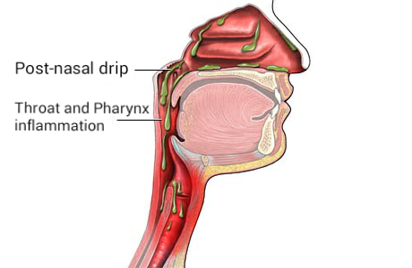
    • Bronchopulmonary Dysplasia Orphanet
      Bronchopulmonary dysplasia is a chronic respiratory disease that results from complications related to lung injury during the treatment of infant acute respiratory distress syndrome (see these terms) in low-birth-weight premature infants or from abnormal lung development in older infants. Clinical signs are tachypnea, tachycardia and signs of respiratory distress such as intercostal recession, grunting and nasal flaring.
  • Post-Nasal Drip Wikipedia
    Annals of Allergy, Asthma & Immunology . 106 (4): 267–74, quiz 275. doi : 10.1016/j.anai.2010.09.004 . PMID 21457874 . ^ Kenn K, Balkissoon R (January 2011). "Vocal cord dysfunction: what do we know?" ... S2CID 12436689 . ^ a b c d e f g h i j k l m Flint PW, Haughey BH, Robbins KT, Thomas JR, Niparko JK, Lund VJ, Lesperance MM (2014). ... OCLC 894112159 . ^ a b Rosenfeld RM, Piccirillo JF, Chandrasekhar SS, Brook I, Ashok Kumar K, Kramper M, et al. (April 2015). "Clinical practice guideline (update): adult sinusitis" . ... PMID 16428694 . ^ a b Segboer C, Gevorgyan A, Avdeeva K, Chusakul S, Kanjanaumporn J, Aeumjaturapat S, et al.
  • Astasis Wikipedia
    Clinical Neurology and Neurosurgery . 9 (111): 766–767. doi : 10.1016/j.clineuro.2009.06.003 . ^ Hirayama K, Nakajima M, Kawamura M, Koguchi Y (1994). ... ISBN 978-1-4200-7973-9 . ^ a b Hirayama, K; Nakajima, M; Kawamura, M; Koguchi, Y (1994). ... PMID 12590833 . ^ Kataoka, H; Sigue, K; Kohara, N; Ueno, S (2006). "Novel Representation of Astasia Associated with Posterior Cingulate Infarction" . ... PMID 17516452 . ^ Okun M; Rodriguez R; Foote K; Fernandez Hubert (2007). "The "Chair Test" to Aid in the Diagnosis of Psychogenic Gait Disorders".
  • Poikiloderma Vasculare Atrophicans Wikipedia
    ISBN 978-1-4160-2999-1 . ^ a b c d e f g h i j k l m n o p q r s t u v w x y z aa ab ac ad Lambert WC, Everett MA (Oct 1981). ... PMID 7026622 . ^ a b Bonvalet, D.; Colau-Gohm, K.; Belaïch, S.; Civatte, J.; Degos, R. ... PMID 843023 . ^ a b c d Kreuter A, Hoffmamm K, Altmeyer P (Apr 2005). "A case of poikiloderma vasculare atrophicans, a rare variant of cutaneous T-cell lymphoma, responding to extracorporeal photopheresis". ... Mosby Co. pp. 936–941. ^ Sehgal, V. N.; Srivastava, G.; Aggarwal, A. K. (Nov–Dec 2007). "Parapsoriasis: a complex issue".
    • Retiform Parapsoriasis Wikipedia
      Retiform parapsoriasis Specialty Dermatology Retiform parapsoriasis is a cutaneous condition, considered to be a type of large-plaque parapsoriasis . [1] It is characterized by widespread, ill-defined plaques on the skin, that have a net-like or zebra-striped pattern. [2] Skin atrophy , a wasting away of the cutaneous tissue , usually occurs within the area of these plaques. [1] See also [ edit ] Parapsoriasis Poikiloderma vasculare atrophicans List of cutaneous conditions References [ edit ] ^ a b Lambert WC, Everett MA (Oct 1981). "The nosology of parapsoriasis". J. Am. Acad. Dermatol . 5 (4): 373–95. doi : 10.1016/S0190-9622(81)70100-2 . PMID 7026622 . ^ Rapini, Ronald P.; Bolognia, Jean L.; Jorizzo, Joseph L. (2007). Dermatology: 2-Volume Set . St. Louis: Mosby. ISBN 1-4160-2999-0 . External links [ edit ] Classification D ICD - 10 : L41.5 ICD - 9-CM : 696.2 v t e Papulosquamous disorders Psoriasis Pustular Generalized pustular psoriasis ( Impetigo herpetiformis ) Acropustulosis / Pustulosis palmaris et plantaris ( Pustular bacterid ) Annular pustular psoriasis Localized pustular psoriasis Other Guttate psoriasis Psoriatic arthritis Psoriatic erythroderma Drug-induced psoriasis Inverse psoriasis Napkin psoriasis Seborrheic-like psoriasis Parapsoriasis Pityriasis lichenoides ( Pityriasis lichenoides et varioliformis acuta , Pityriasis lichenoides chronica ) Lymphomatoid papulosis Small plaque parapsoriasis ( Digitate dermatosis , Xanthoerythrodermia perstans ) Large plaque parapsoriasis ( Retiform parapsoriasis ) Other pityriasis Pityriasis rosea Pityriasis rubra pilaris Pityriasis rotunda Pityriasis amiantacea Other lichenoid Lichen planus configuration Annular Linear morphology Hypertrophic Atrophic Bullous Ulcerative Actinic Pigmented site Mucosal Nails Peno-ginival Vulvovaginal overlap synromes with lichen sclerosus with lupus erythematosis other: Hepatitis-associated lichen planus Lichen planus pemphigoides Other Lichen nitidus Lichen striatus Lichen ruber moniliformis Gianotti–Crosti syndrome Erythema dyschromicum perstans Idiopathic eruptive macular pigmentation Keratosis lichenoides chronica Kraurosis vulvae Lichen sclerosus Lichenoid dermatitis Lichenoid reaction of graft-versus-host disease This dermatology article is a stub . You can help Wikipedia by expanding it . v t e
  • Obsessive–compulsive Spectrum Wikipedia
    Emotions do not affect the behavior but these behaviors are more prevalent in those that suffer with depression. [ citation needed ] Review articles recommend behavioral interventions such as habit reversal training [16] and decoupling . [17] References [ edit ] ^ a b c d e f g h i j k l m McElroy SL; Phillips KA; Keck PE Jr. ... PMID 7961531 . ^ a b Brakoulias, V; Starcevic, V.; Sammut, P.; Berle, D.; Milicevic, D.; Moses, K.; et al. (2011). "Obsessive-compulsive spectrum disorders: a comorbidity and family history perspective". ... PMID 12607216 . Curran S, Matthews K (April 2001). "Response to Yaryura-Tobias et al (2000) negative outcome after neurosurgery for refractory obsessive–compulsive spectrum disorder, World J Biol Psychiatry 1: 197-203". ... "Response to Dr. S. Curran and Dr. K. Matthew's Letter to the editor (World J Biol Psychiatry 2001, 2: 107) concerning Yaryura-Tobias et al (2000) negative outcome after neurosurgery for refractory obsessive–compulsive spectrum disorder, World J Biol Psychiatry 1: 197-203".
  • Leiomyoma Wikipedia
    See also [ edit ] Leiomyosarcoma Elagolix/estradiol/norethindrone acetate References [ edit ] ^ Pedeutour, F.; Quade, B. J.; Sornberger, K.; Tallini, G.; Ligon, A. H.; Weremowicz, S.; Morton, C. ... ISBN 0-7216-2921-0 . ^ Radiologic Pathology Archives: Esophageal Neoplasms: Radiologic-Pathologic Correlation Rachel B. Lewis, Anupamjit K. Mehrotra, Pablo Rodriguez, and Marc S. ... Accessed 2017-07-08 ^ Radiologic Pathology Archives: Esophageal Neoplasms: Radiologic-Pathologic Correlation Rachel B. Lewis, Anupamjit K. Mehrotra, Pablo Rodriguez, and Marc S. ... Archived from the original on 2007-09-27 . Retrieved 2007-03-21 . ^ Patton, K.; Cheng, L.; Papavero, V.; Blum, M.; Yeldandi, A.; Adley, B.; Luan, C.; Diaz, L.; Hui, P.; Yang, X.
    ESR1, SMAD3, TSC2, SFRP1, WNT5B, INHBA, BCL2, PCNA, PARP1, BAX, EZH2, MKI67, TGFBR1, MYC, HMGA2, PGR, IGF1, IGF2, TP53, CYP19A1, TGFB3, FH, MED12, KIT, FN1, CTNNB1, CDKN2A, VCAN, TGFB1, COMT, AR, FGF2, VEGFA, CYP17A1, ACTB, ESR2, EGF, DES, MMRN1, MMP2, KAT6B, EDN1, CD6, GPER1, CXCL8, CYP1A1, MDM2, CCN1, CYP2B6, GSTM1, DNMT1, EGFR, EGR1, PLAG1, CUX1, TNF, MIR29C, MIR21, MIR200C, KANSL1, ALK, CCND1, PRL, COL1A1, PTEN, CD24, GAPDH, GJA1, PRLHR, PBX3, IGFBP2, PTCH1, FMOD, MUC16, HOXA10, HTC2, CD274, IGF1R, PTGS2, SPIN1, SMAD7, MCL1, SMAD4, RBP1, SMAD2, SHBG, KRAS, TERT, TXN, DLEC1, IL4, YWHAG, IL2, PAEP, IGFBP3, SQSTM1, SERPINE1, H3P10, CD34, ACE, EPO, ALDH1A1, DPT, CDH1, DNMT3A, EWSR1, CCN2, EDNRB, CASP3, CYP1B1, CAV1, SLC27A4, DEPDC5, HDAC6, AKT3, ARNT, MVP, ATF3, NCOR2, ABI2, NCOR1, SLC7A8, ADAMTS4, CHST3, TSIX, EBI3, TNRC6B, RFTN1, SDS, ANGPT2, FST, NCOA2, ATG7, PPIG, KHDRBS1, EBP, NFAT5, NES, WASF3, APEX1, FERMT2, AKAP13, SPINT2, ARTN, KLF4, CA2, XRCC1, XPC, XIST, WNT7A, LAT2, VWF, DST, CALD1, XRCC4, TSC1, TP53BP1, SEC62, TIMP2, TIE1, THBS2, THBS1, XRCC3, BMP2, PTTG1, BECN1, SMUG1, SNURF, BAK1, CCN5, IL18R1, TNFSF9, TNFSF10, NCOA1, NR4A3, TP63, IFITM1, KLF11, RECK, TKTL1, SLC7A5, BCL6, ANGPT1, FGFR3, NUP62, SLC27A1, MIR15B, MIR150, MIRLET7C, MIRLET7A1, SERTM2, CASC15, TBPL2, GADL1, TES, HCAR2, AHR, TET3, PLD5, FAM9A, ADAMTS15, DNAJB7, MIR182, MIR197, AGTR2, AGRP, LOC110806263, MTCO2P12, PGR-AS1, RNA5SP202, TRG-GCC5-1, ACTG1, POTEF, POU5F1P4, POU5F1P3, POTEM, MIR146B, MIR93, PLIN2, MIR29A, MIR221, MAGEC3, OSCP1, RXFP2, ADCY10, ATF7IP, MEG3, ANO1, ANGPT4, BET1L, DCTN4, AKR1B1, SLC27A6, PCDH11X, HPGDS, PDCD4, AGO2, FOXP1, SNORD56, CCND2, EDEM2, HDAC8, CYCSP25, RXFP1, GNRHR2, AZIN2, MED8, NACC1, AKT2, LINC00473, ATAD1, NLRC5, ADGRV1, FERMT3, PDCD1LG2, TET1, NANOG, SNIP1, GGCT, PTPN22, TACR3, CCNG1, ATN1, ITGA2, INSR, DRD1, IL18, CXCR1, DRD2, IL6, EDNRA, TFAP2C, IL1RN, IL1B, IKBKB, IGFBP7, EFNA4, EGR2, IGFBP1, ITGAV, ITGB1, KRT19, LAMB1, MME, DAPK1, MEST, MEN1, DBI, MAP2, DFFA, DFFB, DHCR24, SARDH, LTBP2, LTBP1, CYP4F3, LGALS3, LEP, EPHB2, ERBB2, IFNA13, GRB2, GNRHR, GNRH2, GNRH1, GLI1, FBLN1, GHR, GH1, MSTN, FBN1, FZD2, MTOR, EFEMP1, FGF1, FOXO3, FGFR1, F3, GRIA2, IFNA1, NR3C1, IFI27, TNC, ERBB3, HSPA1B, HSPA1A, HSD17B2, HRAS, HPGD, HOXA11, ERCC2, HMGN2, HIF1A, GTF2H1, ETS1, GRM2, MMP1, MPO, MSH2, SLC5A3, SLC3A2, SLC2A4, SLC2A1, SHH, SFRP4, CDK2, SDHB, CCL2, SATB1, SALL1, S100A11, S100A4, RXRA, TRIM27, REST, SMTN, SMARCB1, PLAAT4, SMN1, CD44, TEK, FGR, TACR1, TAC3, TACR2, TAC1, SYP, STK11, SRC, SPINT1, SOX2, SORD, SNRPN, SMN2, CDKN1A, RAD51B, COX2, CPN1, PECAM1, PDGFRA, PCP4, CPN2, CR2, CSF2, PEBP1, OXTR, OGG1, NTS, NOS3, NGF, NEUROG1, MYLK, CTSL, PGAM1, PIK3CA, PTHLH, PIK3CB, CDKN1B, CEBPB, COL3A1, PSMB9, PRLR, COL4A5, MAPK1, PPP4C, PPARG, POU5F1, PLXNA2, PLP1, COL4A6, PIK3CG, PIK3CD, ACP1
  • Borna Disease Wikipedia
    PMID 2244211 . ^ a b c Rott R, Herzog S, Bechter K, Frese K (1991). "Borna disease, a possible hazard for man?". ... J Affect Disord . 90 (1): 43–7. doi : 10.1016/j.jad.2005.10.008 . PMID 16324750 . ^ Fukuda K, Takahashi K, Iwata Y, et al. (February 2001).
    MT3, PARP1, ALDOC, GJB6, CD44, CX3CR1, GJA1, IL16, MIF, PARG, CX3CL1, S100B, CHP1, H3P19, NRSN1, TPPP, CD9, TMED2, MIR146A, MIR155, ACTB, KMO, PENK, OCA2, IRF7, RAB5A
  • Vici Syndrome Wikipedia
    .; Fukushima, Y.; Yamamoto, Y.; Tsunamoto, K.; Nishimura, Y.; Ishida, H.; Toda, T.; Kasubuchi, Y. ... Orphanet Journal of Rare Diseases 11:21 DOI: 10.1186/s13023-016-0399-x ^ Chiyonobu T, Yoshihara T, Fukushima Y, Yamamoto Y, Tsunamoto K et al. (2002) "Sister and brother with Vici syndrome: agenesis of the corpus callosum, albinism, and recurrent infections". ... Nature genetics 45(1): 83-87 ^ Hori I, Otomo T, Nakashima M, Miya F, Negishi Y, Shiraishi H, Nonoda Y, Magara S, Tohyama J, Okamoto N, Kumagai T, Shimoda K, Yukitake Y, Kajikawa D, Morio T, Hattori A, Nakagawa M, Ando N, Nishino I, Kato M, Tsunoda T, Saitsu H, Kanemura Y, Yamasaki M, Kosaki K, Matsumoto N, Yoshimori T, Saitoh S (2017) Defects in autophagosome-lysosome fusion underlie Vici syndrome, a neurodevelopmental disorder with multisystem involvement.
    EPG5, RAB7A, RAB7B, EPGN
    • Vici Syndrome MedlinePlus
      Vici syndrome is a severe disorder that begins early in life and affects many body systems. It is characterized by abnormalities of the brain, immune system, heart, skin, and eyes. Other organs and tissues are less commonly affected. A characteristic feature of Vici syndrome is a brain abnormality called agenesis of the corpus callosum, in which the tissue that connects the left and right halves of the brain (the corpus callosum ) fails to form normally during the early stages of development before birth. Other brain abnormalities can occur in Vici syndrome, including underdevelopment of a region of the brain known as the pons (pontine hypoplasia) and reduced myelin , which is a fatty substance that covers and protects nerve cells. In addition to problems with brain development, breakdown (degeneration) of brain tissue may occur over time, resulting in an unusually small head size (microcephaly ).
    • Vici Syndrome GARD
      Vici syndrome is a multisystem disorder characterized by agenesis (failure to develop) of the corpus callosum, cataracts , hypopigmentation of the eyes and hair, cardiomyopathy, and combined immunodeficiency. Hearing loss, seizures, and delayed motor development have also been reported. Swallowing and feeding difficulties early on may result in a failure to thrive. Recurrent infections of the respiratory, gastrointestinal, and urinary tracts are common. Vici syndrome is caused by mutations in the EPG5 gene and is inherited in an autosomal recessive manner.
    • Vici Syndrome OMIM
      A number sign (#) is used with this entry because Vici syndrome (VICIS) is caused by homozygous or compound heterozygous mutation in the EPG5 gene (615068) on chromosome 18q. Description Vici syndrome is a rare congenital multisystem disorder characterized by agenesis of the corpus callosum (ACC), cataracts, pigmentary defects, progressive cardiomyopathy, and variable immunodeficiency. Affected individuals also have profound psychomotor retardation and hypotonia due to a myopathy (summary by Finocchi et al., 2012). Clinical Features Dionisi Vici et al. (1988) described 2 brothers with a malformation syndrome consisting of agenesis of the corpus callosum, cutaneous hypopigmentation, bilateral cataract, cleft lip and palate, and combined immunodeficiency. The sibs suffered from severe psychomotor retardation, seizures, recurrent severe respiratory infections, and chronic mucocutaneous candidiasis.
    • Vici Syndrome Orphanet
      Vici syndrome is a very rare and severe congenital multisystem disorder characterized by the principal features of agenesis of the corpus callosum, cataracts, oculocutaneous hypopigmentation, cardiomyopathy and combined immunodeficiency. Epidemiology Prevalence is unknown. Only 20 cases have been reported to date. Clinical description Vici syndrome is usually diagnosed in the first years of life. The phenotype is variable but the principal diagnostic features are almost always present at onset or evolve over time. Swallowing and feeding difficulties may be noted early on and may lead to failure to thrive.
  • Seckel Syndrome Wikipedia
    Harper . [6] [7] See also [ edit ] Koo-Koo the Bird Girl References [ edit ] ^ Harsha Vardhan BG, Muthu MS, Saraswathi K, Koteeswaran D (2007). "Bird-headed dwarf of Seckel" . ... Retrieved 7 January 2011 . ^ Jung M, Rai A, Wang L, Puttmann K, Kukreja K, Koh CJ (2018). "Nephrolithiasis in a 17-Year-Old Male With Seckel Syndrome and Horseshoe Kidneys: Case Report and Review of the Literature".
    ATR, CENPJ, PCNT, ATRIP, TRAIP, CENPE, PLK4, CEP152, DNA2, LIG4, WDR4, RBBP8, ORC1, ORC4, IGF1, XRCC4, ORC6, DNMT3A, GMNN, CENPF, CDC6, CDT1, LZTR1, KRAS, PTPN11, RAF1, CEP63, SOS1, RIT1, RTTN, NIN, ATM, AIMP2, GRAP2, BRCA1, CD5L, CD69, MCPH1, CHEK1, LARP7, CRK, MAPK14, DONSON, POLDIP2, DNMT1, RNF19A, FANCA, FANCC, PNKP, FAH, H2AX, AHSA1, MAPK1, TELO2, CEP135, TUBGCP6
    • Seckel Syndrome GARD
      Seckel syndrome is a genetic disorder characterized by growth retardation, very small head (microcephaly( with intellectual disability , and unique facial features such as large eyes, beak-like nose, narrow face, and receding lower jaw. About less than 25% of the patients also have blood abnormalities. Seckel syndrome is inherited in an autosomal recessive fashion. The condition may be divided in 8 different subtypes , according to the specific gene alteration (mutation ). Treatment is supportive.
    • Microcephaly 13, Primary, Autosomal Recessive OMIM
      A number sign (#) is used with this entry because of evidence that autosomal recessive primary microcephaly-13 (MCPH13) is caused by compound heterozygous mutation in the CENPE gene (117143) on chromosome 4q24. One such family has been reported. For a phenotypic description and a discussion of genetic heterogeneity of primary microcephaly, see MCPH1 (251200). Clinical Features Mirzaa et al. (2014) reported a brother and sister, born of unrelated parents of European descent, with microcephaly, poor overall growth, and developmental delay. Both had intrauterine growth retardation and microcephaly apparent on prenatal ultrasound, as well as similar dysmorphic facial features, including sloping forehead, prominent nose, and mild micrognathia. At age 5 years, the older sib, a boy, had microcephaly (-9 SD), short stature (-7 SD), small hands and feet, mild spasticity, and severely delayed psychomotor development with absent speech and poor gross and fine motor skills.
    • Seckel Syndrome Orphanet
      Seckel syndrome is a type of microcephalic primordial dwarfism that is characterized by a proportionate dwarfism of prenatal onset, a severe microcephaly, a typical dysmorphic face (bird-like), and mild to severe intellectual disability.
    • Seckel Syndrome 1 OMIM
      A number sign (#) is used with this entry because Seckel syndrome-1 (SCKL1) is caused by homozygous or compound heterozygous mutation in the ATR gene (601215) on chromosome 3q23. Description Seckel syndrome is a rare autosomal recessive disorder characterized by intrauterine growth retardation, dwarfism, microcephaly with mental retardation, and a characteristic 'bird-headed' facial appearance (Shanske et al., 1997). Genetic Heterogeneity of Seckel Syndrome Other forms of Seckel syndrome include SCKL2 (606744), caused by mutation in the RBBP8 gene (604124) on chromosome 18q11; SCKL4 (613676), caused by mutation in the CENPJ gene (609279) on chromosome 13q12; SCKL5 (613823), caused by mutation in the CEP152 gene (613529) on chromosome 15q21; SCKL6 (614728), caused by mutation in the CEP63 gene (614724) on chromosome 3q22; SCKL7 (614851), caused by mutation in the NIN gene (608684) on chromosome 14q22; SCKL8 (615807), caused by mutation in the DNA2 gene (601810) on chromosome 10q21; SCKL9 (616777), caused by mutation in the TRAIP gene (605958) on chromosome 3p21; and SCKL10 (617253), caused by mutation in the NSMCE2 gene (617246) on chromosome 8q24. The report of a Seckel syndrome locus on chromosome 14q, designated SCKL3, by Kilinc et al. (2003) was found to be in error; see History section. Clinical Features This condition was given the 2 names bird-headed dwarfism and nanocephaly by Virchow.
    • Seckel Syndrome 9 OMIM
      A number sign (#) is used with this entry because of evidence that Seckel syndrome-9 (SCKL9) is caused by homozygous mutation in the TRAIP gene (605958) on chromosome 3p21. For a general phenotypic description and a discussion of genetic heterogeneity of Seckel syndrome, see SCKL1 (210600). Clinical Features Silengo et al. (2001) described 2 Italian sibs with a Seckel-like malformation syndrome characterized by intrauterine growth retardation (IUGR), microcephaly, beaked nose, failure to thrive, and early death. The first sib was a 46,XX girl who at birth underwent surgery for right diaphragmatic hernia. External genitalia appeared normal, with slight clitoral hypertrophy.
    • Microcephaly And Chorioretinopathy, Autosomal Recessive, 2 OMIM
      A number sign (#) is used with this entry because of evidence that autosomal recessive microcephaly and chorioretinopathy-2 (MCCRP2) is caused by homozygous mutation in the PLK4 gene (605031) on chromosome 4q28. Description Microcephaly and chorioretinopathy-2 is an autosomal recessive developmental disorder characterized by delayed psychomotor development, visual impairment, and short stature (summary by Martin et al., 2014). For a discussion of genetic heterogeneity of microcephaly and chorioretinopathy, see MCCRP1 (251270). Clinical Features Martin et al. (2014) reported 7 members of a highly consanguineous Pakistani family with a severe developmental disorder characterized by primary microcephaly (up to -15 SD), profoundly delayed psychomotor development, and growth retardation with dwarfism (up to -8 SD). Many patients had ocular abnormalities, including microphthalmia, microcornea, and cataracts.
    • Seckel Syndrome 2 OMIM
      A number sign (#) is used with this entry because of evidence that Seckel syndrome-2 (SCKL2) is caused by homozygous mutation in the RBBP8 gene (604124) on chromosome 18q11. Jawad syndrome (251255), a microcephaly syndrome involving mental retardation and digital anomalies, is also caused by mutation in the RBBP8 gene. Description Seckel syndrome is a rare autosomal recessive disorder characterized by growth retardation, microcephaly with mental retardation, and a characteristic facial appearance (Borglum et al., 2001). For a general phenotypic description and a discussion of genetic heterogeneity of Seckel syndrome, see SCKL1 (210600). Clinical Features Borglum et al. (2001) studied a consanguineous family of Iraqi descent with Seckel syndrome.
    • Intrauterine Growth Retardation With Increased Mitomycin C Sensitivity OMIM
      Woods et al. (1995) reported the case of an infant with pre- and postnatal microcephaly and growth retardation, a distinctive face, and developmental delay. Seckel syndrome was the initial diagnosis. The infant became pancytopenic at 16 months of age and died soon thereafter. His bone marrow was of normal cellularity but had an infiltration of small lymphocytes. Increased spontaneous chromosome breakage was seen in blood and fibroblasts. Mitomycin C-induced chromosome damage was increased and comparable to that seen in Fanconi anemia.
    • Seckel Syndrome 8 OMIM
      A number sign (#) is used with this entry because of evidence that Seckel syndrome-8 (SCKL8) is caused by homozygous mutation in the DNA2 gene (601810) on chromosome 10q21. One such family has been reported. Description Seckel syndrome is a rare autosomal recessive disorder characterized by intrauterine growth retardation, dwarfism, microcephaly with mental retardation, and a characteristic 'bird-headed' facial appearance (summary by Shanske et al., 1997). For a discussion of genetic heterogeneity of Seckel syndrome, see SCKL1 (210600). Clinical Features Shaheen et al. (2014) studied an uncle and niece, both born of consanguineous marriages, with short stature and strikingly similar facial features consistent with Seckel syndrome. Both uncle and niece had decreased length at birth (-4.9 and -5.1 SD, respectively) and continued to have short stature at ages 18 years and 9 years (-9.5 and -6 SD) as well as severe microcephaly (-7.5 and -11.8 SD).
    • Seckel Syndrome 5 OMIM
      A number sign (#) is used with this entry because Seckel syndrome-5 is caused by homozygous or compound heterozygous mutation in the CEP152 gene (613529) on chromosome 15q21. Description Seckel syndrome is an autosomal recessive disorder characterized by proportionate short stature, severe microcephaly, mental retardation, and a typical 'bird-head' facial appearance (summary by Kalay et al., 2011). For a general phenotypic description and a discussion of genetic heterogeneity of Seckel syndrome, see 210600. Clinical Features Kalay et al. (2011) clinically evaluated 5 consanguineous families with Seckel syndrome originating from an isolated rural area in Turkey. The patients presented with microcephaly, sloping forehead, high nasal bridge, beaked nose, and retrognathia.
    • Seckel Syndrome 4 OMIM
      A number sign (#) is used with this entry because of evidence that Seckel syndrome-4 (SCKL4) is caused by homozygous mutation in the CENPJ gene (609279) on chromosome 13q12. One such family has been reported. Homozygous mutations in the CENPJ gene can also cause primary microcephaly-6 (MCPH6; 608393). Description Seckel syndrome is a rare autosomal recessive disorder characterized by severe pre- and postnatal growth retardation, severe microcephaly with mental retardation, and specific dysmorphic features (Faivre et al., 2002). For a general description and a discussion of genetic heterogeneity of Seckel syndrome, see 210600. Clinical Features Al-Dosari et al. (2010) described a consanguineous Saudi family in which several members had clinical features of Seckel syndrome.
  • Aa Amyloidosis Wikipedia
    . ^ Chapter 5 in: Mitchell, Richard Sheppard; Kumar, Vinay; Abbas, Abul K.; Fausto, Nelson (2007). Robbins Basic Pathology . ... PMID 23171281 . ^ d'Ythurbide, G; Kerrou, K; Brocheriou, I; Hertig, A (2012). ... Retrieved 12 June 2015 . ^ a b Murakami, T; Ishiguro N; Higuchi K (March 2014). "Transmission of Systemic AA Amyloidosis in Animals" .
    SAA1, MEFV, SAA2, TTR, IL1B, CRP, TNFRSF1A, IL1A, APCS, TLR2, IL6, TNF, NLRP3, IGAN1, PYCR1, HERPUD1, TLR4, TARDBP, SAA4, HEY2, CYCS, LPAL2, MOK, AGER, PLG, IAPP, APOA2, B2M, CD14, GSN, HGF, HLA-DRB1, IFNA2, MMP1, IL1RN, IL6R, IL18, LEP, LTF, MAZ, MBL2
    • Amyloidosis Aa GARD
      Amyloidosis is a group of diseases in which a protein, called amyloid, builds up in the body's organs and tissues. Amyloidosis AA is also referred to as Secondary amyloidosis or Inflammatory amyloidosis. This disease is caused by a long-lasting infection or inflammatory disease such as rheumatoid arthritis, familial Mediterranean fever, or osteomyelitis. Infection or inflammation in the body causes an increased amount of a specific protein called serum amyloid A (SAA) protein. In this disease, part of the SAA protein forms deposits called "amyloid fibrils".
    • Aa Amyloidosis Orphanet
      Secondary amyloidosis is a form of amyloidosis (see this term), that complicates chronic inflammatory disorders (mainly rheumatoid arthritis, see this term) and is characterized by the aggregation and deposition of amyloid fibrils composed of serum amyloid A protein, an acute phase reactant. Although spleen, suprarenal gland, liver and gut are frequent sites of amyloid deposition, the clinical picture is dominated by renal involvement.
  • Granulomatous Meningoencephalitis Wikipedia
    PMID 15521442 . ^ Suzuki M, Uchida K, Morozumi M, Yanai T, Nakayama H, Yamaguchi R, Tateyama S (2003). ... Proceedings of the 50° Congresso Nazionale Multisala SCIVAC . Retrieved 2007-02-18 . ^ Greer, K. A.; Wong, A. K.; Liu, H.; Famula, T.
  • Laboratory Animal Allergy Wikipedia
    . ^ a b Lockey, Richard; Ledford, Dennis K. (2008). "Mammalian Allergens". Allergens and Allergen Immunotherapy . ... Informa Health Care. pp. 201–218. ISBN 978-1-4200-6197-0 . ^ Aoyama K, Ueda A, Manda F, Matsushita T, Ueda T, Yamauchi C (January 1992). ... "Suppression of ouabain-insensitive K-ATPase activity in rabbit nephron segments during chronic hyperkalemia".
    IGHG2
  • Hypophosphatemia Wikipedia
    Cardiac monitoring is also advised. [ citation needed ] See also [ edit ] X-linked hypophosphatemia References [ edit ] ^ a b c d e f g h i j k l m n o "Hypophosphatemia" . Merck Manuals Professional Edition . ... Retrieved 23 October 2017 . ^ Shajahan, A., Ajith Kumar, J., Gireesh Kumar, K. P., Sreekrishnan, T. P. and Jismy, K. (2015), Managing hypophosphatemia in critically ill patients: a report on an under-diagnosed electrolyte anomaly.
    SLC34A1, NR1I2, FGFR1, PHEX, FGF23, SLC34A3, DMP1, FAM20C, VDR, SLC9A3R1, SLC2A2, PTH, PTH1R, ALDOB, CDC73, ALG13, TNFSF11, SUCLG1, GCM2, SNX10, OCRL, TCIRG1, CTNS, CYP27B1, PTCHD1-AS, CASR, AP2S1, GNAS, CLCN5, CYP2R1, CLCN7, COL4A3, FBN1, MEN1, GPT, ENPP1, FGF2, SFRP4, PER2, HRAS, KL, AZI2, TAB3, CORO7, NCKAP1, MEPE, ANKH, TRMO, DTL, GGTLC1, NAA25, SRY, NAMPT, NRAS, BCR, CST3, FGF7, FGFR3, CXCL8, NAP1L1, SLC20A2, ACOT8, SMS, AQP2, TCF21, NAPSA, CLOCK, RAPGEF5, LOC102724197
  • Tropical Eosinophilia Wikipedia
    Reported side effects include headache, fever, pruritus and gastrointestinal upset. [15] The eosinophil count often falls dramatically within 7–10 days of starting treatment. [4] [7] References [ edit ] ^ Jha, Suman K.; Karna, Bibek; Mahajan, Kunal (2020), "Tropical Pulmonary Eosinophilia" , StatPearls , Treasure Island (FL): StatPearls Publishing, PMID 32491456 , retrieved 2020-12-01 ^ a b "Pulmonary Eosinophilia" . ... PMID 17908009 . ^ a b c Boggild, A. K.; Keystone, J. S.; Kain, K. C. (2004).
    IL5, GATA3, RAG2, SHARPIN, TLR4, TLR2, MYD88, PTGDS, SERPINA1, SIGLEC8, GGTLC1, IL33, JAK2, IL25, MYDGF, HPGDS, POSTN, PTX3, FOXN1, P2RY6, STAT3, RNASE3, RNASE2, LOC102724197
  • Eales Disease Wikipedia
    Gainesville, Florida: Triad Publishing Company. [ full citation needed ] ^ Deobhakta, Avnish; Chang, Louis K. (2013). "Inflammation in Retinal Vein Occlusion" . ... Retrieved 2019-12-16 . ^ Madhavan, HN; Therese, KL; Doraiswamy, K (March 2002). "Further investigations on the association of Mycobacterium tuberculosis with Eales' disease". ... PMID 23514227 . ^ Comprehensive Ophthalmology, (4th Ed) 2007 by A K Khurana, Professor, Regional Institute of Ophthalmology, Postgraduate Institute of Medical Sciences, Rohtak- 124001, India ^ Eales disease at eMedicine ^ "Eales Disease - an overview | ScienceDirect Topics" . www.sciencedirect.com .
    C3, CRP, HLA-A, HLA-B, HP, IL6, IL10, LGALS1, TNF
    • Eales Disease Orphanet
      A rare ophthalmic disorder characterized by 3 stages: vasculitis, occlusion, and retinal neovascularization, leading to recurrent vitreous hemorrhages and vision loss. Epidemiology The prevalence is unknown. The disorder has been reported worldwide but is more commonly observed in the Indian subcontinent where the estimated annual incidence is 1/135-1/200 ophthalmic patients. Males are predominantly affected. Of late, the disease incidence seems to be reducing. Clinical description The predominant age of onset is 20-30 years (earlier in Asians). The disorder can appear in adolescence. Eales disease (ED) is characterized by 3 sequential vascular responses that determine the course of the disease: inflammation (peripheral retinal perivasculitis); occlusion (peripheral retinal capillary non-perfusion); and neovascularisation of the retina or disk, which often leads to vitreous hemorrhage.
    • Eales Disease GARD
      Eales disease is a rare vision disorder that appears as an inflammation and white haze around the outercoat of the veins in the retina. This condition is most common among young males and normally affects both eyes. In most cases, vision becomes suddenly blurred because the vitreous, the clear jelly that fills the eyeball behind the lens of the eye, seeps out. Treatment includes corticosteroids in the inflammation stage and photocoagulation in the proliferative stage of the disease. Visual prognosis is good if treatment begins early in the course of the disease.
  • Metanephric Adenoma Wikipedia
    . ^ Nagashima Y, Arai N, Tanaka Y, Yoshida S, Sumino K, Ohaki Y, Matsushita K, Morita T, Misugi K 81991) Case record: two cases of renal epithelial tumour resembling immature nephron.
    BRAF, B3GAT1, CCND1, EPHB2, EPO, MAPK1
  • Eem Syndrome Wikipedia
    You can help by adding to it . ( July 2017 ) See also [ edit ] Germ layer Integumentary system Hay-Wells syndrome References [ edit ] ^ a b c Hayakawa M, Yanashima K, Kato K, Nakajima A, Yamauchi H (1989). ... External links [ edit ] Classification D ICD - 10 : Q82.4 ICD - 9-CM : 757.31 OMIM : 225280 MeSH : C536190 SNOMED CT : 720856002 External resources Orphanet : 1897 v t e Congenital malformations and deformations of integument / skin disease Genodermatosis Congenital ichthyosis / erythrokeratodermia AD Ichthyosis vulgaris AR Congenital ichthyosiform erythroderma : Epidermolytic hyperkeratosis Lamellar ichthyosis Harlequin-type ichthyosis Netherton syndrome Zunich–Kaye syndrome Sjögren–Larsson syndrome XR X-linked ichthyosis Ungrouped Ichthyosis bullosa of Siemens Ichthyosis follicularis Ichthyosis prematurity syndrome Ichthyosis–sclerosing cholangitis syndrome Nonbullous congenital ichthyosiform erythroderma Ichthyosis linearis circumflexa Ichthyosis hystrix EB and related EBS EBS-K EBS-WC EBS-DM EBS-OG EBS-MD EBS-MP JEB JEB-H Mitis Generalized atrophic JEB-PA DEB DDEB RDEB related: Costello syndrome Kindler syndrome Laryngoonychocutaneous syndrome Skin fragility syndrome Ectodermal dysplasia Naegeli syndrome / Dermatopathia pigmentosa reticularis Hay–Wells syndrome Hypohidrotic ectodermal dysplasia Focal dermal hypoplasia Ellis–van Creveld syndrome Rapp–Hodgkin syndrome / Hay–Wells syndrome Elastic / Connective Ehlers–Danlos syndromes Cutis laxa ( Gerodermia osteodysplastica ) Popliteal pterygium syndrome Pseudoxanthoma elasticum Van der Woude syndrome Hyperkeratosis / keratinopathy PPK diffuse : Diffuse epidermolytic palmoplantar keratoderma Diffuse nonepidermolytic palmoplantar keratoderma Palmoplantar keratoderma of Sybert Meleda disease syndromic connexin Bart–Pumphrey syndrome Clouston's hidrotic ectodermal dysplasia Vohwinkel syndrome Corneodermatoosseous syndrome plakoglobin Naxos syndrome Scleroatrophic syndrome of Huriez Olmsted syndrome Cathepsin C Papillon–Lefèvre syndrome Haim–Munk syndrome Camisa disease focal : Focal palmoplantar keratoderma with oral mucosal hyperkeratosis Focal palmoplantar and gingival keratosis Howel–Evans syndrome Pachyonychia congenita Pachyonychia congenita type I Pachyonychia congenita type II Striate palmoplantar keratoderma Tyrosinemia type II punctate : Acrokeratoelastoidosis of Costa Focal acral hyperkeratosis Keratosis punctata palmaris et plantaris Keratosis punctata of the palmar creases Schöpf–Schulz–Passarge syndrome Porokeratosis plantaris discreta Spiny keratoderma ungrouped: Palmoplantar keratoderma and spastic paraplegia desmoplakin Carvajal syndrome connexin Erythrokeratodermia variabilis HID / KID Other Meleda disease Keratosis pilaris ATP2A2 Darier's disease Dyskeratosis congenita Lelis syndrome Dyskeratosis congenita Keratolytic winter erythema Keratosis follicularis spinulosa decalvans Keratosis linearis with ichthyosis congenita and sclerosing keratoderma syndrome Keratosis pilaris atrophicans faciei Keratosis pilaris Other cadherin EEM syndrome immune system Hereditary lymphedema Mastocytosis / Urticaria pigmentosa Hailey–Hailey see also Template:Congenital malformations and deformations of skin appendages , Template:Phakomatoses , Template:Pigmentation disorders , Template:DNA replication and repair-deficiency disorder Developmental anomalies Midline Dermoid cyst Encephalocele Nasal glioma PHACE association Sinus pericranii Nevus Capillary hemangioma Port-wine stain Nevus flammeus nuchae Other/ungrouped Aplasia cutis congenita Amniotic band syndrome Branchial cyst Cavernous venous malformation Accessory nail of the fifth toe Bronchogenic cyst Congenital cartilaginous rest of the neck Congenital hypertrophy of the lateral fold of the hallux Congenital lip pit Congenital malformations of the dermatoglyphs Congenital preauricular fistula Congenital smooth muscle hamartoma Cystic lymphatic malformation Median raphe cyst Melanotic neuroectodermal tumor of infancy Mongolian spot Nasolacrimal duct cyst Omphalomesenteric duct cyst Poland anomaly Rapidly involuting congenital hemangioma Rosenthal–Kloepfer syndrome Skin dimple Superficial lymphatic malformation Thyroglossal duct cyst Verrucous vascular malformation Birthmark
    CDH3
    • Ectodermal Dysplasia, Ectrodactyly, And Macular Dystrophy Syndrome OMIM
      A number sign (#) is used with this entry because of evidence that EEM syndrome (EEMS) is caused by homozygous mutation in the CDH3 gene (114021) on chromosome 16q22. Clinical Features In an isolated population on a remote island in Japan, Ohdo et al. (1983) observed a kindred with 6 cases of ectodermal dysplasia, ectrodactyly associated with syndactyly or cleft hand or both, and macular dystrophy which was presumed to be progressive. Because of the parental consanguinity and the occurrence in both sexes, autosomal recessive inheritance was suggested. The presence of macular dystrophy distinguishes it from other syndromes of ectodermal dysplasia and limb malformations. The same disorder was probably reported by Albrectsen and Svendsen (1956) in the son and daughter of first-cousin parents and by Hayakawa et al. (1979) in a 30-year-old woman whose parents were first cousins.
    • Eem Syndrome Orphanet
      A rare ectodermal dysplasia syndrome characterized by the association of ectodermal dysplasia (with hypotrichosis affecting scalp hair, eyebrows, and eyelashes, and partial anodontia), ectrodactyly, and macular dystrophy (appearing as a central geographic atrophy of the retinal pigment epithelium and choriocapillary layer of the macular area with coarse hyperpigmentations and sparing of the larger choroidal vessels). Variable additional limb defects (including absence deformities, polydactyly, syndactyly, or camptodactyly) have also been described, the hands often being more severely affected than the feet.
  • Fowler's Syndrome Wikipedia
    BJU International . 116 (3): 423–431. doi : 10.1111/bju.13042 . PMID 25600712 . ^ K, Jurkat-Rott; H, Lerche; N, Mitrovic; F, Lehmann-Horn (September 1999). ... S2CID 46839550 . ^ Jk, Szymański; A, Słabuszewska-Jóźwiak; K, Zaręba; G, Jakiel (December 2019).
    FLVCR2, TTLL5
    • Proliferative Vasculopathy And Hydranencephaly-Hydrocephaly Syndrome OMIM
      A number sign (#) is used with this entry because of evidence that proliferative vasculopathy and hydranencephaly-hydrocephaly syndrome (PVHH), also known as encephaloclastic proliferative vasculopathy, is caused by homozygous or compound heterozygous mutation in the FLVCR2 gene (610865) on chromosome 14q24. Description The proliferative vasculopathy and hydranencephaly-hydrocephaly syndrome is a rare, autosomal recessive, usually prenatally lethal disorder characterized by hydranencephaly, a distinctive glomerular vasculopathy in the central nervous system and retina, and diffuse ischemic lesions of the brain stem, basal ganglia, and spinal cord with calcifications. It is usually diagnosed by ultrasound between 26 and 33 weeks' gestation (summary by Meyer et al., 2010). Rarely, affected individuals may survive, but are severely impaired with almost no neurologic development (Kvarnung et al., 2016). Clinical Features In identical male twins, Moeschler and Marin-Padilla (1989) described a disorder that had been reported in 5 sibs by Fowler et al. (1972) and in 2 sibs by Harper and Hockey (1983).
    • Fowler Vasculopaty Orphanet
      A rare, genetic neurological disorder characterized by hydranencephaly, distinctive glomeruloid vasculopathy in the central nervous system and retina, polyhydramnios and fetal akinesia with arthrogryposis. The disorder is usually prenatally lethal. In rare reported cases that survived beyond infancy, severe intellectual and neurologic disability with seizures, microcephaly and absence of functional movements were reported.
  • ←
  • 1
  • 2
  • ...
  • 23
  • 24
  • 25
  • 26
  • 27
  • 28
  • 29
  • ...
  • 218
  • 219
  • →

FindZebra

contact@findzebra.com