• FindZebra
  • About
  • Contact
  • Help
  • Login
Advanced
  • Multiple Endocrine Neoplasia Wikipedia
    North Am . 88 (4): 863–95. doi : 10.1016/j.suc.2008.05.001 . PMID 18672144 . ^ a b c d e f g h i Table 4-7 in: Elizabeth D Agabegi; Agabegi, Steven S. (2008). ... External links [ edit ] Endocrine and Metabolic Diseases Information Service Classification D ICD - 10 : E31.20 ICD - 9-CM : 258.0 ICD-O : 8360/1 MeSH : D009377 External resources MedlinePlus : 000398 v t e Disorders involving multiple endocrine glands Autoimmune polyendocrine syndrome APS1 APS2 Carcinoid syndrome Multiple endocrine neoplasia 1 2A 2B Progeria Werner syndrome Acrogeria Metageria Woodhouse–Sakati syndrome v t e Glandular and epithelial cancer Epithelium Papilloma / carcinoma Small-cell carcinoma Combined small-cell carcinoma Verrucous carcinoma Squamous cell carcinoma Basal-cell carcinoma Transitional cell carcinoma Inverted papilloma Complex epithelial Warthin's tumor Thymoma Bartholin gland carcinoma Glands Adenomas / adenocarcinomas Gastrointestinal tract: Linitis plastica Familial adenomatous polyposis pancreas Insulinoma Glucagonoma Gastrinoma VIPoma Somatostatinoma Cholangiocarcinoma Klatskin tumor Hepatocellular adenoma / Hepatocellular carcinoma Urogenital Renal cell carcinoma Endometrioid tumor Renal oncocytoma Endocrine Prolactinoma Multiple endocrine neoplasia Adrenocortical adenoma / Adrenocortical carcinoma Hürthle cell Other/multiple Neuroendocrine tumor Carcinoid Adenoid cystic carcinoma Oncocytoma Clear-cell adenocarcinoma Apudoma Cylindroma Papillary hidradenoma Adnexal and skin appendage sweat gland Hidrocystoma Syringoma Syringocystadenoma papilliferum Cystic, mucinous, and serous Cystic general Cystadenoma / Cystadenocarcinoma Mucinous Signet ring cell carcinoma Krukenberg tumor Mucinous cystadenoma / Mucinous cystadenocarcinoma Pseudomyxoma peritonei Mucoepidermoid carcinoma Serous Ovarian serous cystadenoma / Pancreatic serous cystadenoma / Serous cystadenocarcinoma / Papillary serous cystadenocarcinoma Ductal, lobular, and medullary Ductal carcinoma Mammary ductal carcinoma Pancreatic ductal carcinoma Comedocarcinoma Paget's disease of the breast / Extramammary Paget's disease Lobular carcinoma Lobular carcinoma in situ Invasive lobular carcinoma Medullary carcinoma Medullary carcinoma of the breast Medullary thyroid cancer Acinar cell Acinic cell carcinoma v t e Tumours of endocrine glands Pancreas Pancreatic cancer Pancreatic neuroendocrine tumor α : Glucagonoma β : Insulinoma δ : Somatostatinoma G : Gastrinoma VIPoma Pituitary Pituitary adenoma : Prolactinoma ACTH-secreting pituitary adenoma GH-secreting pituitary adenoma Craniopharyngioma Pituicytoma Thyroid Thyroid cancer (malignant): epithelial-cell carcinoma Papillary Follicular / Hurthle cell Parafollicular cell Medullary Anaplastic Lymphoma Squamous-cell carcinoma Benign Thyroid adenoma Struma ovarii Adrenal tumor Cortex Adrenocortical adenoma Adrenocortical carcinoma Medulla Pheochromocytoma Neuroblastoma Paraganglioma Parathyroid Parathyroid neoplasm Adenoma Carcinoma Pineal gland Pinealoma Pinealoblastoma Pineocytoma MEN 1 2A 2B
    RET, CDKN1B, MEN1, GHRL, GHSR, CALCA, VHL, TICAM2, TMED7-TICAM2, TMED7, IFI27, ZNRD2, CIB1, PTH, PSMD9, PRKAR1A, DCTN6, H3P23, CASR, HPT, GDNF, CDC73, AIP, FGF2, SDHB, GAST, SST, PPY, POMC, FUT4, CDKN2C, SARDH, CXADR, CDKN3, FRS2, SDS, SMUG1, PDE11A, CDKN2A, NR1I3, GFRA4, MEA1, CD109, SMAD3, CXADRP1, CALB1, TRIM13, GNAS, PTEN, SDHD, PTPN11, RENBP, BCL2, RPA2, RYK, SDHC, SLC8A1, PDE8B, SORD, SPG7, GRN, TP53, NF1, ZNF22, ARR3
    • Multiple Endocrine Neoplasia Orphanet
      Multiple endocrine neoplasia (MEN) is a group of rare inherited cancer syndromes characterized by the development of two or more endocrine gland tumors, sometimes with tumor development in other tissues or organs. Epidemiology The overall prevalence and incidence of MEN are not known. The prevalence of MEN1 is estimated to be approximately 1/10,000 to 1/30,000, while the total prevalence of MEN2 variants is approximately 1/35,000. MEN4 is extremely rare. The incidence of MEN1 has been estimated from random postmortem studies to be 0.25%, and to be 1-18% in patients with primary hyperparathyroidism, 16 to 38% in patients with gastrinomas, and less than 3% in patients with pituitary tumors. Clinical description Multiple endocrine neoplasia syndromes can develop in patients of all ages: from infants to elderly patients over 70 years of age.
  • Cowpox Wikipedia
    . ^ "Cowpox infection in US lab worker called a first" . ^ Kurth A, Wibbelt G, Gerber HP, Petschaelis A, Pauli G, Nitsche A (April 2008).
    FLNB, SEC14L2, NXF1, USO1, TNF, XIAP, TAP1, SAMD9L, MTUS1, SAMD9, IL18, CASP1, CXCL8, IL1B, CXCL1, CDS1, CD86, CD80, STING1
  • Foix–chavany–marie Syndrome Wikipedia
    See also [ edit ] Pseudobulbar Palsy Operculum Corticobulbar Tracts Wernicke's Aphasia Broca's Aphasia References [ edit ] ^ a b c d e f g h i j k l m n o Bakar, M; Kirshner, HS; Niaz, F (1998). ... ] [ non-primary source needed ] ^ a b c d e f g h i j k l Lekhjung, Thapa; Raju, Paudel; PVS, Rana (2010).
    • Foix-Chavany-Marie Syndrome Orphanet
      A rare cortico-subcortical suprabulbar or pseudobulbar palsy of the lower cranial nerves, characterized by severe dysarthria and dysphagia associated with bilateral central facio-pharyngo-glosso-masticatory paralysis, with prominent automatic-voluntary dissociation in which involuntary movements of the affected muscles are preserved. Epidemiology Less than 150 cases have been described in the literature so far. Clinical description Foix-Chavany-Marie syndrome (FCMS) can occur at any age and patients present with acute-onset bilateral paresis of the facial, lingual, pharyngeal and masticatory muscles (innervated by the V, VII, IX, X and XII cranial nerves). However, the reflexive, emotional and automatic innervations of these muscles are preserved and smiling, crying or yawning under natural circumstances is possible. Facial appearance is atonic and the mouth is half open. Patients with FCMS have severe speech disturbances and most are mute.
  • Neonatal Meningitis Wikipedia
    It is expected that these numbers are lower than reality due to the difficulty of diagnosing and the healthcare available to underdeveloped countries in Asia and Africa . [2] [3] References [ edit ] ^ a b c d e f g h i j k l m n o p Heath, P T (2003). ... ISBN 978-1-55581-418-2 . ^ a b c d e f g h i j k Sivanandan, S., Soraisham, A.
    SERPINB2, CD47, CRP, FCGR1A, FCGR1B, SERPINE1, ST13, THBS1, HSP90B1
  • Illusory Palinopsia Wikipedia
    Brain : A Journal of Neurology . 91 (2): 321–38. doi : 10.1093/brain/91.2.321 . PMID 5721933 . ^ a b c d e f g h Gersztenkorn, D; Lee, AG (Jul 2, 2014). ... PMID 16643588 . ^ a b c Belcastro, V; Cupini, LM; Corbelli, I; Pieroni, A; D'Amore, C; Caproni, S; Gorgone, G; Ferlazzo, E; Di Palma, F; Sarchielli, P; Calabresi, P (Jul 2011).
  • Neurofibromatosis Wikipedia
    Typically life expectancy is unaffected in those with schwannomatosis. [3] History [ edit ] Descriptions of what is believed to be the condition go back as far back as the 1st century. [6] The conditions were formally described by Friedrich Daniel von Recklinghausen in 1882, after whom it was previously named. [4] References [ edit ] ^ a b c d e f g h i j k l m n o p q r s t u v w x y z aa ab ac "Neurofibromatosis Fact Sheet" . ... This article incorporates text from this source, which is in the public domain . ^ a b c d e f g h "Learning about Neurofibromatosis" .
    NF1, NF2
    • Neurofibromatosis, Type Iv, Of Riccardi OMIM
      Riccardi (1982) described cases of neurofibromatosis that are sufficiently variant that they seem to warrant separation from the classic von Recklinghausen NF I (162200), the acoustic neuroma type, NF II (101000), and the mixed type, NF III (162260). The group still is undoubtedly heterogeneous. Iris Lisch nodules, one of the most specific features of NF I, are usually absent in NF IV. The importance of a separate category for these cases is related to the probable difference in prognosis and genetic counseling and the desirability of avoiding confusion of studies of the natural history and pathogenesis of NF I. Eyes - Iris Lisch nodules usually absent Inheritance - Autosomal dominant - heterogeneous Skin - Atypical neurofibromatosis ▲ Close
  • Vertically Transmitted Infection Wikipedia
    .; Hallmark, R.J.; Frenkel, L.M.; Del Priore, G. (1998). "Maternal syphilis and vertical perinatal transmission of human immunodeficiency virus type-1 infection". ... The Pediatric Infectious Disease Journal . 14 (7): 638–640. doi : 10.1097/00006454-199507000-00028 . PMID 7567307 . ^ Tosone, G.; Maraolo, A.E.; Mascolo, S.; et al. (2014).
  • Gingival Enlargement Wikipedia
    As in humans, it may be seen as a side effect to the use of ciclosporin . [25] References [ edit ] ^ a b c d e f g h Newman MG, Takei HH, Klokkevold PR, Carranza FA, eds. (2012). ... Paul; McNeal, Donald; Smith, Robert G. (1994). "Hypertrophic Oral problems and genetic aspects of individuals with epilepsy".
    KCNK4, AGA, AIMP2, CD96, ABCC9, GTF2IRD1, ADAMTS2, ADAMTS3, ZNHIT3, BAZ1B, CNTNAP1, USP9X, ZBTB16, ABCA5, WNT5A, CLIP2, STAT5B, STAT3, SOS1, SCN8A, SC5D, RMRP, RFC2, RET, EMC1, PIGN, ATP6V1B2, SLC17A5, SH3PXD2B, VPS13B, CCBE1, ANTXR2, PIGS, TBCK, FIP1L1, TBL1XR1, FAT4, NABP1, NXN, TBC1D24, HYMAI, FAM20C, SLC29A3, BCOR, FAM20A, TMCO1, RIN2, OSTM1, TBL2, REST, RARA, PRKAR1A, MGAT2, LIMK1, LBR, KCNJ6, KCNH1, INSR, IDUA, GUSB, GTF2I, GNAQ, GLB1, FZD2, FGFR2, FGF3, ELN, DVL3, DVL1, DHCR7, COL3A1, C1S, C1R, KIF1A, MAN2B1, MMP2, PML, MMP14, PLG, PLAGL1, PIGA, NUMA1, ROR2, NPM1, NFIX, TRNW, TRNS2, TRNS1, TRNQ, TRNL1, TRNH, TRNF, ND6, ND5, ND4, ND1, COX3, COX2, COX1, IRF2BP2
  • Methemoglobinemia Wikipedia
    Retrieved 21 December 2019 . ^ a b c d e f g h i j k l m n o p Ludlow, JT; Wilkerson, RG; Nappe, TM (January 2019). ... PMID 15892615 . ^ Powlson, David S.; Addiscott, Tom M.; Benjamin, Nigel; Cassman, Ken G.; De Kok, Theo M.; Van Grinsven, Hans; l'Hirondel, Jean-Louis; Avery, Alex A.; Van Kessel, Chris (2008).
    CYB5R3, HBB, CYP1A2, HBA1, HBG2, CYB5A, G6PD, DLD, APRT, G6PC, MFAP1, LINC01672
  • Acute Necrotizing Ulcerative Gingivitis Wikipedia
    Many other historical names for this condition (and Vincent's angina) have occurred, including: "acute membranous gingivitis", "fusospirillary gingivitis", " fusospirillosis", "fusospirochetal gingivitis", "phagedenic gingivitis", "Vincent stomatitis", "Vincent gingivitis", and "Vincent infection". [6] In the late 1980s-early 1990s, it was originally thought that some necrotizing periodontal diseases seen in severely affected AIDS patients were strictly a sequela of HIV , and it was even called HIV-associated periodontitis. [7] It is now understood that its association with HIV/AIDS was due to the immunocompromised status of such patients; it also occurs with higher prevalence in association with other diseases in which the immune system is compromised. [4] References [ edit ] ^ a b c d e f g h i j k Karring, edited by Jan Lindhe, Niklaus P. ... CS1 maint: extra text: authors list ( link ) ^ a b c d e f g h i j k l m Scully, Crispian (2008).
    CFB
  • Poems Syndrome Wikipedia
    Primary Care . 43 (4): 677–691. doi : 10.1016/j.pop.2016.07.002 . PMID 27866585 . ^ a b c d e f g h i j k l m n Warsame R, Yanamandra U, Kapoor P (2017). ... PMID 28299525 . S2CID 31324035 . ^ a b c d e f g h i j k Dispenzieri A (2017). "POEMS syndrome: 2017 Update on diagnosis, risk stratification, and management" .
  • B-Cell Prolymphocytic Leukemia Wikipedia
    . ^ Velden, Vincent H. J. van der; Hoogeveen, Patricia G.; Ridder, Dick de; Struijk, Magdalena Schindler-van der; Zelm, Menno C. van; Sanders, Mathijs; Karsch, Dennis; Beverloo, H. ... Retrieved 2016-10-30 . ^ a b c d e f g h Dearden, Claire (2012-07-19). "How I treat prolymphocytic leukemia" .
    TP53, CCND1, LOC102724971, LOC102723407, MYC, IGHV3-69-1, BCL2, TBC1D9, WLS, MARCKSL1, KRT20, IGHV3-23, IGHV3OR16-7, BRAF, CLLS2, MS4A1, RB1, ABCB1, ABCC1, IGH, CTSG, CD38, ZAP70
    • B-Cell Prolymphocytic Leukemia Orphanet
      A rare mature B-cell neoplasm characterized by clonal proliferation of B-cell prolymphocytes, with prolymphocytes constituting more than 55% of lymphoid cells in peripheral blood. IG genes are clonally rearranged. Neoplastic cells are present in the bone marrow, peripheral blood, and spleen. Patients usually present with B symptoms, massive splenomegaly but absent or minimal lymphadenopathy, rapidly increasing lymphocyte count, anemia, and thrombocytopenia. Therapy response is poor.
  • Cardiac Tamponade Wikipedia
    If aggressive treatment is offered immediately and no complications arise (shock, AMI or arrhythmia, heart failure, aneurysm, carditis, embolism, or rupture), or they are dealt with quickly and fully contained, then adequate survival is still a distinct possibility. [ citation needed ] Epidemiology [ edit ] The frequency of tamponade is unclear. [8] One estimate from the United States places it at 2 per 10,000 per year. [3] It is estimated to occur in 2% of those with stab or gunshot wounds to the chest. [25] References [ edit ] ^ a b c d e f g h i j k Spodick, DH (Aug 14, 2003). ... The New England Journal of Medicine . 349 (7): 684–90. doi : 10.1056/NEJMra022643 . PMID 12917306 . ^ a b c d e f g h i j k l m n o Richardson, L (November 2014).
    PLAT, CKM, PCYT1A, SOAT1, THM, TNF, TNFRSF1A, ESCO2
  • Abortion In Iran Wikipedia
    Reproductive Health Matters . 12 (24): 156–157. ^ a b c d e f g Hessini, Leila (2007). "Abortion and Islam: Policies and Practice in the Middle East and North Africa" . ... University of Tehran Journal of Social Science . 11 (22): 1–15. ^ a b c d e f g Erfani, Amir; Kevin McQuillan (2008).
  • Hypohidrotic Ectodermal Dysplasia Wikipedia
    External links [ edit ] GeneReview/NIH/UW entry on Hypohidrotic Ectodermal Dysplasia Hypohidrotic ectodermal dysplasia at NLM Genetics Home Reference Classification D ICD - 10 : Q82.4 ICD - 9-CM : 757.31 OMIM : 305100 224900 , 129490 MeSH : D053358 DiseasesDB : 29810 External resources GeneReviews : Hypohidrotic Ectodermal Dysplasia v t e Congenital malformations and deformations of integument / skin disease Genodermatosis Congenital ichthyosis / erythrokeratodermia AD Ichthyosis vulgaris AR Congenital ichthyosiform erythroderma : Epidermolytic hyperkeratosis Lamellar ichthyosis Harlequin-type ichthyosis Netherton syndrome Zunich–Kaye syndrome Sjögren–Larsson syndrome XR X-linked ichthyosis Ungrouped Ichthyosis bullosa of Siemens Ichthyosis follicularis Ichthyosis prematurity syndrome Ichthyosis–sclerosing cholangitis syndrome Nonbullous congenital ichthyosiform erythroderma Ichthyosis linearis circumflexa Ichthyosis hystrix EB and related EBS EBS-K EBS-WC EBS-DM EBS-OG EBS-MD EBS-MP JEB JEB-H Mitis Generalized atrophic JEB-PA DEB DDEB RDEB related: Costello syndrome Kindler syndrome Laryngoonychocutaneous syndrome Skin fragility syndrome Ectodermal dysplasia Naegeli syndrome / Dermatopathia pigmentosa reticularis Hay–Wells syndrome Hypohidrotic ectodermal dysplasia Focal dermal hypoplasia Ellis–van Creveld syndrome Rapp–Hodgkin syndrome / Hay–Wells syndrome Elastic / Connective Ehlers–Danlos syndromes Cutis laxa ( Gerodermia osteodysplastica ) Popliteal pterygium syndrome Pseudoxanthoma elasticum Van der Woude syndrome Hyperkeratosis / keratinopathy PPK diffuse : Diffuse epidermolytic palmoplantar keratoderma Diffuse nonepidermolytic palmoplantar keratoderma Palmoplantar keratoderma of Sybert Meleda disease syndromic connexin Bart–Pumphrey syndrome Clouston's hidrotic ectodermal dysplasia Vohwinkel syndrome Corneodermatoosseous syndrome plakoglobin Naxos syndrome Scleroatrophic syndrome of Huriez Olmsted syndrome Cathepsin C Papillon–Lefèvre syndrome Haim–Munk syndrome Camisa disease focal : Focal palmoplantar keratoderma with oral mucosal hyperkeratosis Focal palmoplantar and gingival keratosis Howel–Evans syndrome Pachyonychia congenita Pachyonychia congenita type I Pachyonychia congenita type II Striate palmoplantar keratoderma Tyrosinemia type II punctate : Acrokeratoelastoidosis of Costa Focal acral hyperkeratosis Keratosis punctata palmaris et plantaris Keratosis punctata of the palmar creases Schöpf–Schulz–Passarge syndrome Porokeratosis plantaris discreta Spiny keratoderma ungrouped: Palmoplantar keratoderma and spastic paraplegia desmoplakin Carvajal syndrome connexin Erythrokeratodermia variabilis HID / KID Other Meleda disease Keratosis pilaris ATP2A2 Darier's disease Dyskeratosis congenita Lelis syndrome Dyskeratosis congenita Keratolytic winter erythema Keratosis follicularis spinulosa decalvans Keratosis linearis with ichthyosis congenita and sclerosing keratoderma syndrome Keratosis pilaris atrophicans faciei Keratosis pilaris Other cadherin EEM syndrome immune system Hereditary lymphedema Mastocytosis / Urticaria pigmentosa Hailey–Hailey see also Template:Congenital malformations and deformations of skin appendages , Template:Phakomatoses , Template:Pigmentation disorders , Template:DNA replication and repair-deficiency disorder Developmental anomalies Midline Dermoid cyst Encephalocele Nasal glioma PHACE association Sinus pericranii Nevus Capillary hemangioma Port-wine stain Nevus flammeus nuchae Other/ungrouped Aplasia cutis congenita Amniotic band syndrome Branchial cyst Cavernous venous malformation Accessory nail of the fifth toe Bronchogenic cyst Congenital cartilaginous rest of the neck Congenital hypertrophy of the lateral fold of the hallux Congenital lip pit Congenital malformations of the dermatoglyphs Congenital preauricular fistula Congenital smooth muscle hamartoma Cystic lymphatic malformation Median raphe cyst Melanotic neuroectodermal tumor of infancy Mongolian spot Nasolacrimal duct cyst Omphalomesenteric duct cyst Poland anomaly Rapidly involuting congenital hemangioma Rosenthal–Kloepfer syndrome Skin dimple Superficial lymphatic malformation Thyroglossal duct cyst Verrucous vascular malformation Birthmark v t e X-linked disorders X-linked recessive Immune Chronic granulomatous disease (CYBB) Wiskott–Aldrich syndrome X-linked severe combined immunodeficiency X-linked agammaglobulinemia Hyper-IgM syndrome type 1 IPEX X-linked lymphoproliferative disease Properdin deficiency Hematologic Haemophilia A Haemophilia B X-linked sideroblastic anemia Endocrine Androgen insensitivity syndrome / Spinal and bulbar muscular atrophy KAL1 Kallmann syndrome X-linked adrenal hypoplasia congenita Metabolic Amino acid : Ornithine transcarbamylase deficiency Oculocerebrorenal syndrome Dyslipidemia : Adrenoleukodystrophy Carbohydrate metabolism : Glucose-6-phosphate dehydrogenase deficiency Pyruvate dehydrogenase deficiency Danon disease/glycogen storage disease Type IIb Lipid storage disorder : Fabry's disease Mucopolysaccharidosis : Hunter syndrome Purine–pyrimidine metabolism : Lesch–Nyhan syndrome Mineral : Menkes disease / Occipital horn syndrome Nervous system X-linked intellectual disability : Coffin–Lowry syndrome MASA syndrome Alpha-thalassemia mental retardation syndrome Siderius X-linked mental retardation syndrome Eye disorders: Color blindness (red and green, but not blue) Ocular albinism ( 1 ) Norrie disease Choroideremia Other: Charcot–Marie–Tooth disease (CMTX2-3) Pelizaeus–Merzbacher disease SMAX2 Skin and related tissue Dyskeratosis congenita Hypohidrotic ectodermal dysplasia (EDA) X-linked ichthyosis X-linked endothelial corneal dystrophy Neuromuscular Becker's muscular dystrophy / Duchenne Centronuclear myopathy (MTM1) Conradi–Hünermann syndrome Emery–Dreifuss muscular dystrophy 1 Urologic Alport syndrome Dent's disease X-linked nephrogenic diabetes insipidus Bone / tooth AMELX Amelogenesis imperfecta No primary system Barth syndrome McLeod syndrome Smith–Fineman–Myers syndrome Simpson–Golabi–Behmel syndrome Mohr–Tranebjærg syndrome Nasodigitoacoustic syndrome X-linked dominant X-linked hypophosphatemia Focal dermal hypoplasia Fragile X syndrome Aicardi syndrome Incontinentia pigmenti Rett syndrome CHILD syndrome Lujan–Fryns syndrome Orofaciodigital syndrome 1 Craniofrontonasal dysplasia v t e Deficiencies of intracellular signaling peptides and proteins GTP-binding protein regulators GTPase-activating protein Neurofibromatosis type I Watson syndrome Tuberous sclerosis Guanine nucleotide exchange factor Marinesco–Sjögren syndrome Aarskog–Scott syndrome Juvenile primary lateral sclerosis X-Linked mental retardation 1 G protein Heterotrimeic cAMP / GNAS1 : Pseudopseudohypoparathyroidism Progressive osseous heteroplasia Pseudohypoparathyroidism Albright's hereditary osteodystrophy McCune–Albright syndrome CGL 2 Monomeric RAS: HRAS Costello syndrome KRAS Noonan syndrome 3 KRAS Cardiofaciocutaneous syndrome RAB: RAB7 Charcot–Marie–Tooth disease RAB23 Carpenter syndrome RAB27 Griscelli syndrome type 2 RHO: RAC2 Neutrophil immunodeficiency syndrome ARF : SAR1B Chylomicron retention disease ARL13B Joubert syndrome 8 ARL6 Bardet–Biedl syndrome 3 MAP kinase Cardiofaciocutaneous syndrome Other kinase / phosphatase Tyrosine kinase BTK X-linked agammaglobulinemia ZAP70 ZAP70 deficiency Serine/threonine kinase RPS6KA3 Coffin-Lowry syndrome CHEK2 Li-Fraumeni syndrome 2 IKBKG Incontinentia pigmenti STK11 Peutz–Jeghers syndrome DMPK Myotonic dystrophy 1 ATR Seckel syndrome 1 GRK1 Oguchi disease 2 WNK4 / WNK1 Pseudohypoaldosteronism 2 Tyrosine phosphatase PTEN Bannayan–Riley–Ruvalcaba syndrome Lhermitte–Duclos disease Cowden syndrome Proteus-like syndrome MTM1 X-linked myotubular myopathy PTPN11 Noonan syndrome 1 LEOPARD syndrome Metachondromatosis Signal transducing adaptor proteins EDARADD EDARADD Hypohidrotic ectodermal dysplasia SH3BP2 Cherubism LDB3 Zaspopathy Other NF2 Neurofibromatosis type II NOTCH3 CADASIL PRKAR1A Carney complex PRKAG2 Wolff–Parkinson–White syndrome PRKCSH PRKCSH Polycystic liver disease XIAP XIAP2 See also intracellular signaling peptides and proteins v t e Cell surface receptor deficiencies G protein-coupled receptor (including hormone ) Class A TSHR ( Congenital hypothyroidism 1 ) LHCGR ( Luteinizing hormone insensitivity , Leydig cell hypoplasia , Male-limited precocious puberty ) FSHR ( Follicle-stimulating hormone insensitivity , XX gonadal dysgenesis ) GnRHR ( Gonadotropin-releasing hormone insensitivity ) EDNRB ( ABCD syndrome , Waardenburg syndrome 4a , Hirschsprung's disease 2 ) AVPR2 ( Nephrogenic diabetes insipidus 1 ) PTGER2 ( Aspirin-induced asthma ) Class B PTH1R ( Jansen's metaphyseal chondrodysplasia ) Class C CASR ( Familial hypocalciuric hypercalcemia ) Class F FZD4 ( Familial exudative vitreoretinopathy 1 ) Enzyme-linked receptor (including growth factor ) RTK ROR2 ( Robinow syndrome ) FGFR1 ( Pfeiffer syndrome , KAL2 Kallmann syndrome ) FGFR2 ( Apert syndrome , Antley–Bixler syndrome , Pfeiffer syndrome , Crouzon syndrome , Jackson–Weiss syndrome ) FGFR3 ( Achondroplasia , Hypochondroplasia , Thanatophoric dysplasia , Muenke syndrome ) INSR ( Donohue syndrome Rabson–Mendenhall syndrome ) NTRK1 ( Congenital insensitivity to pain with anhidrosis ) KIT ( KIT Piebaldism , Gastrointestinal stromal tumor ) STPK AMHR2 ( Persistent Müllerian duct syndrome II ) TGF beta receptors : Endoglin / Alk-1 / SMAD4 ( Hereditary hemorrhagic telangiectasia ) TGFBR1 / TGFBR2 ( Loeys–Dietz syndrome ) GC GUCY2D ( Leber's congenital amaurosis 1 ) JAK-STAT Type I cytokine receptor : GH ( Laron syndrome ) CSF2RA ( Surfactant metabolism dysfunction 4 ) MPL ( Congenital amegakaryocytic thrombocytopenia ) TNF receptor TNFRSF1A ( TNF receptor associated periodic syndrome ) TNFRSF13B ( Selective immunoglobulin A deficiency 2 ) TNFRSF5 ( Hyper-IgM syndrome type 3 ) TNFRSF13C ( CVID4 ) TNFRSF13B ( CVID2 ) TNFRSF6 ( Autoimmune lymphoproliferative syndrome 1A ) Lipid receptor LRP : LRP2 ( Donnai–Barrow syndrome ) LRP4 ( Cenani–Lenz syndactylism ) LRP5 ( Worth syndrome , Familial exudative vitreoretinopathy 4 , Osteopetrosis 1 ) LDLR ( LDLR Familial hypercholesterolemia ) Other/ungrouped Immunoglobulin superfamily : AGM3, 6 Integrin : LAD1 Glanzmann's thrombasthenia Junctional epidermolysis bullosa with pyloric atresia EDAR ( EDAR hypohidrotic ectodermal dysplasia ) PTCH1 ( Nevoid basal-cell carcinoma syndrome ) BMPR1A ( BMPR1A juvenile polyposis syndrome ) IL2RG ( X-linked severe combined immunodeficiency ) See also cell surface receptors v t e Extracellular ligand disorders Cytokine EDA Hypohidrotic ectodermal dysplasia Camurati–Engelmann disease Ephrin Craniofrontonasal dysplasia WNT Tetra-amelia syndrome TGF OFC 11 Fas ligand Autoimmune lymphoproliferative syndrome 1B Endothelin EDN3 Waardenburg syndrome IVb Hirschsprung's disease 4 Other DHH ( DHH XY gonadal dysgenesis ) BMP15 ( Premature ovarian failure 4 ) TSHB ( Congenital hypothyroidism 4 ) See also intercellular signaling peptides and proteins
    EDA, EDARADD, EDAR, EDA2R, TRAF6, IKBKG, TNF, HAND2, GJB6, WNT10A, NFKBIA, FN1, LEF1, CD38, XIST, MADCAM1, CXCR4, NR4A3, GOLPH3, KDF1, BMS1, ORAI1, TNFRSF1A, TNFRSF13B, TAB2, ACACA, STAT1, GH1, CDC42, CTNNB1, EFNB1, EGFR, ELANE, G6PD, GFI1, IFNG, FAS, IFNGR1, KRT19, LTB, PGK1, PGK1P1, PKP1, PTGS2, RAC1
    • Hypohidrotic Ectodermal Dysplasia GeneReviews
      Summary Clinical characteristics. Hypohidrotic ectodermal dysplasia (HED) is characterized by hypotrichosis (sparseness of scalp and body hair), hypohidrosis (reduced ability to sweat), and hypodontia (congenital absence of teeth). The cardinal features of classic HED become obvious during childhood. The scalp hair is thin, lightly pigmented, and slow-growing. Sweating, although present, is greatly deficient, leading to episodes of hyperthermia until the affected individual or family acquires experience with environmental modifications to control temperature. Only a few abnormally formed teeth erupt, and at a later-than-average age. Physical growth and psychomotor development are otherwise within normal limits.
    • Hypohidrotic Ectodermal Dysplasia GARD
      Hypohidrotic ectodermal dysplasia (HED) is a genetic skin disease. Common symptoms include sparse scalp and body hair, reduced ability to sweat, and missing teeth. HED is caused by mutations in the EDA , EDAR , or EDARADD genes. It may be inherited in an X-linked recessive, autosomal recessive, or autosomal dominant manner depending on the genetic cause of the condition. The X-linked form is the most common form. The forms have similar signs and symptoms, however the the autosomal dominant form tends to be the mildest. Treatment of hypohidrotic ectodermal dysplasia may include special hair care formulas or wigs, measures to prevent overheating, removal of ear and nose concretions, and dental evaluations and treatment (e.g., restorations, dental implants, or dentures).
    • Hypohidrotic Ectodermal Dysplasia MedlinePlus
      Hypohidrotic ectodermal dysplasia is one of more than 100 types of ectodermal dysplasia. Starting before birth, these disorders result in the abnormal development of ectodermal tissues, particularly the skin, hair, nails, teeth, and sweat glands. Most people with hypohidrotic ectodermal dysplasia have a reduced ability to sweat (hypohidrosis) because they have fewer sweat glands than normal or their sweat glands do not function properly. Sweating is a major way that the body controls its temperature; as sweat evaporates from the skin, it cools the body. Reduced sweating can lead to a dangerously high body temperature (hyperthermia), particularly in hot weather.
    • Hypohidrotic Ectodermal Dysplasia Orphanet
      Hypohidrotic ectodermal dysplasia (HED) is a genetic disorder of ectoderm development characterized by malformation of ectodermal structures such as skin, hair, teeth and sweat glands. It comprises three clinically almost indistinguishable subtypes with impaired sweating as the key symptom: Christ-Siemens-Touraine (CST) syndrome (X-linked), autosomal recessive (AR), and autosomal dominant (AD) HED, as well as a fourth rare subtype with immunodeficiency as the key symptom (HED with immunodeficiency) (see these terms). Epidemiology HED has a prevalence of approximately 1/15,000. CST syndrome is the most frequent sub-type (80% of cases) with an incidence in males of 1/50,000 to 1/100,000 births. Clinical description HED is characterized by a triad of signs comprising sparse hair (atrichosis/hypotrichosis), abnormal (e.g. conical) or missing teeth (anodontia/hypodontia), and decreased or absent sudation due to a lack of sweat glands (anhidrosis/hypohidrosis) which leads to heat intolerance and may cause recurrent, potentially life-threatening hyperthermic episodes. The skin is thin, dry and eczematous with regional hyperkeratosis. Most of the patients suffer from ''dry eye'' problems (e.g. chronic conjunctivitis, blepharitis), nasopharyngeal dryness and asthma-like symptoms.
  • Atrial Flutter Wikipedia
    Atrial fibrillation often occurs (30% within 5 years) after catheter ablation for atrial flutter. [1] References [ edit ] ^ a b c d e f g h i j k l Sawhney, NS; Anousheh, R; Chen, WC; Feld, GK (February 2009). ... PMID 18325846 . ^ Kirkland, S; Stiell, I; AlShawabkeh, T; Campbell, S; Dickinson, G; Rowe, BH (July 2014). "The efficacy of pad placement for electrical cardioversion of atrial fibrillation/flutter: a systematic review".
    ACADL, CLIC2, GATA4, GATA6, LMNA, SCN5A, NR2F2, TTN, NUP155, TNNI3K, SCN3B, CRELD1, TTN-AS1
    • Atrial Flutter Mayo Clinic
      Overview In atrial flutter, the heart's upper chambers (atria) beat too quickly. This causes the heart to beat in a fast, but usually regular, rhythm. Atrial flutter is a type of heart rhythm disorder ( arrhythmia ) caused by problems in the heart's electrical system. Atrial flutter is similar to atrial fibrillation , a common disorder that causes the heart to beat in irregular patterns. People with atrial flutter have a heart rhythm that's more organized and less chaotic than that of atrial fibrillation.
  • Urinary Retention Wikipedia
    Prompt urethral catheterization usually resolves the problem. [16] References [ edit ] ^ a b c d e f g h i j k l m n o p q r s t u v w x y z aa ab "Urinary Retention" . ... European Journal of Emergency Medicine . 23 (2): 80–8. doi : 10.1097/MEJ.0000000000000334 . PMID 26479738 . ^ a b c d e f g h i Kowalik, Urszula; Plante, Mark K.
    EDN1, ACTG2, PRNP, VANGL1, BNC2, TMEM94, TGFB1, STAT6, ALDH18A1, NAB2, CACNA1S, SP110, HMBS, GABRA3, EEF1A2, COL7A1, KCNJ18
  • Pseudomyxoma Peritonei Wikipedia
    Semin Diagn Pathol . 21 (2): 134–50. doi : 10.1053/j.semdp.2004.12.002 . PMID 15807473 . ^ Jacquemin G, Laloux P (2005). "Pseudomyxoma peritonei: review on a cluster of peritoneal mucinous diseases". ... PMID 7503361 . ^ Bradley, R.F.; Cortina, G.; Geisinger, K.R. (October 2007).
    MUC2, KRAS, TWIST1, GNAS, MUC5AC, TP53, SERPINA5, MAP2K7, PIK3CB, PIK3CD, PIK3CG, PIK3CA, CXCR6, MUC16, MAPK1, REG4, MAP2K1, PSG2, LPAR2, ACKR3, PTGS2, SSTR4, TOP2A, RNF43, SMUG1, VIM, TLE3, ADRA1A, ADRA2B, COX2, AKT1, ALK, APC, BRS3, CA2, CDH1, CDH2, CEACAM5, CEACAM3, CEACAM7, CTNNB1, EDNRA, EPHB2, FUT8, GPR42, HIF1A, JUN, EPCAM, MCL1, MTCO2P12
    • Pseudomyxoma Peritonei Orphanet
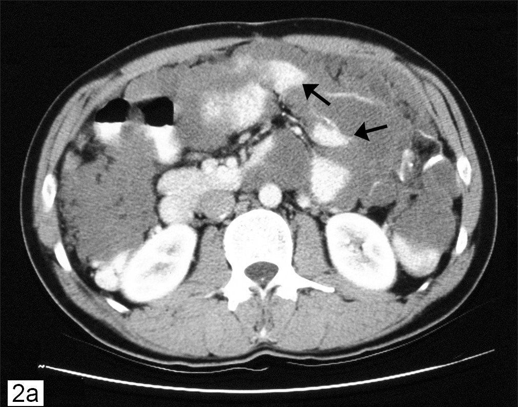
      Pseudomyxoma peritonei is characterized by disseminated intra-peritoneal mucinous tumors and mucinous ascites in the abdomen and pelvis. Epidemiology Annual incidence is estimated at 1/1,000,000 with female predominance. Clinical description The disease is usually diagnosed after the age of 40. In 30 to 50% of cases, patients present with progressive abdominal distension (so-called ``jelly belly''). Diagnosis may follow discovery of an ovarian mass in women or recent development of inguinal hernia, appendicitis or intestinal occlusion.
    • Pseudomyxoma Peritonei GARD
      Pseudomyxoma peritonei (PMP) is a rare disease characterized by the presence of mucin in the abdominal ( peritoneal ) cavity. While the most common cause of PMP is appendix cancer , several types of tumors (including non-cancerous tumors) can cause PMP. Signs and symptoms may include an increase in abdominal size or bloating; inguinal hernia (in men); an ovarian mass that may be felt during a routine pelvic exam (in women); pain or discomfort in the abdomen; and/or appendicitis . Treatment depends on the underlying cause of the condition (the location and type of the original tumor, including whether it is malignant ) and the extent of spreading. A combination of cytoreductive surgery and hyperthermic intraperitoneal chemotherapy is often the most successful treatment.
  • Morvan's Syndrome Wikipedia
    Cranial MRI shows increased signal in the hippocampus . [9] Cerebral spinal fluid (CSF) shows normal protein, glucose , white blood cell , and immunoglobulin G (IgG) levels, but there are weak oligoclonal bands , which are absent in the blood serum . ... Other fatalities without remission have been described by, amongst others, Morvan himself. [2] References [ edit ] ^ a b c d e f Lee, E K; R A Maselli; W G Ellis; M A Agius (1998-06-15). "Morvan's fibrillary chorea: a paraneoplastic manifestation of thymoma" .
    CNTNAP2, LGI1, NTN1, MEFV
    • Morvan Syndrome Orphanet
      Morvan syndrome is a rare, life-threatening, acquired neurologic disease characterized by neuromyotonia, dysautonomia and encephalopathy with severe insomnia. Signs involving central (e.g. hallucinations, confusion, amnesia, myoclonus), autonomic (e.g. variations in blood pressure, hyperhidrosis) and peripheral (e.g. painful cramps, myokymia) hyperactivity, as well as systemic manifestations (such as weight loss, pruritus, fever), are reported. Thymoma is present in some cases.
  • Semantic Dementia Wikipedia
    PMC 3059138 . PMID 21325651 . ^ a b c d e f g Bonner, M.F.; Ash, S.; Grossman, M. ... Warren, "Relatively preserved knowledge of music in semantic dementia," Journal of Neurology, Neurosurgery & Psychiatry 80, no. 7 (2009): doi:10.1136/jnnp.2008.153130. Further reading [ edit ] Gliebus, G. (March 2010). "Primary progressive aphasia: clinical, imaging, and neuropathological findings".
    PSEN1, MAPT, GRN, CHMP2B, TREM2, C9orf72, TMEM106B, SORL1, TOMM40, ABCA7, PSEN2, APP, TARDBP, PCBP2, NOS2, SMUG1, NOS1, NEFL, FUS, APOE
    • Frontotemporal Dementia OMIM
      A number sign (#) is used with this entry because this form of frontotemporal dementia (FTD) is caused by mutation in the gene encoding microtubule-associated protein tau (MAPT; 157140) on chromosome 17q21. Most cases are caused by heterozygous mutation, although rare homozygous mutations have been reported. Description Frontotemporal dementia (FTD) refers to a clinical manifestation of the pathologic finding of frontotemporal lobar degeneration (FTLD). FTD, the most common subtype of FTLD, is a behavioral variant characterized by changes in social and personal conduct with loss of volition, executive dysfunction, loss of abstract thought, and decreased speech output. A second clinical subtype of FTLD is 'semantic dementia,' characterized by specific loss of comprehension of language and impaired facial and object recognition.
    • Pick Disease Of Brain OMIM
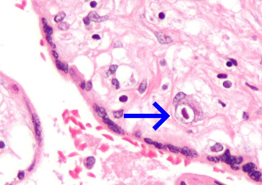
      A number sign (#) is used with this entry because Pick disease, which belongs to a class of neurodegenerative disorders known as frontotemporal dementias (FTD; see 600274), can be caused by heterozygous mutation in the MAPT gene (157140) on chromosome 17q21. Some cases of Pick disease are caused by heterozygous mutation in the presenilin-1 gene (PSEN1; 104311) on chromosome 14q24. Description Pick disease refers to the neuropathologic finding of 'Pick bodies,' which are argyrophilic, intraneuronal inclusions, and 'Pick cells,' which are enlarged neurons. The clinical correlates of Pick disease of brain include those of frontotemporal dementia, which encompass the behavioral variant of FTD, semantic dementia, and progressive nonfluent aphasia (summary by Piguet et al., 2011). Kertesz (2003) suggested the term 'Pick complex' to represent the overlapping syndromes of FTD, primary progressive aphasia (PPA), corticobasal degeneration (CBD), progressive supranuclear palsy (601104), and FTD with motor neuron disease.
    • Semantic Dementia Orphanet
      Semantic dementia (SD) is a form of frontotemporal dementia (FTD; see this term), characterized by the progressive, amodal and profound loss of semantic knowledge (combination of visual associative agnosia, anomia, surface dyslexia or dysgraphia and disrupted comprehension of word meaning) and behavioral abnormalities, attributable to the degeneration of the anterior temporal lobes.
  • ←
  • 1
  • 2
  • ...
  • 218
  • 219
  • 220
  • 221
  • 222
  • 223
  • 224
  • ...
  • 293
  • 294
  • →

FindZebra

contact@findzebra.com