• FindZebra
  • About
  • Contact
  • Help
  • Login
Deep Research Advanced
  • Renpenning's Syndrome Wikipedia
    Am J Med Genet A ^ RENPENNING H, GERRARD JW, ZALESKI WA, TABATA T (November 1962). "Familial sex-linked mental retardation" .
    PQBP1, STS, MAPK1, RNF19A, SIRT1, AHSA1, GRAP2, AIMP2, TNF, IL1B, RUNX2, HSP90AA1, GSTM2, G6PD, FMR1, DLG3, MAPK14, CRK, POLDIP2
    • Renpenning Syndrome GARD
      Renpenning syndrome is a genetic condition which occurs mostly in males. Signs and symptoms include the following: developmental delay, a small head (microcephaly), short stature, and distinctive facial features. Approximately two-thirds of individuals with Renpenning syndrome have moderate to severe intellectual disability. Additional features may include heart defects, muscular atrophy, cleft palate, and eye abnormalities. Renpenning syndrome is caused by mutations in the PQBP1 gene and is inherited in an X-linked recessive manner.
    • Renpenning Syndrome Orphanet
      Renpenning syndrome is an X-linked intellectual disability syndrome (XLMR, see this term) characterized by intellectual deficiency, microcephaly, leanness and mild short stature. Epidemiology Prevalence is unknown. Clinical description The main clinical manifestations of Renpenning syndrome are usually moderate intellectual deficiency, leanness, microcephaly and short stature (relative to familial target measurements) and sometimes small testes (testicular volumes below 15 ml), that are noticed at puberty. Manifestations are expressed only in males, and female carriers show normal facial features, growth development and intelligence. Small head and brain sizes are noted at birth. Characteristic craniofacial features include long triangular faces with upslanting palpebral fissures, half-depilated eyebrows, large ridged or bulbous nose with overhanging columella, short philtrum, and cupped and laterally protruding ears. Patients are thin and show failure to thrive. Delayed motor and language development is noticed in children from an early age.
    • Renpenning Syndrome MedlinePlus
      Renpenning syndrome is a disorder that almost exclusively affects males, causing developmental delay, moderate to severe intellectual disability, and distinctive physical features. Individuals with Renpenning syndrome typically have short stature and a small head size (microcephaly ). Facial features characteristic of this disorder include a long, narrow face ; outside corners of the eyes that point upward (upslanting palpebral fissures ); a long, bulbous nose with a low-hanging separation between the nostrils (overhanging columella ); a shortened space between the nose and mouth (philtrum); and cup-shaped ears . Males with Renpenning syndrome generally have small testes . Seizures and wasting away (atrophy) of muscles used for movement (skeletal muscles) may also occur in this disorder. About 20 percent of individuals with Renpenning syndrome also have other features, which may include a gap or split in structures that make up the eye (coloboma), an opening in the roof of the mouth (cleft palate ), heart abnormalities, or malformations of the anus .
    • Renpenning Syndrome 1 OMIM
      A number sign (#) is used with this entry because Renpenning syndrome is caused by mutation in the PQBP1 gene (300463) on chromosome Xp11. Description Renpenning syndrome is an X-linked mental retardation syndrome with clinically recognizable features. Affected individuals have microcephaly, short stature, small testes, and dysmorphic facies, including tall narrow face, upslanting palpebral fissures, abnormal nasal configuration, cupped ears, and short philtrum. The nose may appear long or bulbous, with overhanging columella. Less consistent manifestations include ocular colobomas, cardiac malformations, cleft palate, and anal anomalies. Stevenson et al. (2005) proposed that the various X-linked mental retardation syndromes due to PQBP1 mutations be combined under the name of Renpenning syndrome.
  • Marfanoid Wikipedia
    ., editors. GeneReviews™ [Internet]. Seattle (WA): University of Washington, Seattle; 1993-2014.
  • Vascular Tumor Wikipedia
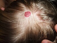
    . ^ a b c Sadick, M; Müller-Wille, R; Wildgruber, M; Wohlgemuth, WA (September 2018). "Vascular Anomalies (Part I): Classification and Diagnostics of Vascular Anomalies" . ... CS1 maint: multiple names: authors list ( link ) ^ a b c Wildgruber, M; Sadick, M; Müller-Wille, R; Wohlgemuth, WA (13 March 2019). "Vascular tumors in infants and adolescents" .
    NDP, PROX1, ALK, CDK4, FLI1, FLII, FOSB, IFNG, CXCL8, SMARCA4, SPARC, TFE3, ZFP36, DLL4, DUX4
  • Uterine Rupture Wikipedia
    See also [ edit ] Uterine perforation References [ edit ] ^ a b c d e f g h i j k l m n o p q r s t Toppenberg, KS; Block WA, Jr (1 September 2002). "Uterine rupture: what family physicians need to know". ... S2CID 22593593 . ^ Chibber R, El-Saleh E, Fadhli RA, Jassar WA, Harmi JA (March 2010). "Uterine rupture and subsequent pregnancy outcome - how safe is it?
    COL3A1, NDP, TGFBR1, TGFBR2
  • Fumarase Deficiency Wikipedia
    "Fumarate Hydratase Deficiency" . GeneReviews . Seattle WA: University of Washington . PMID 20301679 . ^ Online Mendelian Inheritance in Man (OMIM): Fumarase Deficiency - 606812 ^ Devlin, Thomas M. (2006). ... "Hereditary Leiomyomatosis and Renal Cell Cancer" . GeneReviews . Seattle WA: University of Washington . PMID 20301430 .
    FH, CD59
  • Basidiobolomycosis Wikipedia
    Treatment with itraconazole has been described. [3] References [ edit ] ^ van den Berk GE, Noorduyn LA, van Ketel RJ, van Leeuwen J, Bemelman WA, Prins JM (2006). "A fatal pseudo-tumour: disseminated basidiobolomycosis" .
  • Urethral Syndrome Wikipedia
    . ^ a b c d e Brumfitt W, Hamilton-Miller JM, Gillespie WA (July 1991). "The mysterious "urethral syndrome " " .
  • Enamel-Renal Syndrome Wikipedia
    History [ edit ] This condition was first described in 1972. [2] References [ edit ] ^ Jaureguiberry G, De la Dure-Molla M, Parry D, Quentric M, Himmerkus N, Koike T, Poulter J, Klootwijk E, Robinette SL, Howie AJ, Patel V, Figueres ML, Stanescu HC, Issler N, Nicholson JK, Bockenhauer D, Laing C, Walsh SB, McCredie DA, Povey S, Asselin A, Picard A, Coulomb A, Medlar AJ, Bailleul-Forestier I, Verloes A, Le Caignec C, Roussey G, Guiol J, Isidor B, Logan C, Shore R, Johnson C, Inglehearn C, Al-Bahlani S, Schmittbuhl M, Clauss F, Huckert M, Laugel V, Ginglinger E, Pajarola S, Spartà G, Bartholdi D, Rauch A, Addor MC, Yamaguti PM, Safatle HP, Acevedo AC, Martelli-Júnior H, dos Santos Netos PE, Coletta RD, Gruessel S, Sandmann C, Ruehmann D, Langman CB, Scheinman SJ, Ozdemir-Ozenen D, Hart TC, Hart PS, Neugebauer U, Schlatter E, Houillier P, Gahl WA, Vikkula M, Bloch-Zupan A, Bleich M, Kitagawa H, Unwin RJ, Mighell A, Berdal A, Kleta R (2013) Nephrocalcinosis (Enamel Renal Syndrome) caused by autosomal recessive FAM20A Mutations.
    FAM20A, LINC01482, PRKAR1A, SCN5A, KCNJ8, DSP, EGFR, KCND3, KCNE1, CACNA2D1, MYD88, NMT1, DPP6, PRKCE, SCN1B, DDIT3, TLR4, CACNB2, CACNA1C
    • Amelogenesis Imperfecta, Type Ig OMIM
      A number sign (#) is used with this entry because amelogenesis imperfecta type IG (AI1G), also known as enamel-renal syndrome (ERS), is caused by homozygous or compound heterozygous mutation in the FAM20A gene (611062) on chromosome 17q24. Description Amelogenesis imperfecta type IG, also known as enamel-renal syndrome, is characterized by hypoplastic enamel on primary and secondary dentition, pulp stones, delayed or failed eruption of secondary dentition, gingival overgrowth, and nephrocalcinosis. Blood chemistry analyses are typically normal, and nephrocalcinosis, which is found on renal ultrasound, may not appear until later in life (summary by Wang et al., 2013). Clinical Features MacGibbon (1972) reported a brother and sister with absent enamel, nephrocalcinosis, and apparently normal calcium metabolism. Lubinsky et al. (1985) also described an affected brother and sister, aged 11 and 9 years, respectively.
    • Enamel-Renal Syndrome Orphanet
      A extremely rare, genetic malformation syndrome characterized by hypoplastic amelogenesis imperfecta (hypoplastic dental enamel) and nephrocalcinosis (precipitation of calcium salts in renal tissue). Oral manifestations include yellow and misshaped teeth, delayed tooth eruption, and intrapulpal calcifications. Nephrocalcinosis is often asymptomatic but can progress during late childhood or early adulthood to impaired renal function, recurrent urinary infections, renal tubular acidosis, and rarely to end-stage renal failure.
  • Citrullinemia Type I Wikipedia
    (eds.). Citrullinemia Type I . Seattle (WA): University of Washington, Seattle.
    ASS1
    • Citrullinemia, Classic OMIM
      A number sign (#) is used with this entry because of evidence that classic citrullinemia is caused by homozygous or compound heterozygous mutation in the ASS1 gene (603470), which encodes argininosuccinate synthetase, on chromosome 9q34. Clinical Features Severe vomiting spells beginning at the age of 9 months and mental retardation were features of the first reported case, offspring of first-cousin parents; McMurray et al. (1962) found citrulline in very high concentration in serum, spinal fluid, and urine. (The amino acid citrulline gets its name from its high concentration in the watermelon Citrullus vulgaris.) Visakorpi (1962) also described a case of citrullinuria. Ammonia intoxication is another manifestation. The enzyme defect concerns argininosuccinic acid synthetase (EC 6.3.4.5).
    • Citrullinemia Type I Orphanet
      Citrullinemia type I is a rare autosomal recessive urea cycle defect characterized biologically by hyperammonemia and clinically by progressive lethargy, poor feeding and vomiting in the neonatal form (Acute neonatal citrullinemia type I, see this term) and by variable hyperammonemia in the later-onset form (Adult-onset citrullinemia type I, see this term).
  • Intestinal Neuronal Dysplasia Wikipedia
    European Journal of Pediatric Surgery . 18 (1): 59–60. doi : 10.1055/s-2008-1038324 . PMID 18302074 . ^ Fadda B, Maier WA, Meier-Ruge W, Schärli A, Daum R (October 1983).
    FLNA, TLX2, SPRY2
    • Intestinal Pseudoobstruction With Patent Ductus Arteriosus And Natal Teeth OMIM
      Harris et al. (1976) described 2 male sibs with mandibular teeth present at birth, patent ductus arteriosus (see 607411), and intestinal pseudoobstruction evident from birth. Vomiting of bile stain material began soon after birth, and there was no passage of meconium. The older brother died at 5 months of age despite 2 gastrointestinal operations and ligation of the patent ductus arteriosus which had led to cardiac failure. The younger brother died at the age of 6 weeks. Either autosomal or X-linked recessive inheritance is possible.
    • Neuronal Intestinal Pseudoobstruction Orphanet
      Neuronal intestinal pseudoobstruction is a form of chronic intestinal pseudoobstruction caused by a developmental failure of the enteric neurons to differentiate or migrate properly and manifests as a bowel obstruction.
  • Craniodiaphyseal Dysplasia Wikipedia
    .), "Craniometaphyseal Dysplasia, Autosomal Dominant" , GeneReviews® , Seattle (WA): University of Washington, Seattle, PMID 20301634 , retrieved 2021-01-18 External links [ edit ] Craniodiaphyseal dysplasia at orpha.net Classification D ICD - 10 : M85.2 OMIM : 218300 MeSH : C562940 External resources Orphanet : 1513 v t e Congenital malformations and deformations of musculoskeletal system / musculoskeletal abnormality Appendicular limb / dysmelia Arms clavicle / shoulder Cleidocranial dysostosis Sprengel's deformity Wallis–Zieff–Goldblatt syndrome hand deformity Madelung's deformity Clinodactyly Oligodactyly Polydactyly Leg hip Hip dislocation / Hip dysplasia Upington disease Coxa valga Coxa vara knee Genu valgum Genu varum Genu recurvatum Discoid meniscus Congenital patellar dislocation Congenital knee dislocation foot deformity varus Club foot Pigeon toe valgus Flat feet Pes cavus Rocker bottom foot Hammer toe Either / both fingers and toes Polydactyly / Syndactyly Webbed toes Arachnodactyly Cenani–Lenz syndactylism Ectrodactyly Brachydactyly Stub thumb reduction deficits / limb Acheiropodia Ectromelia Phocomelia Amelia Hemimelia multiple joints Arthrogryposis Larsen syndrome RAPADILINO syndrome Axial Skull and face Craniosynostosis Scaphocephaly Oxycephaly Trigonocephaly Craniofacial dysostosis Crouzon syndrome Hypertelorism Hallermann–Streiff syndrome Treacher Collins syndrome other Macrocephaly Platybasia Craniodiaphyseal dysplasia Dolichocephaly Greig cephalopolysyndactyly syndrome Plagiocephaly Saddle nose Vertebral column Spinal curvature Scoliosis Klippel–Feil syndrome Spondylolisthesis Spina bifida occulta Sacralization Thoracic skeleton ribs : Cervical Bifid sternum : Pectus excavatum Pectus carinatum
    SOST, CDKL5, CDA, GRIA2, CXCL8, NPPA, CLIP1
    • Craniodiaphyseal Dysplasia, Autosomal Dominant OMIM
      A number sign (#) is used with this entry because of evidence that autosomal dominant craniodiaphyseal dysplasia (CDD) is caused by heterozygous mutation in the SOST gene (605740) on chromosome 17q21. Sclerosteosis (SOST1; 269500) and van Buchem disease (VBCH; 239100), are allelic disorders that are less severe and show autosomal recessive inheritance. Description Craniodiaphyseal dysplasia is a severe bone dysplasia characterized by massive generalized hyperostosis and sclerosis, especially involving the skull and facial bones. Progressive bony encroachment upon cranial foramina leads to severe neurologic impairment in childhood (summary by Brueton and Winter, 1990). The sclerosis is so severe that the resulting facial distortion is referred to as 'leontiasis ossea' (leonine facies), and the bone deposition results in progressive stenosis of craniofacial foramina (summary by Kim et al., 2011).
    • Craniodiaphyseal Dysplasia Orphanet
      Craniodiaphyseal dysplasia is a rare sclerotic bone disorder with a variable phenotypic expression with massive generalized hyperostosis and sclerosis, particularly of the skull and facial bones, that may lead to severe deformity.
    • Craniodiaphyseal Dysplasia OMIM
      Clinical Features Cranial and facial hyperostosis results in a characteristic clinical and radiographic appearance. The diaphyses of the bones are generally expanded. Halliday (1949) and Stransky et al. (1962) reported isolated cases with similar findings. Facial and cranial thickening and distortion are particularly striking in this form. Most cases have been mentally retarded. Unlike the situation in the craniometaphyseal dysplasias (e.g., 218400), the long bones do not show metaphyseal flaring but show diaphyseal endostosis and are shaped like a policeman's nightstick. Joseph et al. (1958), who first suggested the designation of progressive craniodiaphyseal dysplasia, described a patient with a picture they considered identical to that described by Halliday (1949).
  • Adenoid Cystic Carcinoma Wikipedia
    Cancer Discov . 622 (2): 176–87. doi : 10.1158/2159-8290.CD-15-0859 . PMC 4744535 . PMID 26631070 . ^ Mitani Y, Liu B, Rao PH, Borra VJ, Zafereo M, Weber RS, Kies M, Lozano G, Futreal PA, Caulin C, El-Naggar AK (2016).
    TP53, NOTCH1, PIK3CA, MYBL1, FBXW7, BCOR, HRAS, MYB, NFIB, CREBBP, CDH1, PTEN, CCND1, SOX4, DAPK1, BRCA1, AQP1, IGFBP2, ATM, ESPL1, SRCAP, HOMER3, TLK1, MAGI2, SMARCA2, MORF4L1, KDM6B, MLC1, FGF16, DTX4, H1-4, GUCY1A1, MGA, RBFOX2, GAS6, GAS2, MAGI1, KAT6A, H2AC16, INSRR, SERPINF1, PRKDC, MAP2K2, MYCN, PYGB, ST3GAL4, SMARCE1, SON, SOX11, MARCKS, TOP2A, KDM6A, KRT15, SMC1A, KRT5, ARID1A, ITGB4, ZIM2, BRD1, MYCBP, NSD1, STAG3L1, CNTN6, MIER2, IL17RD, MAML3, CMTR2, ERBIN, SLC24A3, KMT2C, BCORL1, EFHD1, BCL11A, WNT5B, NETO2, ARID5B, ATRX, FOXP2, JMJD1C, JAG1, ASPM, KANSL1, XAGE1A, VCAN, PDZK1, FAT1, PCSK1N, ARID4B, GINS2, SETD2, FOXO3, DTL, UHRF1, FGFR4, IRX4, ISYNA1, FANCA, MARK2, EN1, EP300, KRAS, KIT, ARID2, SF3B1, CYLD, IDH1, EGFR, CTNNB1, CDKN2A, SOX10, CXCR4, ERBB2, HIF1A, VEGFA, ACCS, SOX2, SMUG1, H3P10, NOS2, BCL2, NOS1, RUNX3, FN1, BCL2A1, MIR21, ACACB, SNAI1, MMP2, MIF, MDM2, TBX1, KRT14, ILK, TP63, BECN1, BMS1, ID1, MMP9, CD274, CTSD, ANO1, MYB-AS1, CD44, BTBD7, SLC2A1, ACACA, PCNA, NOTCH4, MAPK1, PSMD7, CTSB, CTAG1B, CCR5, CENPF, CDK2, CDH5, PECAM1, CDH4, PAX3, DNMT1, KRIT1, PA2G4, CCL28, TMPRSS4, ACKR3, CALM3, NDRG2, MTUS1, CALM2, NNMT, EPHA2, BUB1, BBC3, USP22, GPC3, GJA1, GATA1, FZD2, MTOR, SULT4A1, FOLH1, DKK2, LEF1, PIK3CB, FGF2, FABP7, NGF, EWSR1, OBP2A, ERBB3, TMED7, CALM1, CAMKMT, HOXB7, MIR17HG, MIR125A, MIR140, MIR150, MIR181A2, NTF3, MIR222, MIR320A, MIR93, MIR338, SBSN, MIR375, MIR455, ASIC1, MIR1234, TMED7-TICAM2, ADAMTS9-AS2, H3P23, H3P28, MIRLET7B, TICAM2, BSG, ATF1, PDCD1LG2, CD276, BNIP3, NTRK3, REG4, NTRK1, MINDY4, MAML2, AQP5, MACC1, WDR66, AQP3, SKA1, ALCAM, CTAG1A, ADK, ADAM10, ARMH1, SFN, HES1, PSMD9, HSPB1, SRY, STAT3, PPP2R2B, TFE3, TGFB1, ICAM5, TNF, EPCAM, LYZ, TRAF6, TXN, TYMS, LMNA, UVRAG, LGALS3, VEGFC, VIM, NSD2, RPSA, SPN, SMAD4, MCAM, SDC1, PTCH1, MYC, PTGS2, MMP15, RAC1, RPE65, S100A1, S100B, SGTA, PTPA, PROX1, SKP2, NCAM1, SMARCA1, MAPK3, MMP7, NFKB1, PRKD1, PRRX1, KRT7, KIF22, AGR2, IFI27, PIM1, MFN2, AKT3, RABEPK, NOTCH2, HOXB13, ZNRD2, DCTN6, PIK3CD, HSPG2, SMR3B, HSPB2, MLLT11, EBNA1BP2, CKAP4, PTP4A3, RASSF1, ZEB2, IGF1R, PLAG1, NRP2, RECK, GEMIN2, PPM1D, PIN1, PIK3CG, TNFSF10, ADAM9, IL9, PROM1, ATG5, HSPB3, SCAF11, ARHGEF2, IL2, SLC9A3R2, LHX2, DDX23, CCN1, LANCL1
    • Adenoid Cystic Carcinoma GARD
      Adenoid cystic carcinoma (ACC) is a rare form of adenocarcinoma , a type of cancer that begins in glandular tissues. It most commonly arises in the major and minor salivary glands of the head and neck. It can also occur in the breast, uterus, or other locations in the body. Symptoms depend on the tumor's location. Salivary gland tumors may cause painless masses in the mouth or face. Tumors of the lacrimal gland may cause a bulging eye or changes in vision.
  • Cystinosis Wikipedia
    PMID 12110740 . ^ a b Nesterova G, Gahl WA. Cystinosis: the evolution of a treatable disease. Pediatr Nephrol 2012;28:51–9. ^ Gahl WA, Thoene JG, Schneider JA. Cystinosis.
    CTNS, LGALS3, TRPV1, SLC66A1, TFEB, APRT, SCN7A, CCL2, RAB7A, RAB11A, YBX3, PTH, NBAS, NLRP2, RILP, UNC13D, RAB27A, PRKAB1, CASP1, PRKAA2, PRKAA1, NAGLU, MFAP1, LRP2, LDLR, ITGAE, IL18, IL10, IL1B, IFNG, CTSD, RAB7B
    • Cystinosis GeneReviews
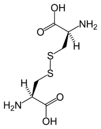
      Summary Clinical characteristics. Cystinosis comprises three allelic phenotypes: Nephropathic cystinosis in untreated children is characterized by renal Fanconi syndrome, poor growth, hypophosphatemic/calcipenic rickets, impaired glomerular function resulting in complete glomerular failure, and accumulation of cystine in almost all cells, leading to cellular dysfunction with tissue and organ impairment. The typical untreated child has short stature, rickets, and photophobia. Failure to thrive is generally noticed after approximately age six months; signs of renal tubular Fanconi syndrome (polyuria, polydipsia, dehydration, and acidosis) appear as early as age six months; corneal crystals can be present before age one year and are always present after age 16 months. Prior to the use of renal transplantation and cystine-depleting therapy, the life span in nephropathic cystinosis was no longer than ten years. With these interventions, affected individuals can survive at least into the mid-forties or fifties with satisfactory quality of life.
  • Fg Syndrome Wikipedia
    .), "MED12-Related Disorders" , GeneReviews , Seattle (WA): University of Washington, Seattle, PMID 20301719 , retrieved 2020-09-01 ^ Lyons, Michael J. (1993), Adam, Margaret P.; Ardinger, Holly H.; Pagon, Roberta A.; Wallace, Stephanie E. (eds.), "MED12-Related Disorders" , GeneReviews , Seattle (WA): University of Washington, Seattle, PMID 20301719 , retrieved 2020-09-01 ^ a b Opitz JM, Smith JF, Santoro L (2008).
    MED12, CASK, FLNA, FGS2, MID2, FGS3, IGAN1, VSX1, OBP2A, AGO2, RCOR1, CCN6, KIF22, IGBP1, HTC2, HPD, GLI3, FMR1, FGS5
    • Opitz-Kaveggia Syndrome OMIM
      A number sign (#) is used with this entry because of evidence that the Opitz-Kaveggia syndrome, also known as FG syndrome-1 (FGS1), is caused by mutation in the MED12 gene (300188) on chromosome Xq13. Description Opitz-Kaveggia syndrome (OKS) is an X-linked recessive mental retardation syndrome characterized by dysmorphic features, including relative macrocephaly, hypertelorism, downslanted palpebral fissures, prominent forehead with frontal hair upsweep, and broad thumbs and halluces. Most have hypotonia, constipation, and partial agenesis of the corpus callosum. Some patients have sensorineural hearing loss and joint laxity evolving into joint contractures. Affected individuals tend to be hyperactive and talkative (summary by Graham et al., 1999).
    • Fg Syndrome Type 1 Orphanet
      A rare X-linked syndromic intellectual disability characterized by developmental delay and intellectual disability, early hypotonia, constipation, feeding problems, imperforate anus, characteristic behavior (affable, eager to please), and dysmorphic craniofacial features (such as relative macrocephaly, prominent forehead with frontal hair upsweep, hypertelorism, downslanting palpebral fissures, and open mouth). Additional manifestations are partial agenesis of the corpus callosum, sensorineural hearing loss, joint laxity, cardiac anomalies, and abnormalities of the fingers and toes, among others.
    • Fg Syndrome GARD
      FG syndrome (FGS) is a genetic condition that affects many parts of the body and occurs almost exclusively in males. "FG" represents the surname initials of the first individuals diagnosed with the disorder. People with FG syndrome frequently have intellectual disability ranging from mild to severe, hypotonia, constipation and/or anal anomalies, a distinctive facial appearance, broad thumbs and great toes, a large head compared to body size (relative macrocephaly), and abnormalities of the corpus callosum. Medical problems including heart defects , seizures, undescended testicle , and an inguinal hernia have also been reported in some affected individuals. Researchers have identified five regions of the X chromosome that are linked to FG syndrome in affected families.
  • Kaufman Oculocerebrofacial Syndrome Wikipedia
    .; Ledbetter, Nikki; Mefford, Heather C. (eds.). GeneReviews . Seattle (WA): University of Washington, Seattle.
    UBE3B, PTPN4, MEG3, WDR20, RTL1
    • Kaufman Oculocerebrofacial Syndrome MedlinePlus
      Kaufman oculocerebrofacial syndrome is a disorder characterized by eye problems (oculo-), intellectual disability (-cerebro-), and a distinctive pattern of facial features (-facial). Most individuals with Kaufman oculocerebrofacial syndrome have an unusually small head size (microcephaly ), and some have structural abnormalities of the brain . Affected individuals have weak muscle tone (hypotonia), and are delayed in developing motor skills such as walking. Intellectual disability is severe or profound. Most affected individuals never acquire the ability to speak. Eye abnormalities and their effect on vision vary among people with Kaufman oculocerebrofacial syndrome.
    • Oculocerebrofacial Syndrome, Kaufman Type Orphanet
      A rare, genetic, syndromic intellectual disability characterized by severe intellectual disability, distinctive craniofacial features and variable multiple congenital anomalies including ocular, brain, urogenital and skeletal abnormalities. Epidemiology To date, 19 molecularly diagnosed cases have been described in the scientific and medical literature. Clinical description The most prominent clinical findings are severe intellectual disability, pre-and postnatal growth retardation, microcephaly, and typical craniofacial features which include non-progressive microcephaly of prenatal onset, prominence of the zygomatic region of the face, full cheeks, prominent frontal tubers, sparse and arched eyebrows, blepharophimosis with epicanthal folds, upslanted palpebral fissures, preauricular skin tags, underdeveloped and abnormally folded ears, wide nasal base, low nasal bridge, anteverted nares, long and flat philtrum and retrognathia. Hypotonia, feeding difficulties, failure to thrive and poor speech development are universal findings. Many patients require tube-feeding. Perceptive language is better than expressive, some patients acquire a few words and basic ambulation skills such as eating and dressing independently.
    • Kaufman Oculocerebrofacial Syndrome GeneReviews
      Summary Clinical characteristics. Kaufman oculocerebrofacial syndrome (KOS) is characterized by severe intellectual disability and distinctive craniofacial features. Most affected children have prenatal-onset microcephaly, failure to thrive, hypotonia, and short stature. Eye abnormalities are common and can include structural abnormalities (microcornea or microphthalmia, coloboma, optic nerve hypoplasia), refractive errors (myopia ± astigmatism, hyperopia), strabismus, and entropion. Less common findings can include: unilateral or bilateral conductive hearing loss or mixed conductive-sensorineural hearing loss of variable severity; congenital heart defects; breathing problems; feeding difficulties; urogenital abnormalities; and/or skeletal abnormalities. Diagnosis/testing. The diagnosis of KOS is established in a proband with developmental delay/intellectual disability and biallelic UBE3B pathogenic variants.
    • Kaufman Oculocerebrofacial Syndrome OMIM
      A number sign (#) is used with this entry because of evidence that Kaufman oculocerebrofacial syndrome (KOS) is caused by homozygous or compound heterozygous mutation in the UBE3B gene (608047) on chromosome 12q24. Clinical Features Kaufman et al. (1971) described a distinctive syndrome in 4 of 7 sibs. Significant positive and negative features included intrauterine and postnatal growth retardation, microcephaly with mental retardation but no gross neurologic abnormalities or seizures, hypertelorism with epicanthi, ptosis of the eyelids, upslanted palpebral fissures, microcornea with pale optic discs, sparse and laterally broad eyebrows, flat philtrum, congenital hypotonia, micrognathia with neonatal respiratory distress, high and narrow palate, lordosis, constipation, and flat feet. Jurenka and Evans (1979) reported a sporadic case, and Garcia-Cruz et al. (1988) described a case. Buntinx and Majewski (1990) reported a child, born to nonconsanguineous parents, with what the authors considered to be a novel phenotype.
  • Lymphangiosarcoma Wikipedia
    . ^ Sher T, Hennessy BT, Valero V, Broglio K, Woodward WA, Trent J, Hunt KK, Hortobagyi GN, Gonzalez-Angulo AM.Primary angiosarcomas of the breast.
    CD34, TSC1, VEGFA, LIAS
  • Cd55 Deficiency Wikipedia
    Kurolap and colleagues treated patients with off-label eculizumab , a humanized anti-C5 monoclonal antibody and complement inhibitor, and it was shown to have beneficial outcomes over an 18-month period. [6] Investigators at Marmara University in Istanbul, Turkey, and the National Institute of Allergy and Infectious Diseases at the US National Institutes of Health in Bethesda, Maryland currently have clinical protocols to study new approaches to the diagnosis and treatment of this disorder. [7] References [ edit ] ^ a b c d e f g h i j k Ozen A, Comrie WA, Ardy RC, Domínguez Conde C, Dalgic B, Beser ÖF, et al.
  • Berdon Syndrome Wikipedia
    . ^ "Ann Arbor boy, 5, overcomes rare diseases: 'He's a fighter ' " . 2015-12-24. ^ Berdon, WE; Baker, DH; Blanc, WA; Gay, B; Santulli, TV; Donovan, C (1976).
    ACTG2, MYH11, MYLK, LMOD1, CHRM3, ACTB, B2M, MYL9, BHLHE23
    • Megacystis-Microcolon-Intestinal Hypoperistalsis Syndrome Overview GeneReviews
      Summary The purpose of this overview is to increase the awareness of clinicians regarding megacystis-microcolon-intestinal hypoperistalsis syndrome (MMIHS) and its genetic causes and management. The following are the goals of this overview: Goal 1. Describe the clinical characteristics of MMIHS. Goal 2. Review the genetic causes of MMIHS. Goal 3. Provide an evaluation strategy to identify the genetic cause of MMIHS in a proband (when possible). Goal 4. Inform genetic counseling of family members of an individual with MMIHS. Goal 5. Review management of MMIHS. Diagnosis Clinical Characteristics Differential Diagnosis Management
    • Megacystis-Microcolon-Intestinal Hypoperistalsis Syndrome Orphanet
      Megacystis microcolon intestinal hypoperistalsis syndrome (MMIHS) is a rare congenital disease characterized by massive abdominal distension caused by a largely dilated non-obstructed urinary bladder (megacystis), microcolon and decreased or absent intestinal peristalsis. Epidemiology MMIHS prevalence is unknown but the disease has been reported in 230 patients, of which 71% are females. Clinical description Enlarged and nonobstructed bladder is the first manifestation of MMIHS and can be detected prenatally. It results in abdominal distension, which is an early constant finding. Usual clinical presentation is similar to other neonatal intestinal obstructions: bile stained vomiting and failure to pass meconium.
    • Megacystis Microcolon Intestinal Hypoperistalsis Syndrome GARD
      Megacystis microcolon intestinal hypoperistalsis syndrome (MMIHS) is a rare congenital condition characterized by abdominal distension caused by a largely dilated non-obstructed urinary bladder (megacystis); very small colon (microcolon); and decreased or absent intestinal movements (intestinal peristalsis). Usual clinical presentation is similar to other neonatal intestinal obstructions: bile stained vomiting and failure to pass meconium (the first bowel movement the baby has). Other intestinal anomalies may be present like intestinal malrotation . Many problems with the urinary tract result from the bladder dysfunction. It is part of a group of conditions caused by changes (mutations) in the ACTG2 gene and is inherited in an autosomal dominant manner.
  • Melorheostosis Wikipedia
    . ^ Kang H, Jha S, Deng Z, Fratzl-Zelman N, Cabral WA, Ivovic A, Meylan F, Hanson EP, Lange E, Katz J, Roschger P, Klaushofer K, Cowen EW, Siegel RM, Marini JC, Bhattacharyya T (April 2018).
    LEMD3, HBB, HBA1, HBA2, HBD, HBG2, MYC, HBG1, SSX2, TGFB1, VIM, MIA, PDCD5, ALAS1, SLC1A1, CD274, PRDM16, CPO, SMIM10L2A, SMIM10L2B, SOX10, NOTCH1, CCL2, PTGS2, MAP2K1, ATP7A, KRAS, HLA-C, HBE1, GATA1, GAGE5, GAGE4, GAGE1, BAGE, SSX2B
    • Melorheostosis MedlinePlus
      Melorheostosis is a rare bone disease. It causes the abnormal growth of new bone tissue on the surface of existing bones. The new bone has a characteristic appearance on x-rays, often described as "flowing" or like dripping candle wax. The excess bone growth typically occurs on the bones in one arm or leg, although it can also affect the pelvis, breastbone (sternum), ribs, or other bones. (The term "melorheostosis" is derived from the Greek words "melos," which means limb; "rheos," which means flow; and "ostosis," which refers to bone formation.) The abnormal bone growth associated with melorheostosis is noncancerous (benign), and it does not spread from one bone to another.
  • Giant Cell Arteritis Wikipedia
    . ^ a b c Mackie, SL; Dejaco, C; Appenzeller, S; Camellino, D; Duftner, C; Gonzalez-Chiappe, S; Mahr, A; Mukhtyar, C; Reynolds, G; de Souza, AWS; Brouwer, E; Bukhari, M; Buttgereit, F; Byrne, D; Cid, MC; Cimmino, M; Direskeneli, H; Gilbert, K; Kermani, TA; Khan, A; Lanyon, P; Luqmani, R; Mallen, C; Mason, JC; Matteson, EL; Merkel, PA; Mollan, S; Neill, L; Sullivan, EO; Sandovici, M; Schmidt, WA; Watts, R; Whitlock, M; Yacyshyn, E; Ytterberg, S; Dasgupta, B (1 March 2020). ... PMID 16987903 . ^ Mackie, SL; Dejaco, C; Appenzeller, S; Camellino, D; Duftner, C; Gonzalez-Chiappe, S; Mahr, A; Mukhtyar, C; Reynolds, G; de Souza, AWS; Brouwer, E; Bukhari, M; Buttgereit, F; Byrne, D; Cid, MC; Cimmino, M; Direskeneli, H; Gilbert, K; Kermani, TA; Khan, A; Lanyon, P; Luqmani, R; Mallen, C; Mason, JC; Matteson, EL; Merkel, PA; Mollan, S; Neill, L; Sullivan, EO; Sandovici, M; Schmidt, WA; Watts, R; Whitlock, M; Yacyshyn, E; Ytterberg, S; Dasgupta, B (1 March 2020). ... OCLC 663444979 . ^ " giant cell arteritis " at Dorland's Medical Dictionary External links [ edit ] Mackie, SL; Dejaco, C; Appenzeller, S; Camellino, D; Duftner, C; Gonzalez-Chiappe, S; Mahr, A; Mukhtyar, C; Reynolds, G; de Souza, AWS; Brouwer, E; Bukhari, M; Buttgereit, F; Byrne, D; Cid, MC; Cimmino, M; Direskeneli, H; Gilbert, K; Kermani, TA; Khan, A; Lanyon, P; Luqmani, R; Mallen, C; Mason, JC; Matteson, EL; Merkel, PA; Mollan, S; Neill, L; Sullivan, EO; Sandovici, M; Schmidt, WA; Watts, R; Whitlock, M; Yacyshyn, E; Ytterberg, S; Dasgupta, B (1 March 2020).
    PTPN22, HLA-DRB1, HLA-DRB9, PLG, P4HA2, RBM45
  • ←
  • 1
  • 2
  • 3
  • 4
  • 5
  • 6
  • ...
  • 13
  • 14
  • →

FindZebra

contact@findzebra.com