Hereditary Ataxia Overview . Seattle (WA): University of Washington, Seattle. ... Spinocerebellar Ataxia Type 2 . Seattle (WA): University of Washington, Seattle. ... Movement Disorders: Genetics and Models . Academic Press. ISBN 9780080470566 .
A clinically and genetically heterogeneous group of neurodegenerative diseases characterized by a slowly progressive ataxia of gait, stance and limbs, dysarthria and/or oculomotor disorder, due to cerebellar degeneration in the absence of coexisting diseases. The degenerative process can be limited to the cerebellum (ADCA type 3) or may additionally involve the retina (ADCA type 2), optic nerve, ponto-medullary systems, basal ganglia, cerebral cortex, spinal tracts or peripheral nerves (ADCA type 1). In ACDA type 4, a cerebellar syndrome is associated with epilepsy.
Autosomal dominant cerebellar ataxia (ADCA) is one of the genetic subtypes of hereditary ataxia . Although the signs and symptoms vary depending on the specific type, the most common symptom of ADCA is poor movement coordination ( ataxia ) especially a jerky, unsteady walking style (gait). Coordination of hands and clearness of speech (dysarthria) are also affected. The area of the brain controlling balance and movement decreases in size ( cerebellar atrophy ). This can be seen on brain imaging . The ataxia usually slowly worsens over time.
Sadikovic et al. (2009) hypothesized that the RUNX2 interactome may be constitutively activated in osteosarcoma, and that the downstream intracellular pathways may be associated with the regulation of osteoblast differentiation and control of cell cycle and apoptosis in osteosarcoma. Animal Model Khanna et al. (2000) developed a murine model of osteosarcoma characterized by tumor growth at appendicular sites, a period of minimal residual disease, spontaneous pulmonary metastasis, and model variants that differ in metastatic potential. The model was developed from a cell line, K12, originating from a spontaneous BALB/c osteogenic sarcoma, and a clonally related cell line, K7M2. Within the model, the K7M2 cell line is aggressive and highly metastatic, whereas the K12 cell line is less aggressive with infrequent pulmonary metastases. ... To define the relevance of ezrin in the biology of metastasis beyond the founding mouse model, Khanna et al. (2004) examined ezrin expression in dogs that naturally developed osteosarcoma.
Osteosarcoma Intermediate-magnification micrograph of an osteosarcoma (center and right of image) adjacent to non-malignant bone (left-bottom of image): The top-right of the image has poorly differentiated tumor. Osteoid with a high density of malignant cells is seen between the non-malignant bone and poorly differentiated tumor ( H&E stain ). Specialty Oncology An osteosarcoma ( OS ) or osteogenic sarcoma ( OGS ) (or simply bone cancer ) is a cancerous tumor in a bone . Specifically, it is an aggressive malignant neoplasm that arises from primitive transformed cells of mesenchymal origin (and thus a sarcoma ) and that exhibits osteoblastic differentiation and produces malignant osteoid . [1] Osteosarcoma is the most common histological form of primary bone sarcoma . [2] It is most prevalent in teenagers and young adults. [3] Contents 1 Signs and symptoms 2 Causes 3 Mechanism 4 Diagnosis 4.1 Variants 5 Treatment 6 Prognosis 7 Epidemiology 8 Other animals 8.1 Risk factors 8.2 Clinical presentation 8.3 Treatment and prognosis 8.4 Cats 9 References 10 Further reading 11 External links Signs and symptoms [ edit ] Many patients first complain of pain that may be worse at night, may be intermittent and of varying intensity and may have been occurring for some time. Teenagers who are active in sports often complain of pain in the lower femur, or immediately below the knee.
Overview Osteosarcoma is a type of bone cancer that begins in the cells that form bones. Osteosarcoma is most often found in the long bones — more often the legs, but sometimes the arms — but it can start in any bone. In very rare instances, it occurs in soft tissue outside the bone. Osteosarcoma tends to occur in teenagers and young adults, but it can also occur in younger children and older adults. Treatment usually involves chemotherapy, surgery and, sometimes, radiation therapy. Doctors select treatment options based on where the osteosarcoma starts, the size of the cancer, the type and grade of the osteosarcoma, and whether the cancer has spread beyond the bone.
Osteosarcoma is a primary malignant tumour of the skeleton characterised by the direct formation of immature bone or osteoid tissue by the tumour cells. Epidemiology Classic osteosarcoma is a rare (0.2% of all malignant tumours) highly malignant tumour, with an estimated incidence of 3 cases/million population/year. Clinical description Osteosarcoma arises predominantly in the long bones and rarely in the soft tissues. The age at presentation ranges from 10 to 25 years of age. Diagnostic methods Plain radiographs, computed tomography, magnetic resonance imaging, angiography and dynamic bone scintigraphy are used for diagnosis, evaluation the extent of tumour involvement and for making decisions about the type of operation and, if necessary, the type of reconstruction required. Management and treatment In the past, all patients with osteosarcoma were treated by amputation but the cure rate was under 10% and almost all patients died within a year from diagnosis.
Osteosarcoma is the most common type of bone cancer. The average age at diagnosis is 15. Boys and girls have a similar incidence of this tumor until late adolescence, at which time boys are more commonly affected. In rare cases, osteosarcoma occurs in adults. Although osteosarcoma tends to occur in the larger bones, such as the shin (near the knee), thigh (near the knee) and upper arm (near the shoulder), it can occur in any bone. A number of variants of osteosarcoma exist, including conventional types (osteoblastic, chondroblastic, and fibroblastic), telangiectatic, multifocal, parosteal, and periosteal. The cause of osteosarcoma is not known. In some cases, it runs in families, and at least one gene has been linked to increased risk.
(March 2019). "Modelling traumatic brain injury and posttraumatic epilepsy in rodents" . ... "Epileptogenesis in experimental models". Epilepsia . 48 (Supplement 2): 13–20. doi : 10.1111/j.1528-1167.2007.01063.x . ... ISBN 0-632-06046-8 . ^ a b c Annegers JF, Hauser WA, Coan SP, Rocca WA (January 1998). ... "Kindling and status epilepticus models of epilepsy: Rewiring the brain". ... (eds.). Neuromethods: Animal Models of Neurological Disease . Totowa, NJ: Humana Press. pp. 153–155.
CS1 maint: multiple names: authors list ( link ) ^ Hallman KB, Nahhas WA, Connelly PJ (September 1991). "Endosalpingiosis as a source of psammoma bodies in a Papanicolaou smear.
You can help by adding to it . ( August 2017 ) Sources [ edit ] Singleton, EB, Merten DF: An unusual syndrome of widened medullary cavities of the metacarpals and phalanges, aortic calcification and abnormal dentition, Pediatric Radiol 1:2, 1973. [1] Resources form the National Institutes of Health [2] WebMD information References [ edit ] ^ Ferreira CR, Crow YJ, Gahl WA, Gardner PJ, Goldbach-Mansky R, Hur S, de Jesús AA, Nehrebecky M, Park JW, Briggs TA (2018) DDX58 and classic Singleton-Merten syndrome.
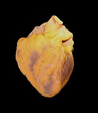
Singleton-Merten syndrome is a very rare disease that affect many organs. The main features are tooth abnormalities with gum infection; calcifications in the aorta artery and in certain valves of the heart (i.e., aortic and mitral valves); and progressive thinning and weakening of the bones (osteoporosis), especially in the upper and back portions of the skull. Other findings may include neurologic problems, generalized short stature, muscle weakness; poor muscle tone (hypotonia); progressive wasting of the muscles ( muscle atrophy ); heart arrhythmia, growth and developmental delay; skin problems such as psoriasis; malformation of the hips and/or feet and limbs or fingers, joint problems, tendon rupture, distinct facial features, and vision problems due to glaucoma. Severe systemic lupus erythematosus can also occur with Singleton-Merten syndrome. Singleton-Merten syndrome is caused by mutations in the IFIH1 gene, and in the DDX58 genes (which causes anatypical form of Singleton-Merten syndrome where there are no teeth problems).
A number sign (#) is used with this entry because of evidence that Singleton-Merten syndrome-2 (SGMRT2) is caused by heterozygous mutation in the DDX58 gene (609631) on chromosome 9p21. Description Singleton-Merten syndrome-2 is characterized by variable expression of glaucoma, aortic calcification, and skeletal abnormalities, without dental anomalies (summary by Jang et al., 2015). For a general phenotypic description and discussion of genetic heterogeneity of Singleton-Merten syndrome, see SGMRT1 (182250). Clinical Features Jang et al. (2015) studied a large 4-generation Korean family with aortic calcification, glaucoma, and skeletal abnormalities. The 56-year-old proband was diagnosed with bilateral glaucoma at 6 years of age and was blind by age 17.
A number sign (#) is used with this entry because of evidence that Singleton-Merten syndrome-1 (SGMRT1) is caused by heterozygous mutation in the IFIH1 gene (606951) on chromosome 2q24. Description Singleton-Merten syndrome (SGMRT) is an uncommon autosomal dominant disorder characterized by abnormalities of blood vessels, teeth, and bone. Calcifications of the aorta and aortic and mitral valves occur in childhood or puberty and can lead to early death. Dental findings include delayed primary tooth exfoliation and permanent tooth eruption, truncated tooth root formation, early-onset periodontal disease, and severe root and alveolar bone resorption associated with dysregulated mineralization, leading to tooth loss. Osseous features consist of osteoporosis, either generalized or limited to distal extremities, distal limb osteolysis, widened medullary cavities, and easy tearing of tendons from bone.
Singleton-Merten dysplasia is characterized by dental dysplasia, progressive calcification of the thoracic aorta with stenosis, osteoporosis and expansion of the marrow cavities in hand bones. Additional features included generalized muscle weakness and atrophy, and chronic psoriasiform skin eruptions. It has been reported in four unrelated patients (male and female) and in a family with multiple affected members (male).
Riccardi (1982) described cases of neurofibromatosis that are sufficiently variant that they seem to warrant separation from the classic von Recklinghausen NF I (162200), the acoustic neuroma type, NF II (101000), and the mixed type, NF III (162260). The group still is undoubtedly heterogeneous. Iris Lisch nodules, one of the most specific features of NF I, are usually absent in NF IV. The importance of a separate category for these cases is related to the probable difference in prognosis and genetic counseling and the desirability of avoiding confusion of studies of the natural history and pathogenesis of NF I. Eyes - Iris Lisch nodules usually absent Inheritance - Autosomal dominant - heterogeneous Skin - Atypical neurofibromatosis ▲ Close
Axial osteomalacia is a rare osteosclerotic disorder first described by Frame et al. (1961). Characteristically, trabecular bone has 'a unique coarsening and spongelike appearance in the x-rays of the axial skeleton.' Radiographically, the skull and appendicular skeleton are normal. Vague chronic axial skeletal pain is the presenting symptom in most patients. Despite osteosclerosis and normal circulating levels of calcium, inorganic phosphate and alkaline phosphatase, bone biopsy specimens show osteomalacia. Until the report of Whyte et al. (1981), 10 cases had been described, all in middle-aged or elderly white men.
Ataxia with vitamin E deficiency is a disorder that impairs the body's ability to use vitamin E obtained from the diet. Vitamin E is an antioxidant, which means that it protects cells in the body from the damaging effects of unstable molecules called free radicals. A shortage (deficiency) of vitamin E can lead to neurological problems, such as difficulty coordinating movements (ataxia) and speech (dysarthria), loss of reflexes in the legs (lower limb areflexia), and a loss of sensation in the extremities (peripheral neuropathy). Some people with this condition have developed an eye disorder called retinitis pigmentosa that causes vision loss. Most people who have ataxia with vitamin E deficiency start to experience problems with movement between the ages of 5 and 15 years.
A number sign (#) is used with this entry because of evidence that ataxia with vitamin E deficiency (AVED) is caused by homozygous or compound heterozygous mutation in the TTPA gene (600415) on chromosome 8q12. Clinical Features Harding et al. (1985) described a young woman with spinocerebellar degeneration thought to be due to a selective defect in vitamin E absorption. There was no evidence of fat malabsorption. Binder et al. (1967) suggested a relationship between neurologic dysfunction and vitamin E deficiency in patients with chronic steatorrhea. This was subsequently confirmed in patients with abetalipoproteinemia (200100), the most severe state of vitamin E deficiency known. When studied at age 23, the proband had no vitamin E in the serum. A progressive neurologic disorder comprising ataxia, areflexia and marked loss of proprioception developed at age 13.
A neurodegenerative disease belonging to the inherited cerebellar ataxias mainly characterized by progressive spino-cerebellar ataxia, loss of proprioception, areflexia, and is associated with a marked deficiency in vitamin E. Epidemiology Global prevalence is not known but population-based studies have been performed and prevalence can be extrapolated at approximately 1/300,000. AVED is the second most frequently inherited cerebellar ataxia in North Africa. As vitamin E deficiency might bring protection against malaria (see this term), it could explain a higher prevalence of AVED in Plasmodium infested areas. Clinical description AVED presents generally between ages 5 and 20 years with variable phenotype and severity.
Summary Clinical characteristics. Ataxia with vitamin E deficiency (AVED) generally manifests in late childhood or early teens between ages five and 15 years. The first symptoms include progressive ataxia, clumsiness of the hands, loss of proprioception, and areflexia. Other features often observed are dysdiadochokinesia, dysarthria, positive Romberg sign, head titubation, decreased visual acuity, and positive Babinski sign. The phenotype and disease severity vary widely among families with different pathogenic variants; age of onset and disease course are more uniform within a given family, but symptoms and disease severity can vary even among sibs. Diagnosis/testing. Presently, no consensus diagnostic criteria for AVED exist; the principal criterion for diagnosis is a Friedreich ataxia-like neurologic phenotype combined with markedly reduced plasma vitamin E (α-tocopherol) concentration and a normal lipoprotein profile in the absence of known causes of malabsorption.
Ataxia with vitamin E deficiency (AVED) is a progressive disease affecting motor control and movement. Symptoms of AVED include slurred speech (dysarthria), difficulty coordinating movements ( ataxia ), numbness in the hands and feet (peripheral neuropathy), and progressive leg weakness. Some affected individuals may experience vision loss due to damage to the back of the eye ( retinitis pigmentosa ). Symptoms typically begin during childhood or adolescence and worsen with age, resulting in the need for a wheelchair by early adulthood. AVED is caused by a mutation to the TTPA gene. When this gene is damaged, vitamin E cannot be distributed throughout the body.
Renpenning syndrome is a genetic condition which occurs mostly in males. Signs and symptoms include the following: developmental delay, a small head (microcephaly), short stature, and distinctive facial features. Approximately two-thirds of individuals with Renpenning syndrome have moderate to severe intellectual disability. Additional features may include heart defects, muscular atrophy, cleft palate, and eye abnormalities. Renpenning syndrome is caused by mutations in the PQBP1 gene and is inherited in an X-linked recessive manner.
Renpenning syndrome is an X-linked intellectual disability syndrome (XLMR, see this term) characterized by intellectual deficiency, microcephaly, leanness and mild short stature. Epidemiology Prevalence is unknown. Clinical description The main clinical manifestations of Renpenning syndrome are usually moderate intellectual deficiency, leanness, microcephaly and short stature (relative to familial target measurements) and sometimes small testes (testicular volumes below 15 ml), that are noticed at puberty. Manifestations are expressed only in males, and female carriers show normal facial features, growth development and intelligence. Small head and brain sizes are noted at birth. Characteristic craniofacial features include long triangular faces with upslanting palpebral fissures, half-depilated eyebrows, large ridged or bulbous nose with overhanging columella, short philtrum, and cupped and laterally protruding ears. Patients are thin and show failure to thrive. Delayed motor and language development is noticed in children from an early age.
Renpenning syndrome is a disorder that almost exclusively affects males, causing developmental delay, moderate to severe intellectual disability, and distinctive physical features. Individuals with Renpenning syndrome typically have short stature and a small head size (microcephaly ). Facial features characteristic of this disorder include a long, narrow face ; outside corners of the eyes that point upward (upslanting palpebral fissures ); a long, bulbous nose with a low-hanging separation between the nostrils (overhanging columella ); a shortened space between the nose and mouth (philtrum); and cup-shaped ears . Males with Renpenning syndrome generally have small testes . Seizures and wasting away (atrophy) of muscles used for movement (skeletal muscles) may also occur in this disorder. About 20 percent of individuals with Renpenning syndrome also have other features, which may include a gap or split in structures that make up the eye (coloboma), an opening in the roof of the mouth (cleft palate ), heart abnormalities, or malformations of the anus .
Noting the substantial phenotypic overlap of the 2 affected males in this family with the previously described patients with PQBP1 mutations, Martinez-Garay et al. (2007) concluded that these allelic X-linked mental retardation syndromes should be combined under the name Renpenning syndrome. Animal Model Ito et al. (2009) generated Pqbp1-knockdown mice using a transgene expressing double-strand RNA that is endogenously cleaved to an siRNA, which inhibited 50% of Pqbp1 expression.
"Additive reductions in zebrafish PRPS1 activity result in a spectrum of deficiencies modeling several human PRPS1-associated diseases" . ... In Pagon, Roberta A.; Adam, Margaret P.; Ardinger, Holly H.; Wallace, Stephanie E.; Amemiya, Anne; Bean, Lora J.H.; Bird, Thomas D.; Ledbetter, Nikki; Mefford, Heather C. (eds.). GeneReviews . Seattle (WA): University of Washington, Seattle.
Genotype-Phenotype Correlations Computer-assisted molecular modeling showed that pathogenic variants causing Arts syndrome and CMTX5 disturb the ATP binding site of PRS-I.
Lethal ataxia with deafness and optic atrophy (also known as Arts syndrome) is characterized by intellectual deficit, early-onset hypotonia, ataxia, delayed motor development, hearing impairment and loss of vision due to optic atrophy. Epidemiology It was initially described in 12 male members from five generations of a Dutch family. Arts syndrome has also been described in one Australian family. Clinical description Other manifestations included floppiness, susceptibility to infections, and later, flaccid tetraplegia and areflexia. Etiology It is caused by missense mutations in the phosphoribosyl pyrophosphate synthetase 1 gene ( PRPS1 ) localized to Xq22.1-q24, leading to impaired purine biosynthesis. Genetic counseling Arts syndrome is transmitted as an X-linked recessive trait.
Arts syndrome is a disorder that causes serious neurological problems in males. Females can also be affected by this condition, but they typically have much milder symptoms. Boys with Arts syndrome have profound sensorineural hearing loss, which is a complete or almost complete loss of hearing caused by abnormalities in the inner ear . Other features of the disorder include weak muscle tone (hypotonia), impaired muscle coordination (ataxia), developmental delay, and intellectual disability. In early childhood, affected boys develop vision loss caused by degeneration of nerves that carry information from the eyes to the brain (optic nerve atrophy).
Both mutations resulted in a loss of PRPS1 activity, as was shown in silico by molecular modeling and in vitro by enzyme assays in erythrocytes and fibroblasts from patients. ... Both the mother and grandmother were heterozygous for the mutation. Molecular modeling predicted that the substitution would disrupt allosteric sites involved in inhibition of PRPS1, resulting in a gain of enzyme function, and the ATP-binding site, resulting in a loss of enzyme function.
Arts syndrome is characterized by sensorineural hearing loss and serious neurological and immune system problems in males. Females can also be affected by this condition, but they typically have much milder symptoms. Arts syndrome is caused by mutations in the PRPS1 gene which is located on the X chromosome. It is inherited in an X-linked recessive manner.
In addition, inherited uni- or bilateral retinoblastomas may be associated with pineoblastoma and other malignant midline supratentorial primitive neuroectodermal tumors (PNETs) with a dismal outcome; retinoblastoma concurrent with a PNET is known as trilateral retinoblastoma . [6] A recent meta-analysis has shown that survival of trilateral retinoblastoma has increased substantially over the last decades. [7] The development of retinoblastoma can be explained by the two-hit model . According to the two-hit model, both alleles need to be affected, so two events are necessary for the retinal cell or cells to develop into tumors. ... Intra-arterial chemotherapy – Chemotherapeutic drugs are administered locally by a thin catheter threaded through the groin, through the aorta, and the neck, directly into the optic vessels. [28] Nanoparticulate chemotherapy – To reduce the adverse effects of systemic therapy, subconjuctival (local) injection of nanoparticle carriers containing chemotherapeutic agents (carboplatin) has been developed, which has shown promising results in the treatment of retinoblastoma in animal models without adverse effects. [29] [30] Chemoreduction is a combined approach using chemotherapy to initially reduce the size of the tumor, and adjuvant focal treatments, such as transpupillary thermotherapy, to control the tumor. [31] [32] Prognosis [ edit ] In the developed world, retinoblastoma has one of the best cure rates of all childhood cancers (95-98%), with more than 90% of sufferers surviving into adulthood. ... "Retinoblastoma". GeneReviews . Seattle, WA: University of Washington. PMID 20301625 . ^ Parsam Ali MJ, Parsam VL, Honavar SG, et al. (2010). ... "Heterogeneity in retinoblastoma: a tale of molecules and models" . Clinical and Translational Medicine . 6 (1). doi : 10.1186/s40169-017-0173-2 .
Summary Clinical characteristics. Retinoblastoma is a malignant tumor of the developing retina that occurs in children, usually before age five years. Retinoblastoma develops from cells that have cancer-predisposing variants in both copies of RB1 . Retinoblastoma may be unifocal or multifocal. About 60% of affected individuals have unilateral retinoblastoma with a mean age of diagnosis of 24 months; about 40% have bilateral retinoblastoma with a mean age of diagnosis of 15 months. Heritable retinoblastoma is an autosomal dominant susceptibility for retinoblastoma. Individuals with heritable retinoblastoma are also at increased risk of developing non-ocular tumors.
Overview Retinoblastoma is an eye cancer that begins in the retina — the sensitive lining on the inside of your eye. Retinoblastoma most commonly affects young children, but can rarely occur in adults. Your retina is made up of nerve tissue that senses light as it comes through the front of your eye. The retina sends signals through your optic nerve to your brain, where these signals are interpreted as images. A rare form of eye cancer, retinoblastoma is the most common form of cancer affecting the eye in children.
Retinoblastoma (RB) is a rare type of eye cancer in the retina that typically develops before the age of 5. It usually affects only one eye, but 1/3 of children with RB develop cancer in both eyes. The first sign is typically a visible whiteness in the pupil called "cat's eye reflex" or leukocoria , which is particularly noticeable in photographs taken with a flash. Other signs and symptoms include strabismus; persistent eye pain, redness or irritation; and blindness or poor vision in the affected eye(s). Retinoblastoma is caused by mutations in the RB1 gene. In about 60% of people with retinoblastoma, mutations are not inherited and occur only in retinal cells.
Retinoblastoma is a rare type of eye cancer that usually develops in early childhood, typically before the age of 5. This form of cancer develops in the retina, which is the specialized light-sensitive tissue at the back of the eye that detects light and color. In children with retinoblastoma, the disease often affects only one eye. However, one out of three children with retinoblastoma develops cancer in both eyes. The most common first sign of retinoblastoma is a visible whiteness in the pupil called "cat's eye reflex" or leukocoria .
They suggested that retinomas represent not the heterozygous state postulated by the Knudson 2-stage model of carcinogenesis but rather the homozygous state occurring in differentiated cell(s). ... Knudson (1971) proposed that a 2-mutation model best fits the data. In this view, a fraction of cases are nonhereditary and result from 2 somatic mutational events in one cell. ... (See review of Knudson (1986) on the 2-mutation model and other aspects of the genetics of human cancer.) Matsunaga (1982) suggested that the almost synchronous appearance of bilateral retinoblastoma argues against the 2-mutation model, which assumes that in the gene carriers the eyes acquire tumors independently. ... The genome imprinting model, on the other hand, assumes that the original cell that gave rise to the tumor bore a genome imprint.
A rare eye tumor disease representing the most common intraocular malignancy in children. It is a life threatening neoplasia but is potentially curable and it can be hereditary or non hereditary, unilateral or bilateral. Epidemiology Retonoblastoma (RB) has an incidence of approximately 1/15-20,000 in Europe. Clinical description RB manifests most often in young children (90% of cases <3 years old). Early clinical signs are leukocoria and strabismus. RB is most often painless and children rarely complain of visual impairment despite its rapid progression towards loss of vision in the affected eye.
Foxp3 decrease is a consequence of unchecked T cell activation, which is secondary to loss of regulatory T cells . [11] Diagnosis [ edit ] The diagnosis of immunodysregulation polyendocrinopathy enteropathy X-linked syndrome is consistent with the following criteria: [1] Clinical examination Family history Laboratory findings Genetic testing Treatment [ edit ] FK506(Tacrolimus) In terms of treatment the following are done to manage the IPEX syndrome in those affected individuals ( corticosteroids are the first treatment that is used): [4] [3] TPN (nutritional purpose) Cyclosporin A and FK506 Sirolimus (should FK506 prove non-effective) Granulocyte colony stimulating factor Bone marrow transplant Rituximab Research [ edit ] There is as well a special mouse model simulating the development and progression of the IPEX syndrome. The model mice are called "scurfy mice" and they have had 2 base pairs inserted within the Foxp3 gene. ... In Pagon, Roberta A.; Adam, Margaret P.; Ardinger, Holly H.; Wallace, Stephanie E.; Amemiya, Anne; Bean, Lora J.H.; Bird, Thomas D.; Ledbetter, Nikki; Mefford, Heather C. (eds.). GeneReviews . Seattle (WA): University of Washington, Seattle.
Summary Clinical characteristics. IPEX ( i mmune dysregulation, p olyendocrinopathy, e nteropathy, X -linked) syndrome is characterized by systemic autoimmunity, typically beginning in the first year of life. Presentation is most commonly the clinical triad of watery diarrhea, endocrinopathy (most commonly insulin-dependent diabetes mellitus), and eczematous dermatitis. Most children have other autoimmune phenomena including cytopenias, autoimmune hepatitis, or nephropathy; lymphadenopathy, splenomegaly, alopecia, arthritis, and lung disease related to immune dysregulation have all been observed. Fetal presentation of IPEX includes hydrops, echogenic bowel, skin desquamation, IUGR, and fetal akinesia. Without aggressive immunosuppression or bone marrow transplantation, the majority of affected males die within the first one to two years of life from metabolic derangements, severe malabsorption, or sepsis; a few with a milder phenotype have survived into the second or third decade of life.
The authors concluded that their data provided evidence for a nonlinked autosomal locus, suggesting genetic heterogeneity. Animal Model 'Scurfy' (sf) is an X-linked recessive mouse mutant that results in lethality in hemizygous males 16 to 25 days after birth and is characterized by overproliferation of CD4+/CD8- T lymphocytes, extensive multiorgan infiltration, and elevation of numerous cytokines (Lyon et al., 1990; Clark et al., 1999).
Immunodysregulation polyendocrinopathy enteropathy x-linked (IPEX) syndrome is a rare autoimmune disease. it affects only males and starts in the first six months of life. The symptoms of IPEX syndrome include severe diarrhea , diabetes , skin conditions (such as eczema , erythroderma , or psoriasis ), and thyroid disease ( thyroiditis ). IPEX syndrome is caused by changes (mutations) of the FOXP3 gene, which is located on the X chromosome. There are several other diseases that are very similar to the IPEX syndrome, caused by mutations in other genes and that affect both males and females. Treatment of IPEX syndrome consists of medications that limit immune system function; a bone marrow transplantation is the only treatment that can cure the disease, but it may have several complications.
Immune dysregulation, polyendocrinopathy, enteropathy, X-linked (IPEX) syndrome primarily affects males and is caused by problems with the immune system. The immune system normally protects the body from foreign invaders, such as bacteria and viruses, by recognizing and attacking these invaders and clearing them from the body. However, the immune system can malfunction and attack the body's own tissues and organs instead, which is known as autoimmunity. IPEX syndrome is characterized by the development of multiple autoimmune disorders in affected individuals. Although IPEX syndrome can affect many different areas of the body, autoimmune disorders involving the intestines, skin, and hormone-producing (endocrine) glands occur most often.
A rare immunodysregulatory disease characterized by refractory diarrhea, endocrinopathies, cutaneous involvement, and infections. Epidemiology Immune dysregulation-polyendocrinopathy-enteropathy-X-linked (IPEX) syndrome prevalence is unknown. The disease has probably been underestimated, and milder clinical phenotypes surviving to adult life are being described. Clinical description IPEX syndrome most commonly develops during the first few days or weeks of life and affects exclusively boys. It classically manifests with the sequential appearance of the triad of enteropathy, autoimmune disease (particularly Type I diabetes mellitus), and cutaneous involvement, but the clinical features and severity of the disease can vary considerably between individuals.
This study confirmed that the disease is not caused by reduced expression of COMP. [29] In 2007, Piròg-Garcia's group generated another mouse model carrying a mutation previously found in a human patient. With this new model, they were able to demonstrate that reduced cell proliferation and increased apoptosis are significant pathological mechanisms involved in MED and PSACH. [30] In 2010, this mouse model allowed a new insight into myopathy and tendinopathy, which are often associated with PSACH and MED. ... PMID 21922596 . ^ "MATN3 review" . ^ "SLC26A2 solute carrier family 26" . ^ Pagon RA, Bird TD, Dolan CR, et al., editors. GeneReviews™ [Internet]. Seattle (WA): University of Washington, Seattle; 1993. ^ Trehan R, Dabbas N, Allwood D, Agarwal M, Kinmont C (2008). ... "Reduced cell proliferation and increased apoptosis are significant pathological mechanisms in a murine model of mild pseudoachondroplasia resulting from a mutation in the C-terminal domain of COMP" . ... PMID 17588960 . ^ Piróg KA, Jaka O, Katakura Y, Meadows RS, Kadler KE, Boot-Handford RP, et al. (2010). "A mouse model offers novel insights into the myopathy and tendinopathy often associated with pseudoachondroplasia and multiple epiphyseal dysplasia" .
Multiple epiphyseal dysplasia (MED) is a group of disorders of cartilage and bone development, primarily affecting the ends of the long bones in the arms and legs (epiphyses). There are two types of MED, which are distinguished by their patterns of inheritance - autosomal dominant and autosomal recessive. Signs and symptoms may include joint pain in the hips and knees; early-onset arthritis; a waddling walk; and mild short stature as adults. Recessive MED may also cause malformations of the hands, feet, and knees; scoliosis; or other abnormalities. Most people are diagnosed during childhood, but mild cases may not be diagnosed until adulthood.
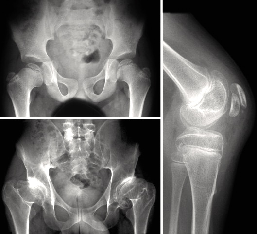
A rare group of primary bone dysplasia disorders characterized by the association of epiphyseal anomalies of long bones causing joint pain early in life, recurrent osteochondritis and early arthrosis. This group contains an heterogeneous group of diseases with variable expression. Common reported clinical signs include waddling gait and pain at onset, and moderate short stature. Some forms are mainly limited to the femoral epiphyses, while several other syndromes are characterized by the association of multiple epiphyseal dysplasia with other clinical manifestations such as myopia, deafness and facial dysmorphism. Diagnosis relies on identification of the radiological features.
. ^ a b c Sadick, M; Müller-Wille, R; Wildgruber, M; Wohlgemuth, WA (September 2018). "Vascular Anomalies (Part I): Classification and Diagnostics of Vascular Anomalies" . ... CS1 maint: multiple names: authors list ( link ) ^ a b c Wildgruber, M; Sadick, M; Müller-Wille, R; Wohlgemuth, WA (13 March 2019). "Vascular tumors in infants and adolescents" .
See also [ edit ] Uterine perforation References [ edit ] ^ a b c d e f g h i j k l m n o p q r s t Toppenberg, KS; Block WA, Jr (1 September 2002). "Uterine rupture: what family physicians need to know". ... S2CID 22593593 . ^ Chibber R, El-Saleh E, Fadhli RA, Jassar WA, Harmi JA (March 2010). "Uterine rupture and subsequent pregnancy outcome - how safe is it?
"Fumarate Hydratase Deficiency" . GeneReviews . Seattle WA: University of Washington . PMID 20301679 . ^ Online Mendelian Inheritance in Man (OMIM): Fumarase Deficiency - 606812 ^ Devlin, Thomas M. (2006). ... "Hereditary Leiomyomatosis and Renal Cell Cancer" . GeneReviews . Seattle WA: University of Washington . PMID 20301430 .
Treatment with itraconazole has been described. [3] References [ edit ] ^ van den Berk GE, Noorduyn LA, van Ketel RJ, van Leeuwen J, Bemelman WA, Prins JM (2006). "A fatal pseudo-tumour: disseminated basidiobolomycosis" .