• FindZebra
  • About
  • Contact
  • Help
  • Login
Deep Research Advanced
  • Psammoma Body Wikipedia
    CS1 maint: multiple names: authors list ( link ) ^ Hallman KB, Nahhas WA, Connelly PJ (September 1991). "Endosalpingiosis as a source of psammoma bodies in a Papanicolaou smear.
  • Singleton Merten Syndrome Wikipedia
    You can help by adding to it . ( August 2017 ) Sources [ edit ] Singleton, EB, Merten DF: An unusual syndrome of widened medullary cavities of the metacarpals and phalanges, aortic calcification and abnormal dentition, Pediatric Radiol 1:2, 1973. [1] Resources form the National Institutes of Health [2] WebMD information References [ edit ] ^ Ferreira CR, Crow YJ, Gahl WA, Gardner PJ, Goldbach-Mansky R, Hur S, de Jesús AA, Nehrebecky M, Park JW, Briggs TA (2018) DDX58 and classic Singleton-Merten syndrome.
    IFIH1, DDX58, PLAAT4, ROBO3, IFNA1, IFNA13, NFATC4, G3BP1
    • Singleton-Merten Syndrome GARD
      Singleton-Merten syndrome is a very rare disease that affect many organs. The main features are tooth abnormalities with gum infection; calcifications in the aorta artery and in certain valves of the heart (i.e., aortic and mitral valves); and progressive thinning and weakening of the bones (osteoporosis), especially in the upper and back portions of the skull. Other findings may include neurologic problems, generalized short stature, muscle weakness; poor muscle tone (hypotonia); progressive wasting of the muscles ( muscle atrophy ); heart arrhythmia, growth and developmental delay; skin problems such as psoriasis; malformation of the hips and/or feet and limbs or fingers, joint problems, tendon rupture, distinct facial features, and vision problems due to glaucoma. Severe systemic lupus erythematosus can also occur with Singleton-Merten syndrome. Singleton-Merten syndrome is caused by mutations in the IFIH1 gene, and in the DDX58 genes (which causes anatypical form of Singleton-Merten syndrome where there are no teeth problems).
    • Singleton-Merten Syndrome 2 OMIM
      A number sign (#) is used with this entry because of evidence that Singleton-Merten syndrome-2 (SGMRT2) is caused by heterozygous mutation in the DDX58 gene (609631) on chromosome 9p21. Description Singleton-Merten syndrome-2 is characterized by variable expression of glaucoma, aortic calcification, and skeletal abnormalities, without dental anomalies (summary by Jang et al., 2015). For a general phenotypic description and discussion of genetic heterogeneity of Singleton-Merten syndrome, see SGMRT1 (182250). Clinical Features Jang et al. (2015) studied a large 4-generation Korean family with aortic calcification, glaucoma, and skeletal abnormalities. The 56-year-old proband was diagnosed with bilateral glaucoma at 6 years of age and was blind by age 17.
    • Singleton-Merten Syndrome 1 OMIM
      A number sign (#) is used with this entry because of evidence that Singleton-Merten syndrome-1 (SGMRT1) is caused by heterozygous mutation in the IFIH1 gene (606951) on chromosome 2q24. Description Singleton-Merten syndrome (SGMRT) is an uncommon autosomal dominant disorder characterized by abnormalities of blood vessels, teeth, and bone. Calcifications of the aorta and aortic and mitral valves occur in childhood or puberty and can lead to early death. Dental findings include delayed primary tooth exfoliation and permanent tooth eruption, truncated tooth root formation, early-onset periodontal disease, and severe root and alveolar bone resorption associated with dysregulated mineralization, leading to tooth loss. Osseous features consist of osteoporosis, either generalized or limited to distal extremities, distal limb osteolysis, widened medullary cavities, and easy tearing of tendons from bone.
    • Singleton-Merten Dysplasia Orphanet
      Singleton-Merten dysplasia is characterized by dental dysplasia, progressive calcification of the thoracic aorta with stenosis, osteoporosis and expansion of the marrow cavities in hand bones. Additional features included generalized muscle weakness and atrophy, and chronic psoriasiform skin eruptions. It has been reported in four unrelated patients (male and female) and in a family with multiple affected members (male).
  • Candida Hypersensitivity Wikipedia
    Because allergic symptoms can be influenced by many factors, including emotions, experiments must be designed to separate the effects of the procedure being tested from the effects of other factors. [4] [6] By 2005, scientists were taking note of "a large pseudoscientific cult" [7] that had developed around the topic of yeast infections , with claims that up to one in three people were affected by yeast-related illnesses including Candida hypersensitivity. [4] Legal action [ edit ] Some practitioners of alternative medicine have promoted dietary supplements as supposed cures for this non-existent illness, rendering themselves liable to prosecution. [4] [8] In 1990, alternative health vendor Nature's Way signed a FTC consent agreement not to misrepresent in advertising any self-diagnostic test concerning yeast conditions or to make any unsubstantiated representation concerning any food or supplement's ability to control yeast conditions, with a fine of US$30,000 payable to the National Institutes of Health for research in genuine candidiasis. [8] See also [ edit ] List of topics characterized as pseudoscience References [ edit ] ^ Crook, William G. (1986).
  • Aneurysm Of Sinus Of Valsalva Wikipedia
    Diagnostic and Interventional Radiology (Ankara, Turkey) . 23 (5): 339–346. doi : 10.5152/dir.2017.16522 . ISSN 1305-3612 . PMC 5602357 . PMID 28814376 . ^ Kenny, Damien; Hijazi, Ziyad M.
    FLNA
    • Aneurysm Of Sinus Of Valsalva Orphanet
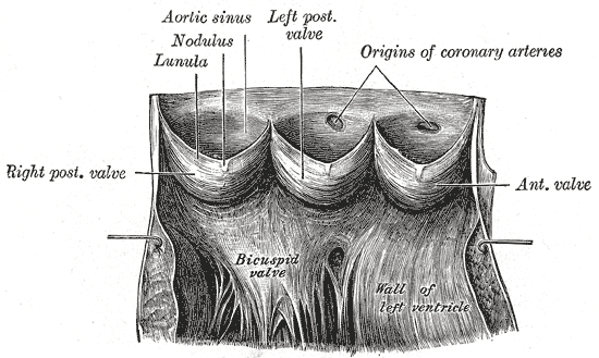
      Sinus of Valsalva aneurysm (SVA) is a rare congenital heart malformation of one or more of the aortic sinuses, consisting of a dilation that when unruptured is usually asymptomatic but when ruptured presents with progressive exertional dyspnea, fatigue, chest pain and that can lead to congestive heart failure if left untreated.
  • Maladjustment Wikipedia
    "Peer Victimization: Cause or Consequence of School Maladjustment?". Child Development . 67 (4): 1305–17. doi : 10.2307/1131701 . ISSN 0009-3920 .
    CCND1, BDNF, CDKN2A, EXT1, MECP2, PLN, TSPAN31, SELPLG, PDLIM7, ARHGAP32, NANS, PLB1, H3P10
  • Axial Osteomalacia Wikipedia
    Find sources: "Axial osteomalacia" – news · newspapers · books · scholar · JSTOR ( March 2010 ) Axial osteomalacia Axial osteomalacia is inherited in an autosomal dominant manner Specialty Orthopedic Axial osteomalacia is a rare osteosclerotic disorder characterized by axial skeleton pain , coarsening of the trabecular bone pattern on radiographs of the axial but not appendicular skeleton . [1] References [ edit ] ^ Whyte MP, Fallon MD, Murphy WA, Teitelbaum SL (December 1981). "Axial osteomalacia.
    • Axial Osteomalacia OMIM
      Axial osteomalacia is a rare osteosclerotic disorder first described by Frame et al. (1961). Characteristically, trabecular bone has 'a unique coarsening and spongelike appearance in the x-rays of the axial skeleton.' Radiographically, the skull and appendicular skeleton are normal. Vague chronic axial skeletal pain is the presenting symptom in most patients. Despite osteosclerosis and normal circulating levels of calcium, inorganic phosphate and alkaline phosphatase, bone biopsy specimens show osteomalacia. Until the report of Whyte et al. (1981), 10 cases had been described, all in middle-aged or elderly white men.
  • Familial Isolated Vitamin E Deficiency Wikipedia
    .; Ledbetter, Nikki; Mefford, Heather C. (eds.). GeneReviews . Seattle (WA): University of Washington, Seattle.
    TTPA, APOB, APOA1, FXN, SH3BP4, ZFP36, SETX, APTX, COQ8A, COPRS, RRS1, AFP, TNF, NOS3, MTHFR, IL6, GNB3, SRR
    • Ataxia With Vitamin E Deficiency MedlinePlus
      Ataxia with vitamin E deficiency is a disorder that impairs the body's ability to use vitamin E obtained from the diet. Vitamin E is an antioxidant, which means that it protects cells in the body from the damaging effects of unstable molecules called free radicals. A shortage (deficiency) of vitamin E can lead to neurological problems, such as difficulty coordinating movements (ataxia) and speech (dysarthria), loss of reflexes in the legs (lower limb areflexia), and a loss of sensation in the extremities (peripheral neuropathy). Some people with this condition have developed an eye disorder called retinitis pigmentosa that causes vision loss. Most people who have ataxia with vitamin E deficiency start to experience problems with movement between the ages of 5 and 15 years.
    • Vitamin E, Familial Isolated Deficiency Of OMIM
      A number sign (#) is used with this entry because of evidence that ataxia with vitamin E deficiency (AVED) is caused by homozygous or compound heterozygous mutation in the TTPA gene (600415) on chromosome 8q12. Clinical Features Harding et al. (1985) described a young woman with spinocerebellar degeneration thought to be due to a selective defect in vitamin E absorption. There was no evidence of fat malabsorption. Binder et al. (1967) suggested a relationship between neurologic dysfunction and vitamin E deficiency in patients with chronic steatorrhea. This was subsequently confirmed in patients with abetalipoproteinemia (200100), the most severe state of vitamin E deficiency known. When studied at age 23, the proband had no vitamin E in the serum. A progressive neurologic disorder comprising ataxia, areflexia and marked loss of proprioception developed at age 13.
    • Ataxia With Vitamin E Deficiency Orphanet
      A neurodegenerative disease belonging to the inherited cerebellar ataxias mainly characterized by progressive spino-cerebellar ataxia, loss of proprioception, areflexia, and is associated with a marked deficiency in vitamin E. Epidemiology Global prevalence is not known but population-based studies have been performed and prevalence can be extrapolated at approximately 1/300,000. AVED is the second most frequently inherited cerebellar ataxia in North Africa. As vitamin E deficiency might bring protection against malaria (see this term), it could explain a higher prevalence of AVED in Plasmodium infested areas. Clinical description AVED presents generally between ages 5 and 20 years with variable phenotype and severity.
    • Ataxia With Vitamin E Deficiency GeneReviews
      Summary Clinical characteristics. Ataxia with vitamin E deficiency (AVED) generally manifests in late childhood or early teens between ages five and 15 years. The first symptoms include progressive ataxia, clumsiness of the hands, loss of proprioception, and areflexia. Other features often observed are dysdiadochokinesia, dysarthria, positive Romberg sign, head titubation, decreased visual acuity, and positive Babinski sign. The phenotype and disease severity vary widely among families with different pathogenic variants; age of onset and disease course are more uniform within a given family, but symptoms and disease severity can vary even among sibs. Diagnosis/testing. Presently, no consensus diagnostic criteria for AVED exist; the principal criterion for diagnosis is a Friedreich ataxia-like neurologic phenotype combined with markedly reduced plasma vitamin E (α-tocopherol) concentration and a normal lipoprotein profile in the absence of known causes of malabsorption.
    • Ataxia With Vitamin E Deficiency GARD
      Ataxia with vitamin E deficiency (AVED) is a progressive disease affecting motor control and movement. Symptoms of AVED include slurred speech (dysarthria), difficulty coordinating movements ( ataxia ), numbness in the hands and feet (peripheral neuropathy), and progressive leg weakness. Some affected individuals may experience vision loss due to damage to the back of the eye ( retinitis pigmentosa ). Symptoms typically begin during childhood or adolescence and worsen with age, resulting in the need for a wheelchair by early adulthood. AVED is caused by a mutation to the TTPA gene. When this gene is damaged, vitamin E cannot be distributed throughout the body.
  • Renpenning's Syndrome Wikipedia
    Am J Med Genet A ^ RENPENNING H, GERRARD JW, ZALESKI WA, TABATA T (November 1962). "Familial sex-linked mental retardation" .
    PQBP1, STS, MAPK1, RNF19A, SIRT1, AHSA1, GRAP2, AIMP2, TNF, IL1B, RUNX2, HSP90AA1, GSTM2, G6PD, FMR1, DLG3, MAPK14, CRK, POLDIP2
    • Renpenning Syndrome GARD
      Renpenning syndrome is a genetic condition which occurs mostly in males. Signs and symptoms include the following: developmental delay, a small head (microcephaly), short stature, and distinctive facial features. Approximately two-thirds of individuals with Renpenning syndrome have moderate to severe intellectual disability. Additional features may include heart defects, muscular atrophy, cleft palate, and eye abnormalities. Renpenning syndrome is caused by mutations in the PQBP1 gene and is inherited in an X-linked recessive manner.
    • Renpenning Syndrome Orphanet
      Renpenning syndrome is an X-linked intellectual disability syndrome (XLMR, see this term) characterized by intellectual deficiency, microcephaly, leanness and mild short stature. Epidemiology Prevalence is unknown. Clinical description The main clinical manifestations of Renpenning syndrome are usually moderate intellectual deficiency, leanness, microcephaly and short stature (relative to familial target measurements) and sometimes small testes (testicular volumes below 15 ml), that are noticed at puberty. Manifestations are expressed only in males, and female carriers show normal facial features, growth development and intelligence. Small head and brain sizes are noted at birth. Characteristic craniofacial features include long triangular faces with upslanting palpebral fissures, half-depilated eyebrows, large ridged or bulbous nose with overhanging columella, short philtrum, and cupped and laterally protruding ears. Patients are thin and show failure to thrive. Delayed motor and language development is noticed in children from an early age.
    • Renpenning Syndrome MedlinePlus
      Renpenning syndrome is a disorder that almost exclusively affects males, causing developmental delay, moderate to severe intellectual disability, and distinctive physical features. Individuals with Renpenning syndrome typically have short stature and a small head size (microcephaly ). Facial features characteristic of this disorder include a long, narrow face ; outside corners of the eyes that point upward (upslanting palpebral fissures ); a long, bulbous nose with a low-hanging separation between the nostrils (overhanging columella ); a shortened space between the nose and mouth (philtrum); and cup-shaped ears . Males with Renpenning syndrome generally have small testes . Seizures and wasting away (atrophy) of muscles used for movement (skeletal muscles) may also occur in this disorder. About 20 percent of individuals with Renpenning syndrome also have other features, which may include a gap or split in structures that make up the eye (coloboma), an opening in the roof of the mouth (cleft palate ), heart abnormalities, or malformations of the anus .
    • Renpenning Syndrome 1 OMIM
      A number sign (#) is used with this entry because Renpenning syndrome is caused by mutation in the PQBP1 gene (300463) on chromosome Xp11. Description Renpenning syndrome is an X-linked mental retardation syndrome with clinically recognizable features. Affected individuals have microcephaly, short stature, small testes, and dysmorphic facies, including tall narrow face, upslanting palpebral fissures, abnormal nasal configuration, cupped ears, and short philtrum. The nose may appear long or bulbous, with overhanging columella. Less consistent manifestations include ocular colobomas, cardiac malformations, cleft palate, and anal anomalies. Stevenson et al. (2005) proposed that the various X-linked mental retardation syndromes due to PQBP1 mutations be combined under the name of Renpenning syndrome.
  • Limb Infarction Wikipedia
    Diagnostic and Interventional Radiology . 16 (1): 79–83. doi : 10.4261/1305-3825.DIR.2654-09.1 . PMID 20044798 . ^ a b Magishi K, Izumi Y, Shimizu N (August 2010).
  • Marfanoid Wikipedia
    ., editors. GeneReviews™ [Internet]. Seattle (WA): University of Washington, Seattle; 1993-2014.
  • Vascular Tumor Wikipedia
    . ^ a b c Sadick, M; Müller-Wille, R; Wildgruber, M; Wohlgemuth, WA (September 2018). "Vascular Anomalies (Part I): Classification and Diagnostics of Vascular Anomalies" . ... CS1 maint: multiple names: authors list ( link ) ^ a b c Wildgruber, M; Sadick, M; Müller-Wille, R; Wohlgemuth, WA (13 March 2019). "Vascular tumors in infants and adolescents" .
    NDP, PROX1, ALK, CDK4, FLI1, FLII, FOSB, IFNG, CXCL8, SMARCA4, SPARC, TFE3, ZFP36, DLL4, DUX4
  • Uterine Rupture Wikipedia
    See also [ edit ] Uterine perforation References [ edit ] ^ a b c d e f g h i j k l m n o p q r s t Toppenberg, KS; Block WA, Jr (1 September 2002). "Uterine rupture: what family physicians need to know". ... S2CID 22593593 . ^ Chibber R, El-Saleh E, Fadhli RA, Jassar WA, Harmi JA (March 2010). "Uterine rupture and subsequent pregnancy outcome - how safe is it?
    COL3A1, NDP, TGFBR1, TGFBR2
  • Fumarase Deficiency Wikipedia
    "Fumarate Hydratase Deficiency" . GeneReviews . Seattle WA: University of Washington . PMID 20301679 . ^ Online Mendelian Inheritance in Man (OMIM): Fumarase Deficiency - 606812 ^ Devlin, Thomas M. (2006). ... "Hereditary Leiomyomatosis and Renal Cell Cancer" . GeneReviews . Seattle WA: University of Washington . PMID 20301430 .
    FH, CD59
  • Steatosis Wikipedia
    Numerous validation studies have demonstrated excellent correlations between the steatosis level quantified at MRI and the steatosis levels semi-quantitavely and quantitatively determined on liver biopsies (reference methods). Several MRI vendors offer automated calculation of percent fat with acquisition sequences no longer than a single breath hold.
  • Basidiobolomycosis Wikipedia
    Treatment with itraconazole has been described. [3] References [ edit ] ^ van den Berk GE, Noorduyn LA, van Ketel RJ, van Leeuwen J, Bemelman WA, Prins JM (2006). "A fatal pseudo-tumour: disseminated basidiobolomycosis" .
  • Urethral Syndrome Wikipedia
    . ^ a b c d e Brumfitt W, Hamilton-Miller JM, Gillespie WA (July 1991). "The mysterious "urethral syndrome " " .
  • Enamel-Renal Syndrome Wikipedia
    History [ edit ] This condition was first described in 1972. [2] References [ edit ] ^ Jaureguiberry G, De la Dure-Molla M, Parry D, Quentric M, Himmerkus N, Koike T, Poulter J, Klootwijk E, Robinette SL, Howie AJ, Patel V, Figueres ML, Stanescu HC, Issler N, Nicholson JK, Bockenhauer D, Laing C, Walsh SB, McCredie DA, Povey S, Asselin A, Picard A, Coulomb A, Medlar AJ, Bailleul-Forestier I, Verloes A, Le Caignec C, Roussey G, Guiol J, Isidor B, Logan C, Shore R, Johnson C, Inglehearn C, Al-Bahlani S, Schmittbuhl M, Clauss F, Huckert M, Laugel V, Ginglinger E, Pajarola S, Spartà G, Bartholdi D, Rauch A, Addor MC, Yamaguti PM, Safatle HP, Acevedo AC, Martelli-Júnior H, dos Santos Netos PE, Coletta RD, Gruessel S, Sandmann C, Ruehmann D, Langman CB, Scheinman SJ, Ozdemir-Ozenen D, Hart TC, Hart PS, Neugebauer U, Schlatter E, Houillier P, Gahl WA, Vikkula M, Bloch-Zupan A, Bleich M, Kitagawa H, Unwin RJ, Mighell A, Berdal A, Kleta R (2013) Nephrocalcinosis (Enamel Renal Syndrome) caused by autosomal recessive FAM20A Mutations.
    FAM20A, LINC01482, PRKAR1A, SCN5A, KCNJ8, DSP, EGFR, KCND3, KCNE1, CACNA2D1, MYD88, NMT1, DPP6, PRKCE, SCN1B, DDIT3, TLR4, CACNB2, CACNA1C
    • Amelogenesis Imperfecta, Type Ig OMIM
      A number sign (#) is used with this entry because amelogenesis imperfecta type IG (AI1G), also known as enamel-renal syndrome (ERS), is caused by homozygous or compound heterozygous mutation in the FAM20A gene (611062) on chromosome 17q24. Description Amelogenesis imperfecta type IG, also known as enamel-renal syndrome, is characterized by hypoplastic enamel on primary and secondary dentition, pulp stones, delayed or failed eruption of secondary dentition, gingival overgrowth, and nephrocalcinosis. Blood chemistry analyses are typically normal, and nephrocalcinosis, which is found on renal ultrasound, may not appear until later in life (summary by Wang et al., 2013). Clinical Features MacGibbon (1972) reported a brother and sister with absent enamel, nephrocalcinosis, and apparently normal calcium metabolism. Lubinsky et al. (1985) also described an affected brother and sister, aged 11 and 9 years, respectively.
    • Enamel-Renal Syndrome Orphanet
      A extremely rare, genetic malformation syndrome characterized by hypoplastic amelogenesis imperfecta (hypoplastic dental enamel) and nephrocalcinosis (precipitation of calcium salts in renal tissue). Oral manifestations include yellow and misshaped teeth, delayed tooth eruption, and intrapulpal calcifications. Nephrocalcinosis is often asymptomatic but can progress during late childhood or early adulthood to impaired renal function, recurrent urinary infections, renal tubular acidosis, and rarely to end-stage renal failure.
  • Citrullinemia Type I Wikipedia
    (eds.). Citrullinemia Type I . Seattle (WA): University of Washington, Seattle.
    ASS1
    • Citrullinemia, Classic OMIM
      A number sign (#) is used with this entry because of evidence that classic citrullinemia is caused by homozygous or compound heterozygous mutation in the ASS1 gene (603470), which encodes argininosuccinate synthetase, on chromosome 9q34. Clinical Features Severe vomiting spells beginning at the age of 9 months and mental retardation were features of the first reported case, offspring of first-cousin parents; McMurray et al. (1962) found citrulline in very high concentration in serum, spinal fluid, and urine. (The amino acid citrulline gets its name from its high concentration in the watermelon Citrullus vulgaris.) Visakorpi (1962) also described a case of citrullinuria. Ammonia intoxication is another manifestation. The enzyme defect concerns argininosuccinic acid synthetase (EC 6.3.4.5).
    • Citrullinemia Type I Orphanet
      Citrullinemia type I is a rare autosomal recessive urea cycle defect characterized biologically by hyperammonemia and clinically by progressive lethargy, poor feeding and vomiting in the neonatal form (Acute neonatal citrullinemia type I, see this term) and by variable hyperammonemia in the later-onset form (Adult-onset citrullinemia type I, see this term).
  • Intestinal Neuronal Dysplasia Wikipedia
    European Journal of Pediatric Surgery . 18 (1): 59–60. doi : 10.1055/s-2008-1038324 . PMID 18302074 . ^ Fadda B, Maier WA, Meier-Ruge W, Schärli A, Daum R (October 1983).
    FLNA, TLX2, SPRY2
    • Intestinal Pseudoobstruction With Patent Ductus Arteriosus And Natal Teeth OMIM
      Harris et al. (1976) described 2 male sibs with mandibular teeth present at birth, patent ductus arteriosus (see 607411), and intestinal pseudoobstruction evident from birth. Vomiting of bile stain material began soon after birth, and there was no passage of meconium. The older brother died at 5 months of age despite 2 gastrointestinal operations and ligation of the patent ductus arteriosus which had led to cardiac failure. The younger brother died at the age of 6 weeks. Either autosomal or X-linked recessive inheritance is possible.
    • Neuronal Intestinal Pseudoobstruction Orphanet
      Neuronal intestinal pseudoobstruction is a form of chronic intestinal pseudoobstruction caused by a developmental failure of the enteric neurons to differentiate or migrate properly and manifests as a bowel obstruction.
  • Craniodiaphyseal Dysplasia Wikipedia
    .), "Craniometaphyseal Dysplasia, Autosomal Dominant" , GeneReviews® , Seattle (WA): University of Washington, Seattle, PMID 20301634 , retrieved 2021-01-18 External links [ edit ] Craniodiaphyseal dysplasia at orpha.net Classification D ICD - 10 : M85.2 OMIM : 218300 MeSH : C562940 External resources Orphanet : 1513 v t e Congenital malformations and deformations of musculoskeletal system / musculoskeletal abnormality Appendicular limb / dysmelia Arms clavicle / shoulder Cleidocranial dysostosis Sprengel's deformity Wallis–Zieff–Goldblatt syndrome hand deformity Madelung's deformity Clinodactyly Oligodactyly Polydactyly Leg hip Hip dislocation / Hip dysplasia Upington disease Coxa valga Coxa vara knee Genu valgum Genu varum Genu recurvatum Discoid meniscus Congenital patellar dislocation Congenital knee dislocation foot deformity varus Club foot Pigeon toe valgus Flat feet Pes cavus Rocker bottom foot Hammer toe Either / both fingers and toes Polydactyly / Syndactyly Webbed toes Arachnodactyly Cenani–Lenz syndactylism Ectrodactyly Brachydactyly Stub thumb reduction deficits / limb Acheiropodia Ectromelia Phocomelia Amelia Hemimelia multiple joints Arthrogryposis Larsen syndrome RAPADILINO syndrome Axial Skull and face Craniosynostosis Scaphocephaly Oxycephaly Trigonocephaly Craniofacial dysostosis Crouzon syndrome Hypertelorism Hallermann–Streiff syndrome Treacher Collins syndrome other Macrocephaly Platybasia Craniodiaphyseal dysplasia Dolichocephaly Greig cephalopolysyndactyly syndrome Plagiocephaly Saddle nose Vertebral column Spinal curvature Scoliosis Klippel–Feil syndrome Spondylolisthesis Spina bifida occulta Sacralization Thoracic skeleton ribs : Cervical Bifid sternum : Pectus excavatum Pectus carinatum
    SOST, CDKL5, CDA, GRIA2, CXCL8, NPPA, CLIP1
    • Craniodiaphyseal Dysplasia, Autosomal Dominant OMIM
      A number sign (#) is used with this entry because of evidence that autosomal dominant craniodiaphyseal dysplasia (CDD) is caused by heterozygous mutation in the SOST gene (605740) on chromosome 17q21. Sclerosteosis (SOST1; 269500) and van Buchem disease (VBCH; 239100), are allelic disorders that are less severe and show autosomal recessive inheritance. Description Craniodiaphyseal dysplasia is a severe bone dysplasia characterized by massive generalized hyperostosis and sclerosis, especially involving the skull and facial bones. Progressive bony encroachment upon cranial foramina leads to severe neurologic impairment in childhood (summary by Brueton and Winter, 1990). The sclerosis is so severe that the resulting facial distortion is referred to as 'leontiasis ossea' (leonine facies), and the bone deposition results in progressive stenosis of craniofacial foramina (summary by Kim et al., 2011).
    • Craniodiaphyseal Dysplasia Orphanet
      Craniodiaphyseal dysplasia is a rare sclerotic bone disorder with a variable phenotypic expression with massive generalized hyperostosis and sclerosis, particularly of the skull and facial bones, that may lead to severe deformity.
    • Craniodiaphyseal Dysplasia OMIM
      Clinical Features Cranial and facial hyperostosis results in a characteristic clinical and radiographic appearance. The diaphyses of the bones are generally expanded. Halliday (1949) and Stransky et al. (1962) reported isolated cases with similar findings. Facial and cranial thickening and distortion are particularly striking in this form. Most cases have been mentally retarded. Unlike the situation in the craniometaphyseal dysplasias (e.g., 218400), the long bones do not show metaphyseal flaring but show diaphyseal endostosis and are shaped like a policeman's nightstick. Joseph et al. (1958), who first suggested the designation of progressive craniodiaphyseal dysplasia, described a patient with a picture they considered identical to that described by Halliday (1949).
  • ←
  • 1
  • 2
  • 3
  • 4
  • 5
  • 6
  • ...
  • 14
  • 15
  • →

FindZebra

contact@findzebra.com