• FindZebra
  • About
  • Contact
  • Help
  • Login
Advanced
  • Chromosome 4q32.1-Q32.2 Triplication Syndrome Omim
    A number sign (#) is used with this entry because it represents a contiguous gene triplication syndrome (chr4:157.35-161.61). Clinical Features Wang et al. (2009) reported a mother and her 3 sons who all had a 4.3-Mb triplication of chromosome 4q32.1-q32.2 associated with delayed psychomotor development and variable mental retardation. ... INHERITANCE - Autosomal dominant HEAD & NECK Head - Macrocephaly Face - Frontal bossing - Long midface - Microretrognathia - Short philtrum - Hypoplastic zygoma Ears - Low-set ears - Small ears - Malformed ears - Squared-off ears Eyes - Downslanting palpebral fissures - Small palpebral fissures - Epicanthal folds - Ptosis Nose - Wide nasal bridge - Short nose ABDOMEN Gastrointestinal - Hirschsprung disease - Constipation NEUROLOGIC Central Nervous System - Hydrocephalus - Delayed psychomotor development - Speech abnormalities Behavioral Psychiatric Manifestations - Autistic features MISCELLANEOUS - One family has been reported (as of October 2010) MOLECULAR BASIS - Contiguous gene syndrome caused by 4.3-Mb triplication of chromosome 4q32.1-q32.2 ▲ Close
  • Hepatoerythropoietic Porphyria Wikipedia
    . ^ " hepatoerythropoietic porphyria " at Dorland's Medical Dictionary External links [ edit ] Classification D ICD - 10 : E80.2 ( ILDS E80.282) ICD - 9-CM : 277.1 OMIM : 176100 MeSH : D017121 DiseasesDB : 29123 SNOMED CT : 111386004 External resources Orphanet : 95159 Hepatoerythropoietic porphyria at NLM Genetics Home Reference Hepatoerythropoietic porphyria at NIH 's Office of Rare Diseases v t e Heme metabolism disorders Porphyria , hepatic and erythropoietic ( porphyrin ) early mitochondrial: ALAD porphyria Acute intermittent porphyria cytoplasmic: Gunther disease/congenital erythropoietic porphyria Porphyria cutanea tarda / Hepatoerythropoietic porphyria late mitochondrial: Hereditary coproporphyria Harderoporphyria Variegate porphyria Erythropoietic protoporphyria Hereditary hyperbilirubinemia ( bilirubin ) unconjugated: Gilbert's syndrome Crigler–Najjar syndrome Lucey–Driscoll syndrome conjugated: Dubin–Johnson syndrome nd sheet Rotor syndrome This article about an endocrine, nutritional, or metabolic disease is a stub .
    UROD, HPSE, C20orf181
    • Hepatoerythropoietic Porphyria Gene_reviews
      Summary Clinical characteristics. Hepatoerythropoietic porphyria (HEP) is characterized by blistering skin lesions, hypertrichosis, and scarring over the affected skin areas. Disease manifestations occur during infancy or childhood and with similar frequency in females and males. Individuals with HEP are not reported to be at increased risk for hepatocellular carcinoma. Diagnosis/testing. The diagnosis of HEP is established in a proband by identification of elevated porphyrins in the urine (predominantly uroporphyrin and heptacarboxylporphyrin) and significantly increased erythrocyte zinc protoporphyrin. Identification of biallelic pathogenic variants in UROD confirms the diagnosis.
    • Hepatoerythropoietic Porphyria Orphanet
      Hepatoerythropioetic porphyria (HEP) is a very rare form of chronic hepatic porphyria (see this term) characterized by bullous photodermatitis. Epidemiology Fewer than 40 cases of HEP have been described. Clinical description The disease starts in childhood. The principle clinical signs include fragile skin, bullous cutaneous lesions that are sometimes erosive, and even mutilating on the surface of the skin exposed to the sun (hands, face). Hepatoerythropioetic porphria corresponds to homozygous and composite heterozygous cases of porphyria cutanea tarda (see this term). Etiology It is caused by a deficiency of uroporphyrinogen decarboxylase (URO-D; the fifth enzyme in the heme biosynthesis pathway) that leads to an accumulation of uroporphrin in the liver.
    • Hepatoerythropoietic Porphyria Gard
      Hepatoerythropoietic porphyria (HEP) affects the skin and is due to a build-up of damaging chemicals in the body. Symptoms usually begin in infancy and include extreme sun sensitivity, extra body hair, discolored teeth, and anemia. Over time, people with HEP may lose skin, bone or develop scarring in sun-exposed areas. HEP is caused by a UROD gene that is not working correctly and is inherited in an autosomal recessive pattern. HEP is diagnosed based on the symptoms, clinical exam, laboratory testing, and confirmed by genetic testing.
  • Severe Early-Childhood-Onset Retinal Dystrophy Orphanet
    Differential diagnosis Differential diagnosis includes LCA, Alström syndrome, autosomal recessive bestrophinopathy, Bardet-Biedl syndrome, achromatopsia, Stargardt disease, Usher syndrome, Senior-Loken syndrome, Saldino-Mainzer syndrome, Joubert syndrome, abetalipoproteinemia, infantile Refsum disease, neonatal adrenoleukodystrophy, Zellweger syndrome and juvenile neuronal ceroid lipofuscinosis.
    ABCA4, RPE65, LRAT, SPATA7, LCA5, ELOVL4, GUCY2D, RDH12, CLTA, CRX, PTPRC, IFT140, CRB1, ADAMTS18
  • Alpha Thalassemia Medlineplus
    The more severe type is known as hemoglobin Bart hydrops fetalis syndrome, which is also called Hb Bart syndrome or alpha thalassemia major. The milder form is called HbH disease. Hb Bart syndrome is characterized by hydrops fetalis, a condition in which excess fluid builds up in the body before birth. ... Thousands of infants with Hb Bart syndrome and HbH disease are born each year, particularly in Southeast Asia. ... The different types of alpha thalassemia result from the loss of some or all of these alleles. Hb Bart syndrome, the most severe form of alpha thalassemia, results from the loss of all four alpha-globin alleles. ... If both parents are missing at least one alpha-globin allele, their children are at risk of having Hb Bart syndrome, HbH disease, or alpha thalassemia trait.
    HBA1, HBA2, HBB, ATRX, SEA, HBG2, G6PD, HBZ, PMCH, UGT1A1, ATR, SCN8A, COL9A2, COL9A3, COMP, HBG1, COL9A1, MYB, HP, HBS1L, RN7SL263P, PSMA5, EPO, DAXX, AP5Z1, OR10A4, KLF1, TP53, DOCK11, BCL11A, UGT1A10, UGT1A6, UGT1A7, UGT1A8, F3, AHSP, FASTK, UGT1A4, SLCO1B1, CD19, LUC7L, RUNX1, HAMP, TSPYL2, NSD1, TMPRSS13, ATAD1, LMLN, FTMT, SCLT1, CDAN1, EPGN, LINC01194, MIR144, ZMPSTE24, NPRL3, RGS6, RNGTT, HBD, DAP, CR1, HFE, HMOX1, CEBPD, LMNA, MSMB, MST1, MTHFR, CD37, PDR, PGD, PKD1, CD36, RAG2, RASA1, CD34, ADCY9, SMARCA2, SMARCA4, TEK, TERC, TFRC, TGFBR3, VCAM1, HBFQTL2, ACTB
    • Hemoglobin H Disease Orphanet
      Hemoglobin H (HbH) disease is a moderate to severe form of alpha-thalassemia (see this term) characterized by pronounced microcytic hypochromic hemolytic anemia. Epidemiology HbH disease is predominantly seen in Southeast Asia, the Middle East and the Mediterranean. Exact prevalence is not known but has been reported to be about 1/1,000,000 for severe forms of alpha-thalassemia in Northern Europe and North America. Clinical description Clinical features are highly variable and generally develop in the first years of life but may not develop until adulthood in some patients. Initial signs may be noticed only during routine hematologic analyses.
    • Alpha-Thalassemia Orphanet
      A rare form called alpha-thalassemia-intellectual deficit syndrome linked to chromosome 16 (16p13.3) has also been identified (see these terms). ... Alpha-thalassemia-intellectual deficit syndrome is characterized by very mild to severe anemia associated with developmental abnormalities. ... An acquired form known as alpha-thalassemia-myelodysplastic syndrome (ATMDS; see this term) has been described mainly in adult males and should also be considered.
    • Alpha-Thalassemia Omim
      The molecular and clinical aspects of the severe alpha-thalassemia syndromes were reviewed by Higgs (1993) and Chui and Waye (1998).
    • Alpha-Thalassemia Gard
      Two types of alpha-thalassemia can cause health problems: the more severe type is known as Hb Bart syndrome; the milder form is called HbH disease. Hb Bart syndrome may be characterized by hydrops fetalis ; severe anemia; hepatosplenomegaly; heart defects; and abnormalities of the urinary system or genitalia. ... No treatment is effective for Hb Bart syndrome. For HbH disease, occasional red blood cell transfusions may be needed.
    • Alpha-Thalassemia Gene_reviews
      Alpha-thalassemia is usually inherited in an autosomal recessive manner. Hb Bart syndrome. At conception, each sib of a proband with Hb Bart syndrome has a 25% chance of having Hb Bart syndrome (e.g., --/--), a 50% chance of having α-thalassemia trait with deletion or inactivation of two α-globin genes in cis (e.g., --/αα), and a 25% chance of being unaffected and not a carrier. ... The severity of the α-thalassemia syndromes depends on the extent of α-globin chain defect (see Genotype-Phenotype Correlations). Hb Bart syndrome is the most severe clinical condition related to α-thalassemia. ... Hemoglobin Bart hydrops fetalis (Hb Bart) syndrome. See Prenatal Testing and Preimplantation Genetic Testing. ... Prevention of Primary Manifestations Hb Bart syndrome. Because of the severity of Hb Bart syndrome and the risk for maternal complications during pregnancy with a fetus with this disorder, prenatal diagnosis and early termination of affected pregnancies is usually considered.
  • Pipecolic Acidemia Wikipedia
    Pipecolic acidemia can also be an associated component of Refsum disease with increased pipecolic acidemia (RDPA), [2] as well as other peroxisomal disorders, including both infantile and adult Refsum disease , [3] [4] [5] and Zellweger syndrome . [6] The disorder is characterized by an increase in pipecolic acid levels in the blood, leading to neuropathy and hepatomegaly . ... "Genetic heterogeneity in the cerebrohepatorenal (Zellweger) syndrome and other inherited disorders with a generalized impairment of peroxisomal functions. ... External links [ edit ] Classification D ICD - 9-CM : 270.7 OMIM : 239400 600964 v t e Inborn error of amino acid metabolism K → acetyl-CoA Lysine /straight chain Glutaric acidemia type 1 type 2 Hyperlysinemia Pipecolic acidemia Saccharopinuria Leucine 3-hydroxy-3-methylglutaryl-CoA lyase deficiency 3-Methylcrotonyl-CoA carboxylase deficiency 3-Methylglutaconic aciduria 1 Isovaleric acidemia Maple syrup urine disease Tryptophan Hypertryptophanemia G G→ pyruvate → citrate Glycine D-Glyceric acidemia Glutathione synthetase deficiency Sarcosinemia Glycine → Creatine : GAMT deficiency Glycine encephalopathy G→ glutamate → α-ketoglutarate Histidine Carnosinemia Histidinemia Urocanic aciduria Proline Hyperprolinemia Prolidase deficiency Glutamate / glutamine SSADHD G→ propionyl-CoA → succinyl-CoA Valine Hypervalinemia Isobutyryl-CoA dehydrogenase deficiency Maple syrup urine disease Isoleucine 2-Methylbutyryl-CoA dehydrogenase deficiency Beta-ketothiolase deficiency Maple syrup urine disease Methionine Cystathioninuria Homocystinuria Hypermethioninemia General BC / OA Methylmalonic acidemia Methylmalonyl-CoA mutase deficiency Propionic acidemia G→ fumarate Phenylalanine / tyrosine Phenylketonuria 6-Pyruvoyltetrahydropterin synthase deficiency Tetrahydrobiopterin deficiency Tyrosinemia Alkaptonuria / Ochronosis Tyrosinemia type I Tyrosinemia type II Tyrosinemia type III / Hawkinsinuria Tyrosine → Melanin Albinism : Ocular albinism ( 1 ) Oculocutaneous albinism ( Hermansky–Pudlak syndrome ) Waardenburg syndrome Tyrosine → Norepinephrine Dopamine beta hydroxylase deficiency reverse: Brunner syndrome G→ oxaloacetate Urea cycle / Hyperammonemia ( arginine aspartate ) Argininemia Argininosuccinic aciduria Carbamoyl phosphate synthetase I deficiency Citrullinemia N-Acetylglutamate synthase deficiency Ornithine transcarbamylase deficiency / translocase deficiency Transport / IE of RTT Solute carrier family : Cystinuria Hartnup disease Iminoglycinuria Lysinuric protein intolerance Fanconi syndrome : Oculocerebrorenal syndrome Cystinosis Other 2-Hydroxyglutaric aciduria Aminoacylase 1 deficiency Ethylmalonic encephalopathy Fumarase deficiency Trimethylaminuria This genetic disorder article is a stub .
    HSD17B4, PIPOX, CAT
  • Poretti-Boltshauser Syndrome Omim
    A number sign (#) is used with this entry because Poretti-Boltshauser syndrome (PTBHS) is caused by homozygous or compound heterozygous mutation in the LAMA1 gene (150320) on chromosome 18p11. Description Poretti-Boltshauser syndrome is an autosomal recessive disorder characterized by cerebellar dysplasia, cerebellar vermis hypoplasia, cerebellar cysts in most patients, high myopia, variable retinal dystrophy, and eye movement abnormalities. ... The sibs in their twenties had normal IQ; 1 had Asperger syndrome. All 7 patients had eye movement abnormalities, including strabismus, oculomotor apraxia, amblyopia, and/or nystagmus, as well as myopia. ... Molecular Genetics In 7 patients from 5 unrelated families with Poretti-Boltshauser syndrome, Aldinger et al. (2014) identified homozygous or compound heterozygous mutations in the LAMA1 gene (see, e.g., 150320.0001-150320.0005).
    LAMA1
    • Ataxia-Intellectual Disability-Oculomotor Apraxia-Cerebellar Cysts Syndrome Orphanet
      A rare neuro-ophthalmological disease characterized by nonprogressive cerebellar ataxia, delayed motor and language development and intellectual disability, in addition to ophthalmological abnormalities (e.g. oculomotor apraxia, strabismus, amblyopia, retinal dystrophy and myopia). Cerebellar cysts, cerebellar dysplasia and cerebellar vermis hypoplasia, seen on magnetic resonance imaging, are also characteristic of the disease.
  • Mitochondrial Complex I Deficiency, Nuclear Type 3 Omim
    Clinical Features Smeitink and van den Heuvel (1999) described 2 brothers with complex I deficiency and features of Leigh syndrome (see 256000) confirmed postmortem. ... Lebon et al. (2007) reported 2 brothers, born of consanguineous Turkish parents, with complex I deficiency and Leigh syndrome resulting in death in infancy. ... Molecular Genetics In 2 brothers with complex I deficiency and features of Leigh syndrome confirmed postmortem, Smeitink and van den Heuvel (1999) identified a missense mutation in the NDUFS7 gene (V122M; 601825.0001). In a girl, born to consanguineous Tunisian parents, with severe complex I defect and Leigh syndrome, Lebon et al. (2007) identified homozygosity for a missense mutation in the NDUFS7 gene (R145H; 601825.0002). ... In 2 brothers, born of consanguineous Turkish parents, with complex I deficiency and Leigh syndrome, Lebon et al. (2007) identified a homozygous splice site mutation in the NDUFS7 gene (601825.0003).
    NDUFS7
  • Mitochondrial Complex I Deficiency, Nuclear Type 17 Omim
    Clinical Features Pagliarini et al. (2008) reported 2 Lebanese sibs, born of consanguineous parents, who presented in infancy with focal seizures, decreased movement and strength, ataxia, lactic acidosis, and neuroimaging results consistent with Leigh syndrome. Biochemical studies showed complex I deficiency in liver, muscle, and fibroblasts. ... Kohda et al. (2016) reported 4 unrelated children with mitochondrial complex I deficiency and Leigh syndrome. Clinical details were limited. Molecular Genetics In 2 Lebanese sibs, born of consanguineous parents, with mitochondrial complex I deficiency nuclear type 17 and Leigh syndrome, Pagliarini et al. (2008) identified a homozygous missense mutation in the NDUFAF6 gene (Q99R; 612392.0001) substitution in a highly conserved residue. ... In a 7-year-old boy, born of unrelated parents, with complex I deficiency and Leigh syndrome, Bianciardi et al. (2016) identified a heterozygous mutation in the NDUFAF6 gene (A178P; 612392.0002). ... Bianciardi et al. (2016) suggested that the second mutational event in the NDUFAF6 gene may be postmeiotic, affecting a nonexonic regulatory element and explaining the different tissue-specific expression, or that it may affect a specific protein In 4 unrelated children with mitochondrial complex I deficiency and Leigh syndrome, Kohda et al. (2016) identified biallelic missense mutations in the NDUFAF6 gene (612392.0003-612392.0007).
    NDUFAF6
  • Mitochondrial Myopathy Wikipedia
    Contents 1 Signs and symptoms 2 Cause 3 Diagnosis 4 Treatment 5 References 6 External links Signs and symptoms [ edit ] Signs and symptoms include (for each of the following causes): Mitochondrial encephalomyopathy, lactic acidosis, and stroke-like syndrome (MELAS) Varying degrees of cognitive impairment and dementia Lactic acidosis Strokes Transient ischemic attacks Hearing loss Weight loss Myoclonic epilepsy and ragged-red fibers (MERRF) Progressive myoclonic epilepsy Clumps of diseased mitochondria accumulate in muscle fibers and appear as "ragged-red fibers" when muscle is stained with modified Gömöri trichrome stain Short stature Kearns–Sayre syndrome (KSS) External ophthalmoplegia Cardiac conduction defects Sensorineural hearing loss Chronic progressive external ophthalmoplegia (CPEO) Progressive ophthalmoparesis Symptomatic overlap with other mitochondrial myopathies Cause [ edit ] Zidovudine, an HIV medication can cause red ragged fiber myopathy. ... External links [ edit ] Classification D ICD - 10 : G71.3 MeSH : D017240 External resources Orphanet : 206966 v t e Diseases of muscle , neuromuscular junction , and neuromuscular disease Neuromuscular- junction disease autoimmune Myasthenia gravis Lambert–Eaton myasthenic syndrome Neuromyotonia Myopathy Muscular dystrophy ( DAPC ) AD Limb-girdle muscular dystrophy 1 Oculopharyngeal Facioscapulohumeral Myotonic Distal (most) AR Calpainopathy Limb-girdle muscular dystrophy 2 Congenital Fukuyama Ullrich Walker–Warburg XR dystrophin Becker's Duchenne Emery–Dreifuss Other structural collagen disease Bethlem myopathy PTP disease X-linked MTM adaptor protein disease BIN1-linked centronuclear myopathy cytoskeleton disease Nemaline myopathy Zaspopathy Channelopathy Myotonia Myotonia congenita Thomsen disease Neuromyotonia / Isaacs syndrome Paramyotonia congenita Periodic paralysis Hypokalemic Thyrotoxic Hyperkalemic Other Central core disease Mitochondrial myopathy MELAS MERRF KSS PEO General Inflammatory myopathy Congenital myopathy v t e Mitochondrial diseases Carbohydrate metabolism PCD PDHA Primarily nervous system Leigh disease LHON NARP Myopathies KSS Mitochondrial encephalomyopathy MELAS MERRF PEO No primary system DAD MNGIE Pearson syndrome Chromosomal OPA1 Kjer's optic neuropathy SARS2 HUPRA syndrome TIMM8A Mohr–Tranebjærg syndrome see also mitochondrial proteins
    FDX2, SLC25A4, SLC25A42, TRNW, RRM2B, TNF, VDAC1, IL1B, IL1A, IL6, CAMKMT, ADCK2, COX10, TK2, POLG, TRNK, CYTB, DNA2, TRNM, ND4, TWNK, TRNL1, COX1, ATP6, TYMP, NDUFV1, PUS1, NDUFA1, TRNF, YARS2, ISCU, COX2, NDUFS4, NDUFS1, NDUFS3, NDUFS2, NDUFS6, NDUFS8, NDUFV2, NDUFAF5, TMEM126B, NDUFB9, AGK, FOXRED1, MSTO1, NDUFB11, POLG2, TIMMDC1, NDUFAF3, NDUFAF4, NDUFAF1, NDUFB10, TRNS2, NDUFB3, ND3, NDUFS7, CAV3, NDUFA11, DGUOK, NDUFAF2, NUBPL, NDUFA6, TRNC, COX3, ND1, ND2, PNPLA8, TRNS1, TRNA, TRNL2, TRNH, TRNV, TRNQ, ND6, ND5, CHCHD10, GDF15, TFAM, FGF21, TBPL1, OPA1, TYRP1, TMEM65, CHCHD2, CHPT1, MIEF2, TMEM70, SLC25A21, PERM1, DHDDS, SARS2, NAT2, TIMM22, MT1B, SLC25A5, BCL2, COX8A, CPOX, CS, FXN, MTOR, GAA, CFH, LIG4, MET, MPV17, MTTP, NFU1, MYH7, OGG1, PPID, SOD1, TAZ, NR2C2, UCN, COX5A, PPIF, IFI30, PPARGC1A, PRPF6, MTCO2P12
  • Pruritus Ani Wikipedia
    Classification D ICD - 10 : L29.0 ICD - 9-CM : 698.0 MeSH : D011538 DiseasesDB : 30154 v t e Diseases of the digestive system Upper GI tract Esophagus Esophagitis Candidal Eosinophilic Herpetiform Rupture Boerhaave syndrome Mallory–Weiss syndrome UES Zenker's diverticulum LES Barrett's esophagus Esophageal motility disorder Nutcracker esophagus Achalasia Diffuse esophageal spasm Gastroesophageal reflux disease (GERD) Laryngopharyngeal reflux (LPR) Esophageal stricture Megaesophagus Esophageal intramural pseudodiverticulosis Stomach Gastritis Atrophic Ménétrier's disease Gastroenteritis Peptic (gastric) ulcer Cushing ulcer Dieulafoy's lesion Dyspepsia Pyloric stenosis Achlorhydria Gastroparesis Gastroptosis Portal hypertensive gastropathy Gastric antral vascular ectasia Gastric dumping syndrome Gastric volvulus Buried bumper syndrome Gastrinoma Zollinger–Ellison syndrome Lower GI tract Enteropathy Small intestine ( Duodenum / Jejunum / Ileum ) Enteritis Duodenitis Jejunitis Ileitis Peptic (duodenal) ulcer Curling's ulcer Malabsorption : Coeliac Tropical sprue Blind loop syndrome Small bowel bacterial overgrowth syndrome Whipple's Short bowel syndrome Steatorrhea Milroy disease Bile acid malabsorption Large intestine ( Appendix / Colon ) Appendicitis Colitis Pseudomembranous Ulcerative Ischemic Microscopic Collagenous Lymphocytic Functional colonic disease IBS Intestinal pseudoobstruction / Ogilvie syndrome Megacolon / Toxic megacolon Diverticulitis / Diverticulosis / SCAD Large and/or small Enterocolitis Necrotizing Gastroenterocolitis IBD Crohn's disease Vascular : Abdominal angina Mesenteric ischemia Angiodysplasia Bowel obstruction : Ileus Intussusception Volvulus Fecal impaction Constipation Diarrhea Infectious Intestinal adhesions Rectum Proctitis Radiation proctitis Proctalgia fugax Rectal prolapse Anismus Anal canal Anal fissure / Anal fistula Anal abscess Hemorrhoid Anal dysplasia Pruritus ani GI bleeding Blood in stool Upper Hematemesis Melena Lower Hematochezia Accessory Liver Hepatitis Viral hepatitis Autoimmune hepatitis Alcoholic hepatitis Cirrhosis PBC Fatty liver NASH Vascular Budd–Chiari syndrome Hepatic veno-occlusive disease Portal hypertension Nutmeg liver Alcoholic liver disease Liver failure Hepatic encephalopathy Acute liver failure Liver abscess Pyogenic Amoebic Hepatorenal syndrome Peliosis hepatis Metabolic disorders Wilson's disease Hemochromatosis Gallbladder Cholecystitis Gallstone / Cholelithiasis Cholesterolosis Adenomyomatosis Postcholecystectomy syndrome Porcelain gallbladder Bile duct / Other biliary tree Cholangitis Primary sclerosing cholangitis Secondary sclerosing cholangitis Ascending Cholestasis / Mirizzi's syndrome Biliary fistula Haemobilia Common bile duct Choledocholithiasis Biliary dyskinesia Sphincter of Oddi dysfunction Pancreatic Pancreatitis Acute Chronic Hereditary Pancreatic abscess Pancreatic pseudocyst Exocrine pancreatic insufficiency Pancreatic fistula Other Hernia Diaphragmatic Congenital Hiatus Inguinal Indirect Direct Umbilical Femoral Obturator Spigelian Lumbar Petit's Grynfeltt-Lesshaft Undefined location Incisional Internal hernia Richter's Peritoneal Peritonitis Spontaneous bacterial peritonitis Hemoperitoneum Pneumoperitoneum v t e Dermatitis and eczema Atopic dermatitis Besnier's prurigo Seborrheic dermatitis Pityriasis simplex capillitii Cradle cap Contact dermatitis ( allergic , irritant ) plants: Urushiol-induced contact dermatitis African blackwood dermatitis Tulip fingers other: Abietic acid dermatitis Diaper rash Airbag dermatitis Baboon syndrome Contact stomatitis Protein contact dermatitis Eczema Autoimmune estrogen dermatitis Autoimmune progesterone dermatitis Breast eczema Ear eczema Eyelid dermatitis Topical steroid addiction Hand eczema Chronic vesiculobullous hand eczema Hyperkeratotic hand dermatitis Autosensitization dermatitis / Id reaction Candidid Dermatophytid Molluscum dermatitis Circumostomy eczema Dyshidrosis Juvenile plantar dermatosis Nummular eczema Nutritional deficiency eczema Sulzberger–Garbe syndrome Xerotic eczema Pruritus / Itch / Prurigo Lichen simplex chronicus / Prurigo nodularis by location: Pruritus ani Pruritus scroti Pruritus vulvae Scalp pruritus Drug-induced pruritus Hydroxyethyl starch-induced pruritus Senile pruritus Aquagenic pruritus Aquadynia Adult blaschkitis due to liver disease Biliary pruritus Cholestatic pruritus Prion pruritus Prurigo pigmentosa Prurigo simplex Puncta pruritica Uremic pruritus Other substances taken internally: Bromoderma Fixed drug reaction Nummular dermatitis Pityriasis alba Papuloerythroderma of Ofuji
    CRP
    • Anal Itching Mayo_clinic
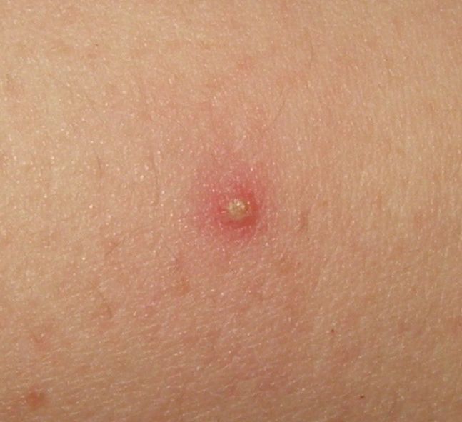
      Overview Anal itching is a common condition. The itch in or around the anus is often intense and can be embarrassing and uncomfortable. Anal itching, also called pruritus ani (proo-RIE-tus A-nie), has several possible causes. They include infections, hemorrhoids and ongoing diarrhea. Skin inflammation, also called dermatitis, is another cause. If the symptoms don't clear up with self-care, talk with your health care provider. With treatment, most people get complete relief. Symptoms Symptoms of anal itching may include intense itching, inflammation, burning and soreness.
  • Folliculitis Wikipedia
    Folliculitis Treatments Malassezia (Pityrosporum) Folliculitis Treatment & Management Classification D ICD - 10 : L73.9 ( ILDS L73.91) ICD - 9-CM : 704.8 MeSH : D005499 DiseasesDB : 31367 External resources MedlinePlus : 000823 eMedicine : derm/159 Patient UK : Folliculitis v t e Diseases of the skin and appendages by morphology Growths Epidermal Wart Callus Seborrheic keratosis Acrochordon Molluscum contagiosum Actinic keratosis Squamous-cell carcinoma Basal-cell carcinoma Merkel-cell carcinoma Nevus sebaceous Trichoepithelioma Pigmented Freckles Lentigo Melasma Nevus Melanoma Dermal and subcutaneous Epidermal inclusion cyst Hemangioma Dermatofibroma (benign fibrous histiocytoma) Keloid Lipoma Neurofibroma Xanthoma Kaposi's sarcoma Infantile digital fibromatosis Granular cell tumor Leiomyoma Lymphangioma circumscriptum Myxoid cyst Rashes With epidermal involvement Eczematous Contact dermatitis Atopic dermatitis Seborrheic dermatitis Stasis dermatitis Lichen simplex chronicus Darier's disease Glucagonoma syndrome Langerhans cell histiocytosis Lichen sclerosus Pemphigus foliaceus Wiskott–Aldrich syndrome Zinc deficiency Scaling Psoriasis Tinea ( Corporis Cruris Pedis Manuum Faciei ) Pityriasis rosea Secondary syphilis Mycosis fungoides Systemic lupus erythematosus Pityriasis rubra pilaris Parapsoriasis Ichthyosis Blistering Herpes simplex Herpes zoster Varicella Bullous impetigo Acute contact dermatitis Pemphigus vulgaris Bullous pemphigoid Dermatitis herpetiformis Porphyria cutanea tarda Epidermolysis bullosa simplex Papular Scabies Insect bite reactions Lichen planus Miliaria Keratosis pilaris Lichen spinulosus Transient acantholytic dermatosis Lichen nitidus Pityriasis lichenoides et varioliformis acuta Pustular Acne vulgaris Acne rosacea Folliculitis Impetigo Candidiasis Gonococcemia Dermatophyte Coccidioidomycosis Subcorneal pustular dermatosis Hypopigmented Tinea versicolor Vitiligo Pityriasis alba Postinflammatory hyperpigmentation Tuberous sclerosis Idiopathic guttate hypomelanosis Leprosy Hypopigmented mycosis fungoides Without epidermal involvement Red Blanchable Erythema Generalized Drug eruptions Viral exanthems Toxic erythema Systemic lupus erythematosus Localized Cellulitis Abscess Boil Erythema nodosum Carcinoid syndrome Fixed drug eruption Specialized Urticaria Erythema ( Multiforme Migrans Gyratum repens Annulare centrifugum Ab igne ) Nonblanchable Purpura Macular Thrombocytopenic purpura Actinic/solar purpura Papular Disseminated intravascular coagulation Vasculitis Indurated Scleroderma / morphea Granuloma annulare Lichen sclerosis et atrophicus Necrobiosis lipoidica Miscellaneous disorders Ulcers Hair Telogen effluvium Androgenic alopecia Alopecia areata Systemic lupus erythematosus Tinea capitis Loose anagen syndrome Lichen planopilaris Folliculitis decalvans Acne keloidalis nuchae Nail Onychomycosis Psoriasis Paronychia Ingrown nail Mucous membrane Aphthous stomatitis Oral candidiasis Lichen planus Leukoplakia Pemphigus vulgaris Mucous membrane pemphigoid Cicatricial pemphigoid Herpesvirus Coxsackievirus Syphilis Systemic histoplasmosis Squamous-cell carcinoma v t e Disorders of skin appendages Nail thickness: Onychogryphosis Onychauxis color: Beau's lines Yellow nail syndrome Leukonychia Azure lunula shape: Koilonychia Nail clubbing behavior: Onychotillomania Onychophagia other: Ingrown nail Anonychia ungrouped: Paronychia Acute Chronic Chevron nail Congenital onychodysplasia of the index fingers Green nails Half and half nails Hangnail Hapalonychia Hook nail Ingrown nail Lichen planus of the nails Longitudinal erythronychia Malalignment of the nail plate Median nail dystrophy Mees' lines Melanonychia Muehrcke's lines Nail–patella syndrome Onychoatrophy Onycholysis Onychomadesis Onychomatricoma Onychomycosis Onychophosis Onychoptosis defluvium Onychorrhexis Onychoschizia Platonychia Pincer nails Plummer's nail Psoriatic nails Pterygium inversum unguis Pterygium unguis Purpura of the nail bed Racquet nail Red lunulae Shell nail syndrome Splinter hemorrhage Spotted lunulae Staining of the nail plate Stippled nails Subungual hematoma Terry's nails Twenty-nail dystrophy Hair Hair loss / Baldness noncicatricial alopecia : Alopecia areata totalis universalis Ophiasis Androgenic alopecia (male-pattern baldness) Hypotrichosis Telogen effluvium Traction alopecia Lichen planopilaris Trichorrhexis nodosa Alopecia neoplastica Anagen effluvium Alopecia mucinosa cicatricial alopecia : Pseudopelade of Brocq Central centrifugal cicatricial alopecia Pressure alopecia Traumatic alopecia Tumor alopecia Hot comb alopecia Perifolliculitis capitis abscedens et suffodiens Graham-Little syndrome Folliculitis decalvans ungrouped: Triangular alopecia Frontal fibrosing alopecia Marie Unna hereditary hypotrichosis Hypertrichosis Hirsutism Acquired localised generalised patterned Congenital generalised localised X-linked Prepubertal Acneiform eruption Acne Acne vulgaris Acne conglobata Acne miliaris necrotica Tropical acne Infantile acne / Neonatal acne Excoriated acne Acne fulminans Acne medicamentosa (e.g., steroid acne ) Halogen acne Iododerma Bromoderma Chloracne Oil acne Tar acne Acne cosmetica Occupational acne Acne aestivalis Acne keloidalis nuchae Acne mechanica Acne with facial edema Pomade acne Acne necrotica Blackhead Lupus miliaris disseminatus faciei Rosacea Perioral dermatitis Granulomatous perioral dermatitis Phymatous rosacea Rhinophyma Blepharophyma Gnathophyma Metophyma Otophyma Papulopustular rosacea Lupoid rosacea Erythrotelangiectatic rosacea Glandular rosacea Gram-negative rosacea Steroid rosacea Ocular rosacea Persistent edema of rosacea Rosacea conglobata variants Periorificial dermatitis Pyoderma faciale Ungrouped Granulomatous facial dermatitis Idiopathic facial aseptic granuloma Periorbital dermatitis SAPHO syndrome Follicular cysts " Sebaceous cyst " Epidermoid cyst Trichilemmal cyst Steatocystoma simplex multiplex Milia Inflammation Folliculitis Folliculitis nares perforans Tufted folliculitis Pseudofolliculitis barbae Hidradenitis Hidradenitis suppurativa Recurrent palmoplantar hidradenitis Neutrophilic eccrine hidradenitis Ungrouped Acrokeratosis paraneoplastica of Bazex Acroosteolysis Bubble hair deformity Disseminate and recurrent infundibulofolliculitis Erosive pustular dermatitis of the scalp Erythromelanosis follicularis faciei et colli Hair casts Hair follicle nevus Intermittent hair–follicle dystrophy Keratosis pilaris atropicans Kinking hair Koenen's tumor Lichen planopilaris Lichen spinulosus Loose anagen syndrome Menkes kinky hair syndrome Monilethrix Parakeratosis pustulosa Pili ( Pili annulati Pili bifurcati Pili multigemini Pili pseudoannulati Pili torti ) Pityriasis amiantacea Plica neuropathica Poliosis Rubinstein–Taybi syndrome Setleis syndrome Traumatic anserine folliculosis Trichomegaly Trichomycosis axillaris Trichorrhexis ( Trichorrhexis invaginata Trichorrhexis nodosa ) Trichostasis spinulosa Uncombable hair syndrome Wooly hair nevus Sweat glands Eccrine Miliaria Colloid milium Miliaria crystalline Miliaria profunda Miliaria pustulosa Miliaria rubra Occlusion miliaria Postmiliarial hypohidrosis Granulosis rubra nasi Ross’ syndrome Anhidrosis Hyperhidrosis Generalized Gustatory Palmoplantar Apocrine Body odor Chromhidrosis Fox–Fordyce disease Sebaceous Sebaceous hyperplasia
    MBTPS2, XIAP, IL10RA, IL10RB, KRT17, IL17RA, EGFR, IL10, IL36RN
    • Folliculitis Mayo_clinic
      It mainly affects people with human immunodeficiency virus (HIV)/acquired immunodeficiency syndrome (AIDS). The cause of this condition isn't fully understood. ... If you have human immunodeficiency virus (HIV)/acquired immunodeficiency syndrome (AIDS), you may see improvement in your eosinophilic folliculitis symptoms after antiretroviral therapy.
  • Hyphema Wikipedia
    External links [ edit ] Classification D ICD - 10 : H21.0 ICD - 9-CM : 364.41 MeSH : D006988 DiseasesDB : 31299 External resources MedlinePlus : 001021 eMedicine : oph/765 Hyphema - Handbook of Ocular Disease Management v t e Diseases of the human eye Adnexa Eyelid Inflammation Stye Chalazion Blepharitis Entropion Ectropion Lagophthalmos Blepharochalasis Ptosis Blepharophimosis Xanthelasma Ankyloblepharon Eyelash Trichiasis Madarosis Lacrimal apparatus Dacryoadenitis Epiphora Dacryocystitis Xerophthalmia Orbit Exophthalmos Enophthalmos Orbital cellulitis Orbital lymphoma Periorbital cellulitis Conjunctiva Conjunctivitis allergic Pterygium Pseudopterygium Pinguecula Subconjunctival hemorrhage Globe Fibrous tunic Sclera Scleritis Episcleritis Cornea Keratitis herpetic acanthamoebic fungal Exposure Photokeratitis Corneal ulcer Thygeson's superficial punctate keratopathy Corneal dystrophy Fuchs' Meesmann Corneal ectasia Keratoconus Pellucid marginal degeneration Keratoglobus Terrien's marginal degeneration Post-LASIK ectasia Keratoconjunctivitis sicca Corneal opacity Corneal neovascularization Kayser–Fleischer ring Haab's striae Arcus senilis Band keratopathy Vascular tunic Iris Ciliary body Uveitis Intermediate uveitis Hyphema Rubeosis iridis Persistent pupillary membrane Iridodialysis Synechia Choroid Choroideremia Choroiditis Chorioretinitis Lens Cataract Congenital cataract Childhood cataract Aphakia Ectopia lentis Retina Retinitis Chorioretinitis Cytomegalovirus retinitis Retinal detachment Retinoschisis Ocular ischemic syndrome / Central retinal vein occlusion Central retinal artery occlusion Branch retinal artery occlusion Retinopathy diabetic hypertensive Purtscher's of prematurity Bietti's crystalline dystrophy Coats' disease Sickle cell Macular degeneration Retinitis pigmentosa Retinal haemorrhage Central serous retinopathy Macular edema Epiretinal membrane (Macular pucker) Vitelliform macular dystrophy Leber's congenital amaurosis Birdshot chorioretinopathy Other Glaucoma / Ocular hypertension / Primary juvenile glaucoma Floater Leber's hereditary optic neuropathy Red eye Globe rupture Keratomycosis Phthisis bulbi Persistent fetal vasculature / Persistent hyperplastic primary vitreous Persistent tunica vasculosa lentis Familial exudative vitreoretinopathy Pathways Optic nerve Optic disc Optic neuritis optic papillitis Papilledema Foster Kennedy syndrome Optic atrophy Optic disc drusen Optic neuropathy Ischemic anterior (AION) posterior (PION) Kjer's Leber's hereditary Toxic and nutritional Strabismus Extraocular muscles Binocular vision Accommodation Paralytic strabismus Ophthalmoparesis Chronic progressive external ophthalmoplegia Kearns–Sayre syndrome palsies Oculomotor (III) Fourth-nerve (IV) Sixth-nerve (VI) Other strabismus Esotropia / Exotropia Hypertropia Heterophoria Esophoria Exophoria Cyclotropia Brown's syndrome Duane syndrome Other binocular Conjugate gaze palsy Convergence insufficiency Internuclear ophthalmoplegia One and a half syndrome Refraction Refractive error Hyperopia Myopia Astigmatism Anisometropia / Aniseikonia Presbyopia Vision disorders Blindness Amblyopia Leber's congenital amaurosis Diplopia Scotoma Color blindness Achromatopsia Dichromacy Monochromacy Nyctalopia Oguchi disease Blindness / Vision loss / Visual impairment Anopsia Hemianopsia binasal bitemporal homonymous Quadrantanopia subjective Asthenopia Hemeralopia Photophobia Scintillating scotoma Pupil Anisocoria Argyll Robertson pupil Marcus Gunn pupil Adie syndrome Miosis Mydriasis Cycloplegia Parinaud's syndrome Other Nystagmus Childhood blindness Infections Trachoma Onchocerciasis v t e Disorders of bleeding and clotting Coagulation · coagulopathy · Bleeding diathesis Clotting By cause Clotting factors Antithrombin III deficiency Protein C deficiency Activated protein C resistance Protein S deficiency Factor V Leiden Prothrombin G20210A Platelets Sticky platelet syndrome Thrombocytosis Essential thrombocythemia DIC Purpura fulminans Antiphospholipid syndrome Clots Thrombophilia Thrombus Thrombosis Virchow's triad Trousseau sign of malignancy By site Deep vein thrombosis Bancroft's sign Homans sign Lisker's sign Louvel's sign Lowenberg's sign Peabody's sign Pratt's sign Rose's sign Pulmonary embolism Renal vein thrombosis Bleeding By cause Thrombocytopenia Thrombocytopenic purpura : ITP Evans syndrome TM TTP Upshaw–Schulman syndrome Heparin-induced thrombocytopenia May–Hegglin anomaly Platelet function adhesion Bernard–Soulier syndrome aggregation Glanzmann's thrombasthenia platelet storage pool deficiency Hermansky–Pudlak syndrome Gray platelet syndrome Clotting factor Hemophilia A/VIII B/IX C/XI von Willebrand disease Hypoprothrombinemia/II Factor VII deficiency Factor X deficiency Factor XII deficiency Factor XIII deficiency Dysfibrinogenemia Congenital afibrinogenemia Signs and symptoms Bleeding Bruise Hematoma Petechia Purpura Nonthrombocytopenic purpura By site head Epistaxis Hemoptysis Intracranial hemorrhage Hyphema Subconjunctival hemorrhage torso Hemothorax Hemopericardium Pulmonary hematoma abdomen Gastrointestinal bleeding Hemobilia Hemoperitoneum Hematocele Hematosalpinx joint Hemarthrosis
    COL2A1, ATOH7
  • Microcephaly Wikipedia
    Unsourced material may be challenged and removed. ( February 2016 ) ( Learn how and when to remove this template message ) Isolated Familial ( autosomal recessive ) microcephaly [13] Autosomal dominant microcephaly [14] [15] X-linked microcephaly [13] Chromosomal (balanced rearrangements and ring chromosome) Syndromes Chromosomal Poland syndrome [16] Down syndrome [17] Edward syndrome [18] Patau syndrome [19] Unbalanced rearrangements Contiguous gene deletion 4p deletion ( Wolf–Hirschhorn syndrome ) 5p deletion ( Cri-du-chat ) 7q11.23 deletion ( Williams syndrome ) 22q11 deletion ( DiGeorge syndrome ) Single gene defects Smith–Lemli–Opitz syndrome Seckel syndrome Cornelia de Lange syndrome Holoprosencephaly Primary microcephaly 4 [20] Wiedemann-Steiner syndrome Acquired Disruptive injuries Ischemic stroke [21] Hemorrhagic stroke [21] Death of a monozygotic twin Vertically transmitted infections Congenital cytomegalovirus infection [22] Toxoplasmosis [22] Congenital rubella syndrome [22] Congenital Varicella Syndrome [22] Zika virus (see Zika fever#Microcephaly ) [23] Drugs Fetal hydantoin syndrome [22] Fetal alcohol syndrome [22] Other Radiation exposure to mother Maternal malnutrition [22] Maternal phenylketonuria [22] Poorly controlled gestational diabetes Hyperthermia Maternal hypothyroidism Placental insufficiency Craniosynostosis [22] Postnatal onset [ edit ] This section needs additional citations for verification . ... Microcephaly is a feature common to several different genetic disorders arising from a deficiency in the cellular DNA damage response. [64] Individuals with the following DNA damage response disorders exhibit microcephaly: Nijmegen breakage syndrome , ATR- Seckel syndrome , MCPH1 -dependent primary microcephaly disorder, xeroderma pigmentosum complementation group A deficiency, Fanconi anemia , ligase 4 deficiency syndrome and Bloom syndrome . ... Retrieved 2019-07-30 . ^ "Poland syndrome" . Genetic and Rare Diseases Information Center . ... Retrieved 2019-08-01 . ^ "Zellweger syndrome" . Orphanet . Retrieved 2019-08-01 . ^ Reference, Genetics Home. ... PMID 28737626 . ^ "Galloway-Mowat Syndrome" . NORD (National Organization for Rare Disorders) .
    TRAIP, CEP63, DYRK1A, FOXG1, WDR62, CASK, SLC2A1, PNKP, LIG4, MFSD2A, PQBP1, SLC9A6, KIF2A, EIF2S3, ERCC6, CENPJ, UFM1, KIF14, ORC1, DONSON, GPT2, DNM1L, PLK4, NSD2, MIR17HG, NUP214, PUS7, CENPF, BUB1B, KIFBP, TSEN34, ZNHIT3, TRRAP, ZC4H2, MCPH1, FARSB, TCF4, DYNC1I2, TSEN54, ASPM, IARS1, SIN3A, UFC1, KIF5C, NCAPH, PUS3, STAG2, CDK5RAP2, TUBB, CIT, AGMO, METTL5, COPB2, TUBG1, PCDH12, STAMBP, ZNF292, DYNC1H1, DNMT3A, TMX2, H4C3, CCDC88A, ZMIZ1, LSS, ATP1A2, NTNG2, GOT2, TSEN2, TBCD, PISD, BRD4, CARS1, SLC5A6, KIF20B, NDE1, MECP2, KIF11, NBN, STIL, TRAPPC9, EFTUD2, IGF1R, ATR, TP53, ZBTB18, WDR73, TRMT10A, NHEJ1, HNRNPU, MYCN, KATNB1, TUBB4A, QARS1, IGF1, DIAPH1, KNL1, BRAT1, VPS13B, RTTN, PYCR2, POMT2, SLC25A19, ASXL3, TRIP13, COG6, PRUNE1, TUBA1A, ZNF335, NSD1, RBBP8, SHH, RPL10, ZEB2, PCNT, PAH, ORC4, PPP1R15B, SPATA5, TUBB2B, RNU4ATAC, CDC6, KAT6A, ANKLE2, POMT1, CTNNB1, AUTS2, PUF60, PHGDH, TUBGCP4, WWOX, NSUN2, ALDH18A1, POLG, MAPK1, PTCH1, RAC1, LINGO1, PEX6, RAD51, DDX59, WDR4, RPS23, POLE, DLL1, TLK2, PAX6, TPRKB, GPKOW, NEUROD2, NDP, TUBGCP6, UBE3B, TP53RK, SASS6, L1CAM, WDFY3, ERCC6L2, C2CD3, SCN2A, POMGNT1, MED25, EPG5, NUP133, TBC1D23, MAP11, SEC24C, MBD5, LINS1, SNAP29, NCAPG2, FMN2, SMC3, VARS2, DYM, KCNT1, ARID1A, LAGE3, OSGEP, NAA10, PRMT7, DEAF1, AIMP2, ZIC2, MTHFS, BCL11A, TOP3A, UBA5, TBCE, FBXO11, TAF13, CDKL5, SOX11, AP4B1, AARS1, BRCA1, GATA4, DHCR7, FKTN, CLTC, ATRX, DDX11, BPTF, CREBBP, COX7B, DPP6, ACTB, DLD, RPGRIP1L, KAT6B, WASHC4, SZT2, ADNP, SPIDR, GRIP1, CDCA7, AP4E1, ATP5F1A, SLC35A3, DLAT, HAAO, SATB2, DPAGT1, NCAPD3, DNM1, TMEM107, RAB3GAP1, RAB18, PHF6, CEP152, DNA2, CNOT1, POMK, TNIK, BRIP1, FAR1, IQSEC2, RNASEH2C, SPART, POGZ, ATRIP, DENND5A, LEMD3, ATP6V0A2, DSTYK, ORC6, FRRS1L, TBL2, ATP6V1A, CYFIP2, ATP7A, LAS1L, B9D1, DDB2, ACE, DCPS, OSTM1, NDUFAF4, DAG1, UBE2T, ANKRD11, PSMC3IP, CYB5A, GMPPB, TINF2, VPS33B, DDX3X, POC1A, DKC1, TRAK1, RAB3GAP2, TXN2, NIPBL, COG4, POLR1A, CYB5R3, TCTN3, NDUFAF3, SAMHD1, DHCR24, PARS2, MMACHC, NECAP1, DGUOK, DHX30, COG8, CNKSR2, HUWE1, NCAPD2, FANCC, MAFB, ARVCF, SLC12A6, DGCR2, FANCA, EXT2, EXT1, NTNG1, RAD50, BCAP31, ALG3, CD96, SF3B4, ERCC5, ERCC4, RXYLT1, FIG4, TECPR2, TELO2, ARSL, EIF2AK3, TBX4, TECR, MPDU1, GABBR2, GTF2IRD1, GFM1, SELENOI, PIGY, FANCE, ZNF592, CEP135, CEP57, SEMA3E, HDAC4, FANCD2, DISP1, WARS2, MICU1, TUBB3, KIF1C, PHC1, CPLX1, RERE, MYO18B, HES7, MAPRE2, COL25A1, SLX4, LIAS, B4GAT1, DPYD, AP4S1, MAN1B1, USP18, TREX1, RNF13, CTSD, KDM5B, RAI1, ERCC3, EEF1A2, EMG1, MAD2L2, COG5, ERCC2, ERCC1, FBLN5, RNASEH2A, SLC35A1, EP300, ELN, POMGNT2, CTCF, FARS2, DLL3, EGF, YME1L1, EFNB1, GMPPA, CTNND2, SLC25A24, CCDC47, KLHL7, SMG9, ALG1, ENTPD1, MRPS22, MFF, FAM20C, KIF15, TCTN2, CHRNA7, CSPP1, RPGRIP1, BLM, C12orf4, NUP107, SLC25A20, CACNA1B, THOC2, ALG13, HDAC8, TMEM126B, CDH11, CHD7, DHDDS, FLVCR2, CHN1, NHP2, PEX26, PACS1, CHKB, HHAT, ZC3H14, CENPE, NGLY1, CDK6, BDNF, CDK5, MCTP2, VPS11, MCOLN1, MYORG, TBC1D24, VIPAS39, FAT4, PRDM16, EFL1, XYLT1, IFIH1, RNASEH2B, SIL1, TMEM231, ARV1, PORCN, PLEKHG2, ACD, THOC6, FKRP, MBOAT7, NUP37, BMP4, PIEZO2, TRAPPC11, SHROOM4, ALX4, ARID1B, EHMT1, ALG9, CACNA1A, ARHGAP31, HACE1, CC2D2A, DOCK6, FBXO31, SLC25A22, NARS2, FANCM, PALB2, BUB1, TBL1XR1, BRCA2, KMT2C, PGAP1, TRMT1, PSAT1, CRKL, PIGP, TMEM216, B9D2, TIMMDC1, RSRC1, AUH, MBTPS2, ACTL6B, LARS1, FRAS1, TDP2, LARP7, COASY, OTUD6B, SUFU, CPT2, RTEL1, PUS1, NIN, CRBN, RLIM, IER3IP1, MED23, CDT1, GEMIN4, SLC45A1, NSDHL, CDON, CTBP1, TRIM8, GMNN, WDPCP, YARS2, CLPB, CSNK2A1, LMAN2L, NDUFAF1, TRAPPC12, KIF1A, INPP5K, TUBA8, COX15, AGGF1, RBM28, RFWD3, PNPO, CEP55, CEP290, CHUK, SETD5, FANCI, CHRNG, SLC35C1, IFT74, NOP10, EDC3, DHTKD1, SLC30A10, FOXRED1, RNPC3, FANCL, TTI2, FGFRL1, NUBPL, RIPK4, NANS, DGCR8, COMT, NDUFB11, GTPBP2, TRIT1, CNNM2, COL4A1, BCR, CC2D1A, QRICH1, BCOR, CLCN4, ERCC8, MKS1, TMEM70, NDUFAF5, ARCN1, CRIPT, PMM2, PEX14, AGT, PIGC, NALCN, PTF1A, STAC3, AGTR1, PLP1, EXOSC9, PEX12, POLA1, BRF1, GTF2I, POR, PPP3CA, PRKD1, PRKDC, GTF2E2, PEX13, PEX10, HMGB3, UNC80, NSMCE2, PRDX1, HCFC1, PAK3, PARN, PAX3, HCCS, SUMF1, HBA2, PEX7, ADSL, SLC13A5, PDGFB, PDGFRB, PDHA1, AGA, PEX1, HBA1, PHACTR1, MASP1, RELN, GNAO1, FLCN, IBA57, REN, ACSF3, DPF2, RFC2, LDHD, RPS6KA3, RREB1, JMJD1C, CLIP1, UBR1, SALL1, SARS1, SBF1, MSMO1, SC5D, GLI2, RB1, RARS1, STT3B, RAP1B, PSPH, GRIN2D, SAMD9L, AHCY, PTS, PEX19, PEX2, PEX5, PYCR1, GRIK2, GRIA4, QDPR, TAPT1, RAD21, GP1BB, RAD51C, RAP1A, NAT8L, SIX6, PEX16, HSPG2, KIT, LIPT2, ADAR, LETM1, LFNG, NDST1, LIMK1, LRP5, LTC4S, KIF5A, SMAD4, NDUFS7, MCM4, MCM5, HCN1, MEIS2, MGAT2, KMT2A, MYMK, SP110, NTRK2, ITGB6, RBPJ, OCLN, CRPPA, ALG11, ACTG1, INSR, GTF2H5, ITGA3, STT3A, KCNJ11, ITPA, ACTG2, KCNA1, KCNA2, KCNA4, KCNB1, KCNJ2, KCNJ6, ALDH6A1, MOCS1, MOCS2, NDUFS2, HNMT, HMGCL, NDUFA1, NDUFA6, NDUFB3, NDUFB9, NDUFB10, NDUFS1, NDUFS3, CTU2, NDUFV1, NDUFS4, NDUFS6, NDUFS8, NDUFV2, CYP26C1, NF1, NODAL, MYH3, FREM2, MVK, TRNW, ADD3, COX1, COX2, COX3, MTHFR, ND1, ND2, ND3, ND4, ND5, ND6, TRNF, TRNH, TRNL1, TRNQ, TRNS1, TRNS2, SCN3A, ARID2, SCN8A, ESS2, SMC1A, FLII, FH, ADAMTSL1, FGFR3, CDC45, MGME1, G6PC3, FGFR1, GNPAT, CUL4B, OGT, OFD1, GORAB, PRSS12, PEX3, CNTNAP1, KDM5C, DGCR6, IKBKG, AP3B2, XRCC4, ASXL1, NACC1, FUT8, PCGF2, ZBTB16, ZNF148, RNF113A, NR5A1, FSHR, TUSC3, ANK1, PIGS, PDHX, KMT2D, AAAS, HMGA2, KCNAB2, AP3B1, TOE1, SEMA5A, ATPAF2, AP4M1, ARHGEF2, BUB3, BMP15, XPR1, LARGE1, RAB11B, AIMP1, FBN1, FANCG, ACADSB, FANCF, FANCB, TMEM67, PLAA, RSPRY1, PIGQ, BAZ1B, CDK10, NEK9, FGF12, DCHS1, NDUFAF2, CRADD, PEX11B, SUCLA2, DPM1, SYNGAP1, SYNJ1, ST3GAL5, RFT1, SGPL1, FGF8, AP1S2, FOXH1, KYNU, AP3D1, XRCC2, YWHAG, XPC, GBA, SMARCE1, ESCO2, KCTD7, SNRPB, CKAP2L, SOX2, SOX4, GJA1, SPR, SIK1, B3GALNT2, SSR4, MESP2, B3GLCT, VPS37A, STXBP1, ABCC8, TAF1, MPLKIP, SMARCC2, SMARCB1, SMARCA4, SIM1, ARX, AHSG, SET, RNF168, ALDH3A2, GJB3, ST3GAL3, GJA8, SIX3, SMARCA2, SKI, SLC1A2, NAGS, GJA5, SLC6A8, SLC16A2, SLC20A2, SNAI2, XPA, TAF6, TAT, GABRG2, TBX1, TRPS1, GABRB2, TUFM, HIRA, NDUFA11, UFD1, SLC35A2, KDM6A, METTL23, EZR, CLIP2, WHCR, KDSR, NELFA, NUS1, WT1, GABRD, TRIO, GJB4, TFAP2A, RIPPLY2, GAS1, TBX6, GAD1, TDGF1, TERT, FTO, NADK2, TGIF1, NKX2-1, GLYCTK, TBC1D20, ARFGEF2, AKT3, SLC1A4, ASNS, FLNA, NLRP2, VRK1, EPRS1, ATM, WDR81, ERVK-32, RBM8A, SNX3, PAFAH1B1, CDH1, TNF, HTC2, VARS1, CHMP1A, CCL2, EOMES, FOXL2, ERVK-6, RXRG, STAT3, SRF, SPTAN1, SNRPE, RMDN2, SMN2, SCN7A, CXCL6, SMN1, AURKA, SLC1A3, ITSN1, SFRP1, TRAPPC2, AMPD2, CPLANE1, GDNF, CNTROB, MTOR, ZIC1, XRCC1, TUBGCP5, WNT7A, COX20, TSEN15, VEGFA, TAF2, TWIST1, NR2E1, ZNF513, TLE1, TGFBR2, UPF1, CNTN2, GOLGA2, RARA, RBP1, HMGN2, MSI1, MRE11, HES1, MCM2, ICAM1, NOTCH2NLA, IFI16, KARS1, ITGA2B, CXCL10, IL10, CXCL8, IL6, MIR1304, IL4, NOTCH2NLC, NOTCH2NLB, HPGD, MNX1, GOLGA4, PBX1, FOXP2, MLNR, PTPN11, PTEN, PSMD12, MAPK8, PLK1, PIK3R1, HEPACAM2, KCTD13, PIK3CB, PIGH, PGD, PDPK1, CDK16, SLC9C2, PCDH8, FOLH1, FZD4, APC, CDK12, SMPD3, TRPV6, RMDN3, KIF26B, BCS1L, TARS2, FEZF2, APTX, PIAS4, COLQ, SLC19A3, FZR1, CSF3, RMDN1, GPR63, SEPSECS, NDEL1, NOSIP, TASP1, SMPD4, RMI1, SYBU, UPF3B, DCLRE1C, BMP6, BLVRB, LSM2, FAM160B1, CHD8, DHX37, VPS51, AARS2, TTC21B, MRTFB, PARP11, CAT, RARS2, TDP1, CHEK1, EXOSC3, EFEMP2, DDT, NES, EMX2, TLK1, CEP170, RGS6, ARG1, CELSR1, MAD2L1BP, MED17, NRXN1, IL33, SLC9A3R2, SCAF11, CH25H, ARHGEF7, COG7, GEMIN2, PCDH15, IVNS1ABP, TUBGCP2, LAT, AFG3L2, TPK1, NUFIP1, SCRIB, POFUT1, ADGRL2, KATNAL1, DMD, CDK19, KIF1B, MYO16, MAST1, WDR47, IGHG3, EDNRB, IMMT, ATP1A3, CLP1, IGFALS
    • Isolated Congenital Microcephaly Orphanet
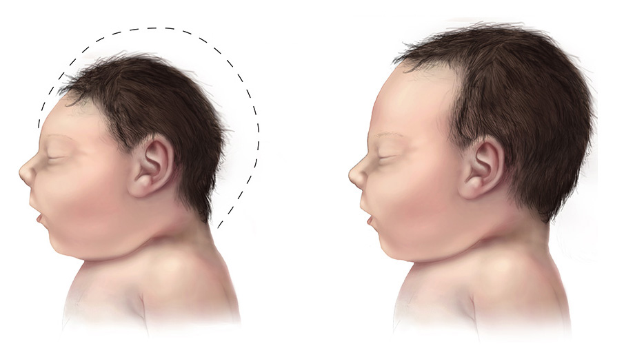
      A rare neurological disorder characterized by a reduced head circumference at birth with no gross anomalies of brain structure. It can be an isolated finding or it can be associated with seizures, developmental delay, intellectual disability, balance disturbances, hearing loss or vision problems.
    • Microcephaly Mayo_clinic
      This surgery relieves pressure on the brain, giving it enough space to grow and develop. Genetic changes. Down syndrome and other conditions may result in microcephaly.
    • Microcephaly Gard
      Microcephaly is a rare neurological condition in which a person's head is significantly smaller than expected based on standardized charts. Some cases of microcephaly are detected at birth, while others develop in the first few years of life. Some children with microcephaly have normal intelligence and development. However, microcephaly can be associated with seizures; developmental delay; intellectual disability; problems with movement and balance; feeding difficulties; hearing loss; and/or vision problems depending on the severity of the condition. Because the growth of the skull is determined by brain growth, the condition often occurs when the brain fails to grow at a normal rate.
  • Deafness-Small Bowel Diverticulosis-Neuropathy Syndrome Orphanet
    A rare neurologic disease characterized by progressive sensorineural deafness, progressive sensory neuropathy and gastrointestinal abnormalities, including progressive loss of gastric motility and small bowel diverticulosis and ulcerations, resulting in cachexia. Additonal neurological manifestations may include dysarthria and absent tendon reflexes, as well as ptosis and external ophthalmoplegia. There have been no further descriptions in the literature since 1985.
    • Deafness, Nerve Type, With Mesenteric Diverticula Of Small Bowel And Progressive Sensory Neuropathy Omim
      This appears to be an entity distinct from others such as Refsum syndrome (266500) and hereditary sensory radicular neuropathy (162400).
  • Antecubital Pterygium Syndrome Orphanet
    A rare, genetic, dermis disorder characterized by bilateral, fairly symmetrical, antecubital webbing extending from distal third of humerus to proximal third of forearm, associated with musculoskeletal abnormalities (i.e. absent long head of triceps, bilateral posterior dislocation of the radial head and hypoplasia of the olecranon processes) and absent skin creases over the terminal interphalangeal joints of fingers, clinically manifesting with moderate to severe elbow extension and supination limitation.
    PSD3, CHRNG, MYH3
    • Pterygium, Antecubital Omim
      Description Antecubital pterygium syndrome is an autosomal dominant disorder characterized by a fleshy web extending across the anterior aspect of the cubital fossa, absence of the long head of the triceps, limitation of full elbow extension, and missing skin creases over the terminal interphalangeal joints of the fingers (summary by Wallis et al., 1988). ... Inheritance The presence of affected persons of both sexes in 5 generations of the family reported by Wallis et al. (1988) indicated that the antecubital pterygium syndrome is inherited as an autosomal dominant trait.
    • Antecubital Pterygium Gard
      This condition is sometimes found as a symptom of nail-patella syndrome .
  • Familial Hyperphosphatemic Tumoral Calcinosis/hyperphosphatemic Hyperostosis Syndrome Orphanet
    Familial tumoral calcinosis (FTC) refers to a rare autosomal recessive disorder characterized by the occurrence of cutaneous and subcutaneous calcified masses, usually adjacent to large joints, such as hips, shoulders and elbows. FTC can occur in the setting of hyperphosphatemia or normophosphatemia, depending on the type of gene mutation involved.
    GALNT3, KL, FGF23, GLUD1, CHSY3, IL1A, IL1B, IL6, MTG1
    • Hyperphosphatemic Familial Tumoral Calcinosis Medlineplus
      A similar condition called hyperphosphatemia-hyperostosis syndrome (HHS) results in increased levels of phosphate in the blood, excessive bone growth, and bone lesions.
    • Hyperphosphatemic Familial Tumoral Calcinosis Gene_reviews
      Summary Clinical characteristics. Hyperphosphatemic familial tumoral calcinosis (HFTC) is characterized by: Ectopic calcifications (tumoral calcinosis) typically found in periarticular soft tissues exposed to repetitive trauma or prolonged pressure (e.g., hips, elbows, and shoulders); and Painful swellings (referred to as hyperostosis) in the areas overlying the diaphyses of the tibiae (and less often the ulna, metacarpal bones, and radius). The dental phenotype unique to HFTC includes enamel hypoplasia, short and bulbous roots, obliteration of pulp chambers and canals, and pulp stones. Less common are large and small vessel calcifications that are often asymptomatic incidental findings on radiologic studies but can also cause peripheral vascular insufficiency (e.g., pain, cold extremities, and decreased peripheral pulses). Less frequently reported findings include testicular microlithiasis and angioid streaks of the retina. Diagnosis/testing. HFTC results from a relative deficiency of – or resistance to – the phosphate-regulating hormone, fibroblast growth factor 23 (FGF23).
    • Tumoral Calcinosis, Hyperphosphatemic, Familial, 3 Omim
      The term 'hyperostosis-hyperphosphatemia syndrome' is sometimes used when the disorder is characterized by involvement of the long bones associated with the radiographic findings of periosteal reaction and cortical hyperostosis.
  • Cranioacrofacial Syndrome Omim
    Grosse (1974) described associated cardiac, craniofacial and hand anomalies in a father and daughter. The cardiac defect was combined ventricular septal defect and pulmonic stenosis. The craniofacial 'defect' consisted mainly of narrow head and face. Very minor abnormalities were present in the hands, e.g., Dupuytren contractures in the father. Cardiac - Ventricular septal defect - Pulmonic stenosis Limbs - Minor hand anomalies - Dupuytren contractures Facies - Narrow head and face Inheritance - Autosomal dominant ▲ Close
  • Cirrhosis-Dystonia-Polycythemia-Hypermanganesemia Syndrome Orphanet
    A rare disorder of manganese transport characterized by childhood onset of extrapyramidal movement disorder (including dystonia, tremor, and bradykinesia), liver cirrhosis, polycythemia, and hypermanganesemia. Cases with spastic paraparesis without extrapyramidal dysfunction have also been reported. Cognitive functions are preserved. Brain imaging findings are consistent with deposition of manganese in the basal ganglia, dentate nucleus, brain stem, and anterior pituitary.
    SLC30A10, ATP2C1
    • Hypermanganesemia With Dystonia 1 Omim
      A number sign (#) is used with this entry because hypermanganesemia with dystonia-1 (HMNDYT1) is caused by homozygous mutation in the SLC30A10 gene (611146) on chromosome 1q41. Description Hypermanganesemia with dystonia-1 is an autosomal recessive metabolic disorder characterized by increased serum manganese, motor neurodegeneration with extrapyramidal features, polycythemia, and hepatic dysfunction, which leads to cirrhosis in some cases. Intellectual function is preserved (summary by Tuschl et al., 2012 and Quadri et al., 2012). Genetic Heterogeneity of Hypermanganesemia With Dystonia See also HMNDYT2 (617013), caused by mutation in the SLC39A14 gene (608736) on chromosome 8p21. Clinical Features Tuschl et al. (2008) reported an Arabic girl with a constellation of clinical features consisting of hypermanganesemia, liver cirrhosis, an extrapyramidal motor disorder, and polycythemia.
    • Hypermanganesemia With Dystonia Medlineplus
      Hypermanganesemia with dystonia is an inherited disorder in which excessive amounts of the element manganese accumulate in the body (hypermanganesemia). One place manganese builds up in particular is in a region of the brain responsible for the coordination of movement, causing neurological problems that make controlling movement difficult. Consequently, the condition is characterized by involuntary, sustained muscle contractions (dystonia) and other uncontrolled movements. Two types of hypermanganesemia with dystonia, called hypermanganesemia with dystonia, polycythemia, and cirrhosis (HMDPC) and hypermanganesemia with dystonia 2, have been identified. They are distinguished by their genetic causes and certain specific features.
  • Hemangiomas Of Small Intestine Omim
    One patient had mucocutaneous pigment spots precisely like those of the Peutz-Jeghers syndrome. See blue rubber nevus syndrome (112200). Inheritance - Autosomal dominant GI - Cavernous hemangiomas of small intestine Skin - Mucocutaneous pigment spots as in Peutz-Jeghers syndrome ▲ Close
  • Senior-Boichis Syndrome Orphanet
    A rare ciliopathy characterized by the association of nephronophthisis and liver fibrosis. Renal manifestations include chronic renal failure, polyuria, polydipsia, anemia, as well as increased echogenicity on renal ultrasound and interstitial fibrosis and tubular dilation on biopsy. Hepatic involvement manifests as hepatosplenomegaly with extensive fibrosis, destruction of the bile ducts, and cholestasis. Mild psychomotor retardation and ocular symptoms, such as strabismus, nystagmus, retinal degeneration, and anisocoria, have been reported in some patients.
    TMEM67
    • Nephronophthisis 11 Omim
      Mutations in the TMEM67 gene can also cause Meckel syndrome-3 (MKS3; 607361), Joubert syndrome-6 (JBTS6; 610688), and COACH syndrome (216360), all of which show more severe yet overlapping clinical features with NPHP.
  • ←
  • 1
  • 2
  • ...
  • 195
  • 196
  • 197
  • 198
  • 199
  • 200
  • 201
  • ...
  • 556
  • 557
  • →

FindZebra

contact@findzebra.com