.; Scharf, Richard (2018), "Keratoacanthoma" , StatPearls , StatPearls Publishing, PMID 29763106 , retrieved 17 September 2018 ^ a b c d Joseph A. Regezi; James Sciubba; Richard C. K. Jordan (2012). "6. Neoplasms" . Oral Pathology - E-Book: Clinical Pathologic Correlations .
WMO Tech Note 99. 143 pp. Hovmøller, M. S., Sørensen, C. K., Walter, S., Justesen, A. F. (2011) Diversity of Puccinia striiformis on cereals and grasses.
Mosby-YearBook, Inc. 1994. p. 907. ^ a b c d e f g h i j k l m n o p q Ibrahim, Abdisamad M.; Siddique, Momin S. (2020), "Libman Sacks Endocarditis" , StatPearls , StatPearls Publishing, PMID 30422459 , retrieved 2020-05-30 ^ a b Roldan, C.
. ^ Thomas, J. J., Vartanian, L. R., & Brownell, K. D. (2009). "The relationship between eating disorder not otherwise specified (EDNOS) and officially recognized eating disorders: Meta-analysis and implications for DSM".
Acrocyanosis can also return in a newborn if a baby is cold, such as after a bath, and is considered normal as well. [10] See also [ edit ] Pernio (Chilblains) Cyanosis Peripheral artery occlusive disease Raynaud's phenomenon References [ edit ] ^ a b c d e f g h i j k Kurklinsky AK, Miller VM, Rooke TW.
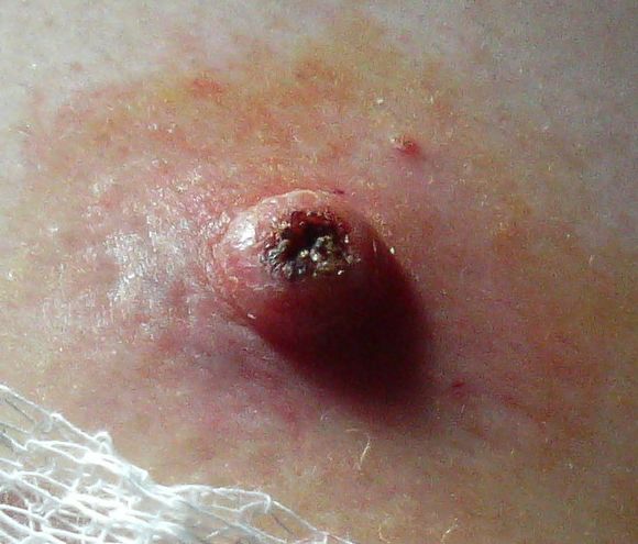
Hailey-Hailey disease is a genetic skin disease that causes blistering. Signs and symptoms include a painful rash and blistering in skin folds, such as the armpits, groin, neck, under the breasts, and between the buttocks. Secondary bacterial infections are not uncommon. Symptoms are often worse in summer months due to heat, sweating, and friction. Hailey-Hailey disease is caused by mutations in the ATP2C1 gene and is inherited in an autosomal dominant manner. Treatment focuses on reducing symptoms and preventing flares, and may include topical medication, laser, and other procedures.
Hailey-Hailey disease, also known as benign chronic pemphigus, is a rare skin condition that usually appears in early adulthood. The disorder is characterized by red, raw, and blistered areas of skin that occur most often in skin folds, such as the groin, armpits, neck, and under the breasts. These inflamed areas can become crusty or scaly and may itch and burn. The skin problems tend to worsen with exposure to moisture (such as sweat), friction, and hot weather. The severity of Hailey-Hailey disease varies from relatively mild episodes of skin irritation to widespread, persistent areas of raw and blistered skin that interfere with daily activities.
A number sign (#) is used with this entry because of evidence that Hailey-Hailey disease is caused by heterozygous mutation in the ATP2C1 gene (604384) on chromosome 3q22. Description Hailey-Hailey disease, also known as benign chronic pemphigus, is a rare autosomal dominant cutaneous disorder that usually becomes manifest in the third or fourth decade of life with erythema, vesicles, and erosions involving the body folds, particularly the groin and axillary regions. Other sites of the body, such as the neck, perianal, and submammary regions, may likewise be affected (summary by Poblete-Gutierrez et al., 2004). This disorder was first described by the dermatologist brothers Hailey and Hailey (1939). Clinical Features Loewenthal (1959) thought that pyogenic bacteria act as a precipitating factor.
Benign chronic familial pemphigus of Hailey-Hailey is characterized by rhagades mostly located in the armpits, inguinal and perineal folds (scrotum, vulva). Epidemiology Prevalence is unknown. Clinical description Skin lesions appear during adolescence or more often at the age of 30-40 years; they are relapsing and recurrent. Lesions can be complicated by heat, rubbing or superinfections. Etiology Mutations in the ATP2C1 gene (localised to 3q21-q24), encoding a calcium pump, cause the disease by impairing epidermal keratinocyte adhesion. Diagnostic methods Histopathological analysis of the lesions shows suprabasal acantholysis of epidermal cells. Genetic counseling Benign chronic familial pemphigus is transmitted as a dominant trait, with incomplete penetrance.
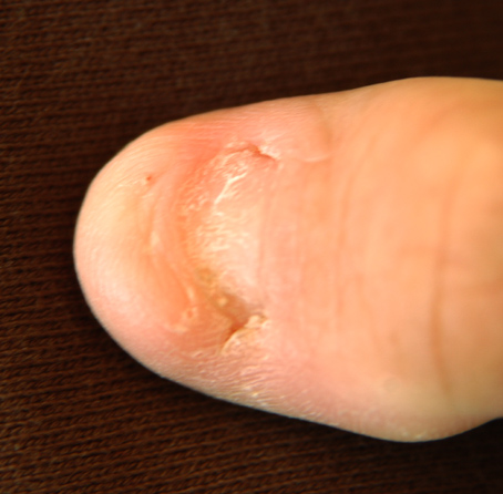
Nail-patella syndrome causes changes in the nails, elbows, kneecaps (patellae), and hip bone. The most common symptom of the syndrome is having missing or underdeveloped fingernails and toenails. Other symptoms may include having small or missing kneecaps, underdeveloped elbows, and an extra small piece of bone on both sides of the hip (called iliac horns). People with nail-patella syndrome are at an increased risk for developing high fluid pressure in the eye (glaucoma) and kidney disease. Nail-patella syndrome is caused by genetic changes (pathogenic variants or mutations) in the LMX1B gene.
A rare hereditary patellar dysostosis characterized by nail hypoplasia or aplasia, aplastic or hypoplastic patellae, elbow dysplasia, and the presence of iliac horns as well as renal and ocular anomalies. Epidemiology The reported prevalence is 1/50,000; however, epidemiological studies are lacking. Clinical description Nail-patella syndrome (NPS) is a multisystemic disorder characterized by significant inter- and intrafamilial variability in clinical manifestations and severity of the disease. Cases can range from mild with no functional impact to severe leading to disability. The classical tetrad involves nails dysplasia, absent or hypoplastic patellae, presence of iliac horns, and elbow deformities.
A number sign (#) is used with this entry because of evidence that nail-patella syndrome (NPS) is caused by heterozygous mutation in the LIM-homeodomain protein LMX1B (602575) on chromosome 9q33. Clinical Features Dysplasia of the nails and absent or hypoplastic patellae are the cardinal features but others are iliac horns, abnormality of the elbows interfering with pronation and supination, and in some cases nephropathy. Nephropathy was an associated abnormality in the family of Hawkins and Smith (1950). The renal change resembles glomerulonephritis. It is relatively benign although fatality at a young age from this complication has been described (Leahy, 1966). The renal disorder in the case of Simila et al. (1970) took the appearance of congenital nephrosis; 8 persons in the family had nail-patella syndrome, of whom 5 also had renal disease.
Summary Clinical characteristics. Nail-patella syndrome (NPS) (previously referred to as Fong's disease), encompasses the classic clinical tetrad of changes in the nails, knees, and elbows, and the presence of iliac horns. Nail changes are the most constant feature of NPS. Nails may be absent, hypoplastic, or dystrophic; ridged longitudinally or horizontally; pitted; discolored; separated into two halves by a longitudinal cleft or ridge of skin; and thin or (less often) thickened. The patellae may be small, irregularly shaped, or absent. Elbow abnormalities may include limitation of extension, pronation, and supination; cubitus valgus; and antecubital pterygia. Iliac horns are bilateral, conical, bony processes that project posteriorly and laterally from the central part of the iliac bones of the pelvis. Renal involvement, first manifest as proteinuria with or without hematuria, occurs in 30%-50% of affected individuals; end-stage renal disease occurs up to 15% of affected individuals.
PMID 24728303 . S2CID 3100453 . ^ Yee, TJ; Swong, K; Park, P (March 2020). "Complications of anterior cervical spine surgery: a systematic review of the literature" .
Cerebrospinal fluid (CSF) otorrhea is the leakage of cerebrospinal fluid (CSF) though the ear. It is a rare but very serious condition that requires rapid intervention. Symptoms include leak of clear fluid through the ear, inflammation of the membranes that cover the brain (meningitis), hearing loss, and seizures. The cause of a spinal fluid leak through the ear is a defect of the bone and meningeal layers covering the brain that separate the subarachnoid space of the brain from the middle ear and mastoid bone (located just behind the ear). The leaks occur after a surgery in the base of the skull, temporal bone fractures, congenital defects of the inner ear , trauma, or they may be spontaneous.
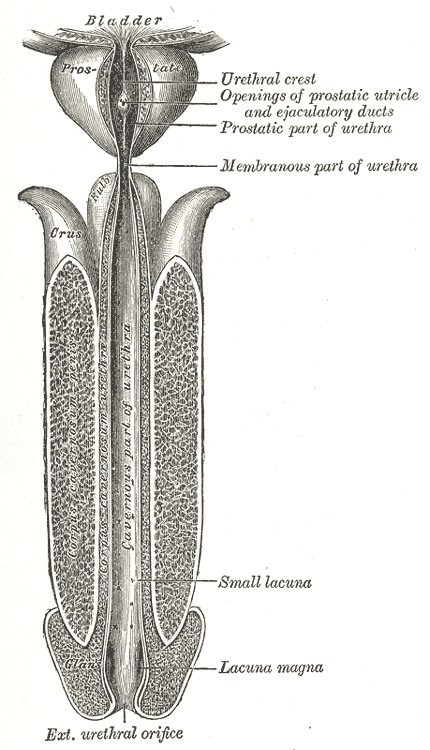
Overview A urethral (u-REE-thrul) stricture involves scarring that narrows the tube that carries urine out of your body (urethra). A stricture restricts the flow of urine from the bladder and can cause a variety of medical problems in the urinary tract, including inflammation or infection. Symptoms Signs and symptoms of urethral stricture include: Decreased urine stream Incomplete bladder emptying Spraying of the urine stream Difficulty, straining or pain when urinating Increased urge to urinate or more-frequent urination Urinary tract infection Causes Scar tissue, which can narrow the urethra, can be due to: A medical procedure that involves inserting an instrument, such as an endoscope, into the urethra Intermittent or long-term use of a tube inserted through the urethra to drain the bladder (catheter) Trauma or injury to the urethra or pelvis An enlarged prostate or previous surgery to remove or reduce an enlarged prostate gland Cancer of the urethra or prostate Sexually transmitted infections Radiation therapy Urethral stricture is much more common in males than in females. Often the cause is unknown. Clinical trials Explore Mayo Clinic studies testing new treatments, interventions and tests as a means to prevent, detect, treat or manage this condition.
Summary Clinical characteristics. Glycogen storage disease type III (GSD III) is characterized by variable liver, cardiac muscle, and skeletal muscle involvement. GSD IIIa is the most common subtype, present in about 85% of affected individuals; it manifests with liver and muscle involvement. GSD IIIb, with liver involvement only, comprises about 15% of all GSD III. In infancy and early childhood, liver involvement presents as ketotic hypoglycemia, hepatomegaly, hyperlipidemia, and elevated hepatic transaminases. In adolescence and adulthood, liver disease becomes less prominent. Hypertrophic cardiomyopathy develops in the majority of those with GSD IIIa, usually during childhood.
Glycogen storage disease type 3 (GSDIII) is an inherited disorder caused by the buildup of glycogen in the body's cells. This buildup impairs the function of certain organs and tissues, especially the liver and muscles. Symptoms typically begin in infancy and may include hypoglycemia, hyperlipidemia (excess of fats in the blood), and elevated blood levels of liver enzymes; later symptoms may include hepatomegaly , chronic liver disease ( cirrhosis ) and liver failure later in life. Some individuals have short stature and noncancerous (benign) tumors called adenomas in the liver. GSDIII is cause by mutations in the AGL gene and is inherited in an autosomal recessive manner.
Glycogen storage disease type III (also known as GSDIII or Cori disease) is an inherited disorder caused by the buildup of a complex sugar called glycogen in the body's cells. The accumulated glycogen is structurally abnormal and impairs the function of certain organs and tissues, especially the liver and muscles. GSDIII is divided into types IIIa, IIIb, IIIc, and IIId, which are distinguished by their pattern of signs and symptoms. GSD types IIIa and IIIc mainly affect the liver and muscles, and GSD types IIIb and IIId typically affect only the liver . It is very difficult to distinguish between the types of GSDIII that affect the same tissues.
A number sign (#) is used with this entry because glycogen storage disease III (GSD3) is caused by homozygous or compound heterozygous mutation in the AGL gene (610860), which encodes the glycogen debrancher enzyme, on chromosome 1p21. Description Glycogen storage disease III is an autosomal recessive metabolic disorder caused by deficiency of the glycogen debrancher enzyme and associated with an accumulation of abnormal glycogen with short outer chains. Most patients are enzyme-deficient in both liver and muscle (IIIa), but about 15% are enzyme-deficient in liver only (IIIb) (Shen et al., 1996). These subtypes have been explained by differences in tissue expression of the deficient enzyme (Endo et al., 2006). In rare cases, selective loss of only 1 of the 2 debranching activities, glucosidase or transferase, results in type IIIc or IIId, respectively.
Glycogen debranching enzyme (GDE) deficiency, or glycogen storage disease type 3 (GSD 3), is a form of glycogen storage disease characterized by severe muscle weakness and hepatopathy. Epidemiology Estimated prevalence is approximately 1/100,000 births (it may be higher among North Africans). Clinical description GSD 3 commonly occurs in early childhood. Children present with hepatomegaly, growth retardation and occasional seizures related to hypoglycemia. Hepatomegaly may disappear with adulthood. Muscle weakness is slowly progressive. Other frequently associated signs include muscular hypotonia and hypertrophic cardiomyopathy.
A number sign (#) is used with this entry because glycogen storage disease type IV (GSD4) is caused by homozygous or compound heterozygous mutation in the GBE1 gene (607839), which encodes the glycogen branching enzyme, on chromosome 3p12. Mutation in the GBE1 gene causes an allelic disorder, adult polyglucosan body neuropathy (APBN; 263570). Clinical Features Glycogen storage disease type IV is a clinically heterogeneous disorder. The typical 'classic' hepatic presentation is liver disease of childhood, progressing to lethal cirrhosis. The neuromuscular presentation of GSD IV is distinguished by age at onset into 4 groups: perinatal, presenting as fetal akinesia deformation sequence (FADS) and perinatal death; congenital, with hypotonia, neuronal involvement, and death in early infancy; childhood, with myopathy or cardiomyopathy; and adult, with isolated myopathy or adult polyglucosan body disease (Bruno et al., 2004).
Without active treatment, symptoms usually last two to seven days. [16] Statistics [ edit ] In the United States, there are about 2.4 million emergency department visits with throat-related complaints per year. [3] References [ edit ] ^ a b c d e f g h i j k l m n o p q Cohen, Jérémie F.; Pauchard, Jean-Yves; Hjelm, Nils; Cohen, Robert; Chalumeau, Martin (June 2020).
Overview A sore throat is pain, scratchiness or irritation of the throat that often worsens when you swallow. The most common cause of a sore throat (pharyngitis) is a viral infection, such as a cold or the flu. A sore throat caused by a virus resolves on its own. Strep throat (streptococcal infection), a less common type of sore throat caused by bacteria, requires treatment with antibiotics to prevent complications. Other less common causes of sore throat might require more complex treatment. Symptoms Symptoms of a sore throat can vary depending on the cause. Signs and symptoms might include: Pain or a scratchy sensation in the throat Pain that worsens with swallowing or talking Difficulty swallowing Sore, swollen glands in your neck or jaw Swollen, red tonsils White patches or pus on your tonsils A hoarse or muffled voice Throat anatomy The throat includes the esophagus; windpipe, also known as the trachea; voice box, also known as the larynx; tonsils; and epiglottis.