• FindZebra
  • About
  • Contact
  • Help
  • Login
Advanced
  • N-Acetylglutamate Synthase Deficiency Wikipedia
    External links [ edit ] GeneReviews/NCBI/NIH/UW entry on Urea Cycle Disorders Overview Classification D OMIM : 237310 MeSH : C536109 DiseasesDB : 29823 External resources eMedicine : ped/10 v t e Inborn error of amino acid metabolism K → acetyl-CoA Lysine /straight chain Glutaric acidemia type 1 type 2 Hyperlysinemia Pipecolic acidemia Saccharopinuria Leucine 3-hydroxy-3-methylglutaryl-CoA lyase deficiency 3-Methylcrotonyl-CoA carboxylase deficiency 3-Methylglutaconic aciduria 1 Isovaleric acidemia Maple syrup urine disease Tryptophan Hypertryptophanemia G G→ pyruvate → citrate Glycine D-Glyceric acidemia Glutathione synthetase deficiency Sarcosinemia Glycine → Creatine : GAMT deficiency Glycine encephalopathy G→ glutamate → α-ketoglutarate Histidine Carnosinemia Histidinemia Urocanic aciduria Proline Hyperprolinemia Prolidase deficiency Glutamate / glutamine SSADHD G→ propionyl-CoA → succinyl-CoA Valine Hypervalinemia Isobutyryl-CoA dehydrogenase deficiency Maple syrup urine disease Isoleucine 2-Methylbutyryl-CoA dehydrogenase deficiency Beta-ketothiolase deficiency Maple syrup urine disease Methionine Cystathioninuria Homocystinuria Hypermethioninemia General BC / OA Methylmalonic acidemia Methylmalonyl-CoA mutase deficiency Propionic acidemia G→ fumarate Phenylalanine / tyrosine Phenylketonuria 6-Pyruvoyltetrahydropterin synthase deficiency Tetrahydrobiopterin deficiency Tyrosinemia Alkaptonuria / Ochronosis Tyrosinemia type I Tyrosinemia type II Tyrosinemia type III / Hawkinsinuria Tyrosine → Melanin Albinism : Ocular albinism ( 1 ) Oculocutaneous albinism ( Hermansky–Pudlak syndrome ) Waardenburg syndrome Tyrosine → Norepinephrine Dopamine beta hydroxylase deficiency reverse: Brunner syndrome G→ oxaloacetate Urea cycle / Hyperammonemia ( arginine aspartate ) Argininemia Argininosuccinic aciduria Carbamoyl phosphate synthetase I deficiency Citrullinemia N-Acetylglutamate synthase deficiency Ornithine transcarbamylase deficiency / translocase deficiency Transport / IE of RTT Solute carrier family : Cystinuria Hartnup disease Iminoglycinuria Lysinuric protein intolerance Fanconi syndrome : Oculocerebrorenal syndrome Cystinosis Other 2-Hydroxyglutaric aciduria Aminoacylase 1 deficiency Ethylmalonic encephalopathy Fumarase deficiency Trimethylaminuria
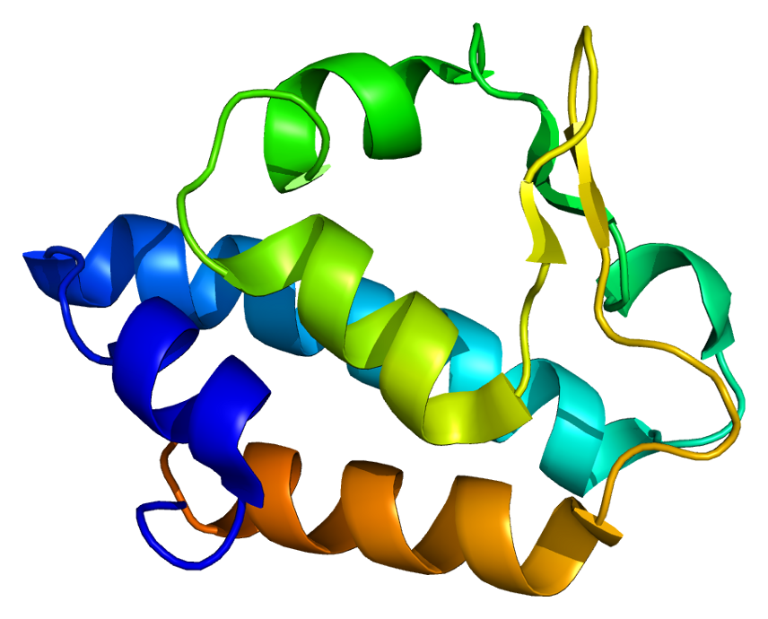
    NAGS
    • Hyperammonemia Due To N-Acetylglutamate Synthase Deficiency Orphanet
      N-acetylglutamate synthase (NAGS) deficiency is a urea cycle disorder leading to hyperammonaemia. Epidemiology The disorder is very rare but the prevalence is unknown. Clinical description Onset occurs at any age, but neonatal presentation appears to be the most frequent. The clinical manifestations are variable but common features include vomiting, hyperactivity or lethargy, diarrhoea, poor feeding, seizures, hypotonia, delayed psychomotor development and respiratory distress. The hyperammonaemia is often severe and may lead to hyperammonaemic coma.
    • N-Acetylglutamate Synthase Deficiency Gard
      N-acetylglutamate synthase deficiency (NAGS) is type of metabolic disorder that affects the processing of proteins and removal of ammonia from the body. When proteins are processed by the body, ammonia is formed. Individuals with NAGS are not able to remove ammonia from the body and have symptoms due to toxic levels of ammonia that build up in the blood. NAGS and other similar disorders are a type of metabolic condition known as a urea cycle disorder . Signs and symptoms in newborns with NAGS may include a lack of energy, unwillingness to eat, seizures, unusual body movements, and poorly controlled breathing or body temperature. Complications may include coma, developmental delay, and learning disability.
    • N-Acetylglutamate Synthase Deficiency Medlineplus
      N-acetylglutamate synthase deficiency is a disorder that causes abnormally high levels of ammonia to accumulate in the blood. Ammonia, which is formed when proteins are broken down in the body, is toxic if the levels become too high. The brain is especially sensitive to the effects of excess ammonia. The signs and symptoms of N-acetylglutamate synthase deficiency often become evident in the first few days of life. An infant with this condition may be lacking in energy (lethargic) or unwilling to eat, and have difficulty controlling his or her breathing rate or body temperature. Severely affected babies may experience seizures or unusual body movements, or go into a coma.
    • N-Acetylglutamate Synthase Deficiency Omim
      A number sign (#) is used with this entry because N-acetylglutamate synthase deficiency (NAGSD) is caused by homozygous or compound heterozygous mutation in the NAGS gene (608300) on chromosome 17q21. Description N-acetylglutamate synthase deficiency is an autosomal recessive disorder of the urea cycle. The clinical and biochemical features of the disorder are indistinguishable from carbamoyl phosphate synthase I deficiency (237300), since the CPS1 enzyme (608307) has an absolute requirement for NAGS (Caldovic et al., 2007). Clinical Features Bachmann et al. (1981) reported hyperammonemia due to deficiency of N-acetylglutamate synthetase in a newborn male and presumably in 2 of his sibs who died in the neonatal period. Autopsy in 1 of the sibs suggested hyperammonemia. The authors suggested that a deficiency of N-acetylglutamate synthetase should be considered in cases of hyperammonemia without increased excretion of orotic acid.
  • Toxic Multinodular Goitre Wikipedia
    Bioscientifica . 161 (5): 771–777. doi : 10.1530/EJE-09-0286 . PMID 19671708 . ^ Krohn, K; Fuhrer, D; Bayer, Y; Eszlinger, M; Brauer, V; Neumann, S; Paschke, R (2005).
    CACNA1S, GABRA3, KCNJ18, TSHR, GNAS, THRB, TTN
  • Gaffkaemia Wikipedia
    ISBN 978-0-8138-1844-3 . ^ a b c d e f g h i j k l Jeffrey D. Shields; Fran J. Stephens; Brian Jones (2006).
  • Red Thread Disease Wikipedia
    References [ edit ] ^ Buczacki, S. and Harris, K., Pests, Diseases and Disorders of Garden Plants , HarperCollins, 1998, p484 External links [ edit ] http://www.lawnandmower.com/red-thread-disease.aspx http://www.grassclippings.co.uk/RedThread.pdf
  • Aortoiliac Occlusive Disease Wikipedia
    Following treatment the 30-year-old was able to walk without pain and maintain an erection. [7] See also [ edit ] Claudication Peripheral arterial disease References [ edit ] ^ a b F. Charles Brunicardi; Dana K. Andersen; Timothy R. Billiar (5 June 2014).
  • Kufs Disease Wikipedia
    . ^ Benitez BA, Alvarado D, Cai Y, Mayo K, Chakraverty S, Norton J, Morris JC, Sands MS, Goate A, et al. (2011).
    CLN6, TPP1, CLN3, ATP13A2, CLCN3, CLCN6, CLN5, CTSD, CLN8, PPT1, MFSD8, CTSF, PSEN1, DNAJC5, SCARB2, NCL
    • Ceroid Lipofuscinosis, Neuronal, 13 Omim
      A number sign (#) is used with this entry because neuronal ceroid lipofuscinosis-13 (CLN13) is caused by homozygous or compound heterozygous mutation in the CTSF gene (603539) on chromosome 11q13. Description Neuronal ceroid lipofuscinosis-13 is an autosomal recessive neurodegenerative disorder characterized by adult onset of progressive cognitive decline and motor dysfunction leading to dementia and often early death. Some patients develop seizures. Neurons show abnormal accumulation of autofluorescent material (summary by Smith et al., 2013). Adult-onset neuronal ceroid lipofuscinosis is sometimes referred to as Kufs disease. For a discussion of genetic heterogeneity of neuronal ceroid lipofuscinosis (CLN), see CLN1 (256730).
    • Ceroid Lipofuscinosis, Neuronal, 11 Omim
      A number sign (#) is used with this entry because neuronal ceroid lipofuscinosis-11 (CLN11) is caused by homozygous mutation in the GRN gene (138945) on chromosome 17q. Heterozygous mutation in the GRN gene causes frontotemporal lobar degeneration with TDP43-inclusions (607485). Description Neuronal ceroid lipofuscinosis-11 is an autosomal recessive neurologic disorder characterized by rapidly progressive visual loss due to retinal dystrophy, seizures, cerebellar ataxia, and cerebellar atrophy. Cognitive decline may also occur (summary by Smith et al., 2012). For a general phenotypic description and a discussion of genetic heterogeneity of CLN, see CLN1 (256730). Clinical Features Smith et al. (2012) reported 2 Italian sibs with young-adult onset of neuronal ceroid lipofuscinosis.
    • Ceroid Lipofuscinosis, Neuronal, 4b, Autosomal Dominant Omim
      A number sign (#) is used with this entry because neuronal lipofuscinosis-4B (CLN4B) is caused by heterozygous mutation in the DNAJC5 gene (611203) on chromosome 20q13. Description Neuronal ceroid lipofuscinosis-4B is an autosomal dominant neurodegenerative disorder characterized by onset of symptoms in adulthood. It belongs to a group of progressive neurodegenerative diseases characterized by accumulation of intracellular autofluorescent lipopigment storage material in the brain and other tissues. Several different forms have been described according to age of onset (see, e.g., CLN3, 204200). Individuals with the adult form, sometimes referred to as Kufs disease, develop psychiatric manifestations, seizures, cerebellar ataxia, and cognitive decline.
    • Adult Neuronal Ceroid Lipofuscinosis Gard
      Adult neuronal ceroid lipofuscinosis is a rare condition that affects the nervous system. Signs and symptoms usually begin around age 30, but they can develop anytime between adolescence and late adulthood. There are two forms of adult neuronal ceroid lipofuscinosis that are differentiated by their underlying genetic cause, mode of inheritance and certain symptoms: Type A is characterized by a combination of seizures and uncontrollable muscle jerks (myoclonic epilepsy); dementia; difficulties with muscle coordination (ataxia); involuntary movements such as tremors or tics; and dysarthria. It is caused by changes (mutations) in the CLN6 or PPT1 gene and is inherited in an autosomal recessive manner. Type B shares many features with type A; however, affected people also experience behavioral abnormalities and do not develop myoclonic epilepsy or dysarthria.
    • Adult Neuronal Ceroid Lipofuscinosis Orphanet
      A genetically heterogeneous group of neuronal ceroid lipofuscinoses (NCLs) with onset during the third decade of life, characterized by dementia, seizures and loss of motor capacities, and sometimes associated with visual loss caused by retinal degeneration. Epidemiology Prevalence is unknown. Clinical description The clinical picture is characterized by onset with progressive myoclonic epilepsy or behavioral disturbances, dementia and extrapyramidal motor symptoms that appear at the age of 20-30 years. Vision loss is an uncommon feature and depends on the underlying genetic cause. Etiology The ANCL phenotype was originally designated as CLN4 disease, although the causative gene has not yet been identified. CLN4 may be inherited in an autosomal recessive (CLN4A) or autosomal dominant (CLN4B) manner.
    • Ceroid Lipofuscinosis, Neuronal, 4a, Autosomal Recessive Omim
      A number sign (#) is used with this entry because autosomal recessive neuronal ceroid lipofuscinosis-4A (CLN4A) is caused by homozygous or compound heterozygous mutation in the CLN6 gene (606725) on chromosome 15q23. Mutation in the CLN6 gene can also cause earlier onset of neuronal ceroid lipofuscinosis (CLN6; 601780) with ocular involvement. Description Adult-onset neuronal ceroid lipofuscinosis, also known as Kufs disease, is a neurodegenerative disorder without retinal involvement. There are 2 overlapping phenotypes: type A, characterized by progressive myoclonic epilepsy, and type B, characterized by dementia and a variety of motor-system signs (summary by Arsov et al., 2011). In general, the neuronal ceroid lipofuscinoses (NCL; CLN) are a clinically and genetically heterogeneous group of neurodegenerative disorders characterized by the intracellular accumulation of autofluorescent lipopigment storage material in different patterns ultrastructurally.
  • Sertoli Cell Tumour Wikipedia
    . ^ Leach S, Heatley JJ, Pool RR, Spaulding K (December 2008). "Bilateral testicular germ cell-sex cord-stromal tumor in a pekin duck (Anas platyrhynchos domesticus)".
    DICER1, AR, SRY, TNFRSF8, CTNNB1, INHA, SOX9, STK11, WT1
    • Malignant Sertoli-Leydig Cell Tumor Of The Ovary Orphanet
      Malignant Sertoli-Leydig cell tumor of ovary is a rare malignant sex cord stromal tumor of ovary (see this term) occuring typically in young women and characterized by manifestations of androgen excess (hirsutism, hair loss, amenorrhea, or oligomenorrhea), when functional.
  • Osteochondrosis Wikipedia
    .; Ekman, S.; Ellermann, J.; Olstad, K.; Ytrehus, B.; Carlson, C. S. (2013).
    ACTB, ACTG1, FGFR1, KRAS, BMP2, COL9A2, GABRA1, HMGA2, ECA1
  • Pusher Syndrome Wikipedia
    Steps to follow: A guide to the treatment of adult hemiplegia : Based on the concept of K. and B. Bobath . New York: Springer-Verlag. ^ O'Sullivan, S. (2007).
  • Wiedemann–rautenstrauch Syndrome Wikipedia
    . ^ Rautenstrauch, T; Snigula, F; Krieg, Thomas; Gay, Steffen; Müller, P. K. (1977). "Progeria: A cell culture study and clinical report of familial incidence".
    POLR3A, LMNA, POLR3GL
    • Wiedemann-Rautenstrauch Syndrome Orphanet
      Wiedemann-Rautenstrauch syndrome is a very rare disorder with features of premature aging recognizable at birth, decreased subcutaneous fat, hypotrichosis, relative macrocephaly and dysmorphism. Epidemiology More than 30 patients have been reported. Clinical description Dysmorphism includes triangular old-looking face, relatively large skull with large anterior fontanelle and prominent veins especially on the scalp, sparse scalp hair, decreased eyebrows and eyelashes, and micrognathia. Natal teeth represent a common, but variable finding. The clinical spectrum is broad but intrauterine growth retardation and decreased subcutaneous fat have been reported as cardinal features. Mild to moderate intellectual deficit is common. The syndrome is usually lethal by seven months but, on rare occasions, patients have survived into the teens. Etiology Etiology remains unknown. An increased chromosomal breakage, observed in some cases, suggests that DNA repair defects could be involved in the pathogenesis of this disorder.
    • Wiedemann-Rautenstrauch Syndrome Omim
      A number sign (#) is used with this entry because of evidence that Wiedemann-Rautenstrauch syndrome (WDRTS) is caused by compound heterozygous mutation in the POLR3A gene (614258) on chromosome 10q22. Description Wiedemann-Rautenstrauch syndrome (WDRTS) is a rare autosomal recessive neonatal progeroid disorder characterized by intrauterine growth retardation, failure to thrive, short stature, a progeroid appearance, hypotonia, and variable mental impairment (summary by Toriello, 1990). Average survival in WDRTS is 7 months, although survival into the third decade of life has been reported (Akawi et al., 2013). Clinical Features Based on the observation of 2 sisters reported by Rautenstrauch et al. (1977) and 2 unrelated patients of his own, Wiedemann (1979) suggested the existence of a distinct neonatal progeroid syndrome showing autosomal recessive inheritance. Snigula and Rautenstrauch (1981) gave follow-up information on a patient, then 4 years old, who had been reported by Rautenstrauch et al. (1977).
    • Neonatal Progeroid Syndrome Gard
      Neonatal progeroid syndrome is a rare genetic syndrome characterized by an aged appearance at birth. Other signs and symptoms include intrauterine growth restriction , feeding difficulties, distinctive craniofacial features, hypotonia, developmental delay and mild to severe intellectual disability. In most cases, affected infants pass away before age 7 months, but rare reports exist of survival into the teens or early 20s. Although the exact underlying cause of neonatal progeroid syndrome is unknown, it is likely a genetic condition that is inherited in an autosomal recessive manner. Treatment is symptomatic and supportive.
    • Wiedemann-Rautenstrauch Syndrome Medlineplus
      Wiedemann-Rautenstrauch syndrome is a type of progeria, which is a group of genetic conditions characterized by the dramatic, rapid appearance of aging earlier in life than expected. Signs and symptoms of Wiedemann-Rautenstrauch syndrome begin before birth. Affected individuals do not grow and gain weight at the expected rate before and after birth. People with this condition have distinctive facial features that give the appearance of old age. They often have a large head, a triangular face with a prominent forehead and pointed chin, a small mouth with a thin upper lip , low-set ears, and abnormal lower eyelids.
  • Ngly1 Deficiency Wikipedia
    In Adam MP, Ardinger HH, Pagon RA, Wallace SE, Bean LJ, Stephens K, Amemiya A, Lam C, Wolfe L, Need A, Shashi V, Enns G (eds.).
    NGLY1, PGM1, ENGASE
    • Congenital Disorder Of Deglycosylation Omim
      A number sign (#) is used with this entry because congenital disorder of deglycosylation (CDDG) is caused by homozygous or compound heterozygous mutation in the NGLY1 gene (610661) on chromosome 3p24. Description Congenital disorder of deglycosylation is an autosomal recessive multisystem disorder characterized by global developmental delay, hypotonia, abnormal involuntary movements, and alacrima or poor tear production. Other common features include microcephaly, intractable seizures, abnormal eye movements, and evidence of liver dysfunction. Liver biopsy shows cytoplasmic accumulation of storage material in vacuoles (summary by Enns et al., 2014). For a discussion of the classification of congenital disorders of glycosylation, see CDG1A (212065).
    • Deficiency Of N-Glycanase 1 Gard
      Deficiency of N-glycanase 1 (NGLY1 deficiency) is a complex neurological syndrome in which there is a deficiency of an enzyme known as N-glycanase 1 (NGLY1). This enzyme normally helps the body remove proteins that are not functioning properly. The typical features of NGLY1 deficiency include abnormal tear production, a movement disorder (choreoathetosis), and liver disease. Additional features may include developmental delay, hypotonia (weak muscle tone), peripheral neuropathy , EEG abnormalities, and a small head size (microcephaly). The condition is caused by mutations in the N-glycanase 1 gene ( NGLY1 gene ) and is inherited in an autosomal recessive manner.
    • Ngly1-Congenital Disorder Of Deglycosylation Medlineplus
      NGLY1 -congenital disorder of deglycosylation ( NGLY1 -CDDG) is an inherited condition that affects many parts of the body. The severity of the signs and symptoms varies widely among people with the condition. Individuals with NGLY1 -CDDG typically develop features of the condition during infancy. They often have delayed development of speech and motor skills, such as sitting and walking, and weak muscle tone (hypotonia). Many affected individuals have movement abnormalities, such as uncontrolled movements of the limbs (choreoathetosis), and some develop seizures that are difficult to treat.
    • Alacrimia-Choreoathetosis-Liver Dysfunction Syndrome Orphanet
      A rare, genetic, inborn error of metabolism disorder characterized by global developmental delay, hypotonia, choreoathetosis, hypo-/alacrimia, and liver dysfunction which manifests with elevated liver transanimases and hepatocyte cytoplasmic storage material or vacuolization on liver biposy. Additional features reported include acquired microcephaly, hypo-/areflexia, seizures, peripheral neuropathy, intellectual and language/speech disability, additional ocular anomalies and EEG and brain imaging abnomalities.
    • Ngly1-Related Congenital Disorder Of Deglycosylation Gene_reviews
      Summary Clinical characteristics. Individuals with NGLY1 -related congenital disorder of deglycosylation (NGLY1-CDDG) typically display a clinical tetrad of developmental delay / intellectual disability in the mild to profound range, hypo- or alacrima, elevated liver transaminases that may spontaneously resolve in childhood, and a complex hyperkinetic movement disorder that can include choreiform, athetoid, dystonic, myoclonic, action tremor, and dysmetric movements. About half of affected individuals will develop clinical seizures. Other findings may include obstructive and/or central sleep apnea, oral motor defects that affect feeding ability, auditory neuropathy, constipation, scoliosis, and peripheral neuropathy. Diagnosis/testing. The diagnosis of NGLY1-CDDG is established in a proband by the identification of biallelic pathogenic variants in NGLY1 on molecular genetic testing. Typical serum screening tests for congenital disorders of glycosylation (i.e., analysis of serum transferrin glycoforms, N and O glycan profiling) will NOT reliably detect NGLY1-CDDG. Management. Treatment of manifestations: Lubricating eye drops and/or bland ointments for hypolacrima; feeding therapy and/or supplemental tube feeding for those with oromotor deficits and feeding difficulties; adequate access to water and a cool environment (including a cooling vest for those who live in hot climates) for hypohydrosis; vitamin D supplementation for those with vitamin D deficiency; evaluation by a developmental pediatrician and supportive therapies for developmental and cognitive issues; standard treatment for hearing loss, sleep apnea, constipation, scoliosis, and seizure disorder; consideration of referral to a hematologist for abnormal hematologic studies; consideration of referral to a gastroenterologist for elevated liver transaminases.
  • Mystical Psychosis Wikipedia
    Deikman considered that all-encompassing unity opened in mysticism can be all-encompassing unity of reality. [15] See also [ edit ] Altered state of consciousness Depersonalization and Derealization Existential crisis Dhyāna in Buddhism Dhyāna in Hinduism Jerusalem syndrome Mental health Moksha Mirror neurons Mysticism Monomyth Near-death experience Posttraumatic stress disorder Religious experience Spiritualism Spirituality Spiritual crisis Wujud References [ edit ] ^ Whitney, E. (1998). " Personal accounts: Mania as spiritual emergency " Psychiatric Services 49 : 1547–1548 ^ Jackson, M., & Fulford, K.W.M., K. W. M.; Jackson, Mike (1997). "Spiritual experience and psychopathology".
  • Salla Disease Wikipedia
    PMID 420628 . [ permanent dead link ] ^ Autio-Harmainen H, Oldfors A, Sourander P, Renlund M, Dammert K, Simila S (1988). "Neuropathology of Salla disease".
    SLC17A5, GNE, CEACAM5, MATN1, VEGFA, RENBP, CD274, HPGDS, SGCB, TAF2, TGFB1, ZFPM2-AS1, TNF, EFL1, PCGF2, ZBTB16, PER2, ADIPOQ, KDM5A, DLL3, BACE1, RETN, LAMP3, APP, PIK3CD, PIK3CG, ATP7A, CALB2, CASP3, CD28, CDKN2B, CHIT1, DBH, DCC, DCX, EPO, ERBB2, GH1, HSP90AA1, IFNG, IDO1, LEP, LSAMP, NPY, PIK3CA, PIK3CB, LOC108281177
  • Gourmand Syndrome Wikipedia
    PMID 18502182 . ^ a b c d e f g h i j k Regard, Marianne; Landis, Theodor (1997). " " Gourmand syndrome": Eating passion associated with right anterior lesions".
  • Duodenal Lymphocytosis Wikipedia
    . ^ a b c d Hammer, Suntrea T. G.; Greenson, Joel K. (2013). "The Clinical Significance of Duodenal Lymphocytosis With Normal Villus Architecture".
  • Perineal Hernia Wikipedia
    Hernia . 10 (4): 360–3. doi : 10.1007/s10029-006-0092-4 . PMID 16705361 . ^ Abhinav K, Shaaban M, Raymond T, Oke T, Gullan R, Montgomery AC (April 2008).
  • Pacific Coast Tick Fever Wikipedia
    ., Robinson, L.K., White, F.H., Slater, K., Karpathy, S.E., Eremeeva, M.E. and Dasch, G.A.
  • Nodular Fasciitis Wikipedia
    Because recurrence is rare, even when there is incomplete excision, in case of recurrence initial diagnosis of nodular fasciitis should be revisited. [5] See also [ edit ] Leiomyosarcoma List of cutaneous conditions Inflammatory myofibroblastic tumour References [ edit ] ^ a b c Freedberg IM, Eisen AZ, Wolff K, Austen KF, Goldsmith LA, Katz SI, eds. (2003).
    USP6, MYH9, PPP6R3, CTNNB1, EPHA4, SEC31A, COL1A1, CCN4, AXIN2, TWIST1, TP53, TCF20, SYK, PTPRF, PITX2, OAS1, CD34, MITF, LYN, GNAZ, FOLH1, FGF7, EYA2, EPHB3, BIRC3
    • Nodular Fasciitis Orphanet
      A rare soft tissue tumor characterized by a solitary mass-forming fibrous proliferation that usually occurs in the subcutaneous tissue, composed of uniform fibroblastic/myofibroblastic cells displaying a loose growth pattern. Upper extremities, trunk, and head and neck are most frequently affected. The lesion typically grows rapidly and almost always measures less than five centimeters in diameter. Macroscopically, it may appear circumscribed or infiltrative but is not encapsulated. Recurrence after excision is very rare, and metastasis does not occur.
  • Aschoff Body Wikipedia
    History [ edit ] The Aschoff bodies were discovered independently by the German pathologist Ludwig Aschoff 1904 [1] [2] and one year later by Paul Rudolf Geipel . [3] References [ edit ] ^ Aschoff-Geipel bodies at Who Named It? ^ K. A. L. Aschoff. Zur Myocarditisfrage.
  • Damping Off Wikipedia
    . ^ a b c d e f g Buczacki, S., and Harris, K., Pests, Diseases and Disorders of Garden Plants , Collins, 1998, pp. 481–2. ^ a b c "Damping off" .
  • ←
  • 1
  • 2
  • ...
  • 112
  • 113
  • 114
  • 115
  • 116
  • 117
  • 118
  • ...
  • 218
  • 219
  • →

FindZebra

contact@findzebra.com