• FindZebra
  • About
  • Contact
  • Help
  • Login
Advanced
  • Simple Cryoglobulinemia Orphanet
    Patients with simple cryoglobulinemia lack the typical vasculitic manifestations and serological findings (RF positivity and low complement C4) that characterize MC patients.
  • Vertebral Anomalies And Variable Endocrine And T-Cell Dysfunction OMIM
    The son and daughter had thymus aplasia or hypoplasia, with absent or low T cell numbers, whereas their mother had low naive T cells. ... Endocrine anomalies in this family included autoimmune hypothyroidism in the son, hypoparathyroidism in the daughter, and low-normal PTH in the mother. The son showed average intelligence with attention-deficit/hyperactivity disorder and autistic behaviors, whereas the daughter showed mild developmental delay; their mother had normal intelligence. ... INHERITANCE - Autosomal dominant GROWTH Height - Short stature HEAD & NECK Head - Brachycephaly Face - Triangular face - Low anterior hairline - Glabellar hemangioma Ears - Low-set ears - Cupped ears - Overfolded helices Eyes - Hypertelorism - Epicanthal folds - Corectopia Nose - Depressed nasal bridge - Depressed nasal tip - Broad nasal tip Mouth - Cleft palate - High-arched narrow palate - Cleft lip Neck - Short neck - Webbed neck CARDIOVASCULAR Heart - Double-outlet right ventricle - Pulmonary valve stenosis - Atrial septal defect Vascular - Patent ductus arteriosus CHEST Ribs Sternum Clavicles & Scapulae - Sprengel deformity - Fusion of 4th and 5th ribs SKELETAL Spine - Klippel-Feil anomaly (fusion C2-C4) - Congenital fusions of thoracic spine - Open laminae posteriorly - Hemivertebrae at T10-T11 - Scoliosis - Kyphosis Hands - Camptodactyly 3rd and 4th digits NEUROLOGIC Central Nervous System - Developmental delay, mild - Attention-deficit hyperactivity disorder (ADHD) Behavioral Psychiatric Manifestations - Autistic behaviors ENDOCRINE FEATURES - Hashimoto thyroiditis - Hypoparathyroidism - Borderline low parathyroid hormone (PTH) - Growth hormone deficiency IMMUNOLOGY - Thymus aplasia or hypoplasia - Low or no functional T cells - Abnormal B cells - Very low naive T cells (CD4 and CD8) MISCELLANEOUS - Variable features may be present MOLECULAR BASIS - Caused by mutation in the T-box 2 gene (TBX2, 600747.0001 ) ▲ Close
    TBX2
  • Spondylocostal Dysostosis 6, Autosomal Recessive OMIM
    The proband had failure of formation of the posterior elements of C1 to C4 with descent of the occipital bone, causing spinal canal stenosis and spinal cord compression. ... His brother had deficiency of the posterior elements of C1 to C3, left hemivertebrae at C4 and T9, and a right hemivertebra at T4, causing marked cervical kyphosis at the C2/C3 level with associated cord compression.
    DLL3, TBX6, RIPPLY2, HES7, MESP2, LFNG, PAX1, ROR2, PAX9, SOX9, SLC35A3, GDF6
    • Spondylocostal Dysostosis GARD
      Spondylocostal dysostosis is a group of conditions characterized by abnormal development of the bones in the spine and ribs. In the spine, the vertebrae are misshapen and fused. Many people with this condition have an abnormal side-to-side curvature of the spine (scoliosis). The ribs may be fused together or missing. These bone malformations lead to short, rigid necks and short midsections. Infants with spondylocostal dysostosis have small, narrow chests that cannot fully expand. This can lead to life-threatening breathing problems. Males with this condition are at an increased risk for inguinal hernia , where the diaphragm is pushed down, causing the abdomen to bulge out.
    • Spondylocostal Dysostosis 3, Autosomal Recessive OMIM
      A number sign (#) is used with this entry because of evidence that autosomal recessive spondylocostal dysostosis-3 (SCDO3) is caused by homozygous or compound heterozygous mutation in the LFNG gene (602576) on chromosome 7p22. For a phenotypic description and a discussion of genetic heterogeneity of spondylocostal dysostosis, see 277300. Clinical Features Sparrow et al. (2006) reported a proband of Lebanese background who presented with extensive congenital vertebral anomalies; long, slender fingers; and camptodactyly of the left index finger. X-ray and magnetic resonance imaging (MRI) scans showed multiple vertebral ossification centers in the thoracic spine, which showed fitted angular shapes similar to those seen in the patient with spondylocostal dysostosis (SCDO2; see 608681) caused by mutation in the MESP2 gene (605195) (Whittock et al., 2004). Severe foreshortening of the spine was emphasized by comparison of the patient's arm span (186.5 cm) with adult height (155 cm; lower segment 92.5 cm).
    • Spondylocostal Dysostosis 2, Autosomal Recessive OMIM
      A number sign (#) is used with this entry because autosomal recessive spondylocostal dysostosis-2 (SCDO2) is caused by homozygous or compound heterozygous mutation in the MESP2 gene (605195) on chromosome 15q26. Description Spondylocostal dysostosis is a term given to a heterogeneous group of disorders characterized by abnormal vertebral segmentation. For a general phenotypic description and a discussion of genetic heterogeneity of the disorder, see SCDO1 (277300). Clinical Features Whittock et al. (2004) studied a consanguineous Lebanese Arab family in which 2 offspring were affected with spondylocostal dysostosis. Affected individuals presented with trunkal shortening and short necks but no other abnormalities.
    • Spondylocostal Dysostosis 1, Autosomal Recessive OMIM
      A number sign (#) is used with this entry because autosomal recessive spondylocostal dysostosis-1 (SCDO1) is caused by homozygous or compound heterozygous mutation in the DLL3 gene (602768) on chromosome 19q13. Description The spondylocostal dysostoses are a heterogeneous group of axial skeletal disorders characterized by multiple segmentation defects of the vertebrae (SDV), malalignment of the ribs with variable points of intercostal fusion, and often a reduction in rib number. The term 'spondylocostal dysostosis' is best applied to those phenotypes with generalized SDV and a broadly symmetric thoracic cage (summary by Gucev et al., 2010). Genetic Heterogeneity of Spondylocostal Dysostosis Other forms of SCDO include SCDO2 (608681), caused by mutation in the MESP2 gene (605195) on chromosome 15q26; SCDO3 (609813), caused by mutation in the LFNG gene (602576) on chromosome 7p22; SCDO4 (613686), caused by mutation in the HES7 gene (608059) on chromosome 17p13; SCDO5 (122600), caused by mutation in the TBX6 gene (602427) on chromosome 16p11; and SCDO6 (616566), caused by mutation in the RIPPLY2 gene (609891) on chromosome 6q14. Clinical Features Lavy et al. (1966) observed 4 of 7 offspring of a third-cousin marriage who had characteristic vertebral anomalies including hemivertebrae and block vertebrae accompanied by deformity of the ribs.
    • Spondylocostal Dysostosis MedlinePlus
      Spondylocostal dysostosis is a group of conditions characterized by abnormal development of bones in the spine and ribs. The bones of the spine (vertebrae) are misshapen and abnormally joined together (fused). Many people with this condition have abnormal side-to-side curvature of the spine (scoliosis ) due to malformation of the vertebrae. In addition to spinal abnormalities, some of the rib bones may be fused together or missing. Affected individuals have short, rigid necks and short torsos because of the bone malformations.
    • Spondylocostal Dysostosis Wikipedia
      Axial skeleton growth disorder Jarcho-Levin Syndrome Radiograph depicting typical skeletal features of Jarcho-Levin syndrome, subtype spondylothoracic dysplasia . Note fanlike configuration of the ribs, with extensive posterior fusion, along with multiple vertebral segmentation defects. Specialty Medical genetics Spondylocostal dysostosis , also known as Jarcho-Levin syndrome (JLS) , is a rare, heritable axial skeleton growth disorder. It is characterized by widespread and sometimes severe malformations of the vertebral column and ribs , shortened thorax , and moderate to severe scoliosis and kyphosis . Individuals with Jarcho-Levin typically appear to have a short trunk and neck, with arms appearing relatively long in comparison, and a slightly protuberant abdomen .
    • Autosomal Recessive Spondylocostal Dysostosis Orphanet
      A rare condition of variable severity associated with vertebral and rib segmentation defects and characterised by a short neck with limited mobility, winged scapulae, a short trunk, and short stature with multiple vertebral anomalies at all levels of the spine. Epidemiology The incidence and prevalence are unknown. The disease seems to be more frequent in the Puerto Rican population. Clinical description Autosomal recessive spondylocostal dysostosis (ARSD) is usually diagnosed in the neonatal period. The main skeletal malformations include fusion of the vertebrae, hemivertebrae, and rib fusion with other rib malformations. Deformity of the chest and spine (severe scoliosis, kyphoscoliosis and lordosis) is a natural consequence of these malformations and leads to a dwarf-like appearance.
    • Spondylothoracic Dysostosis GARD
      Spondylothoracic dysostosis (STD) is a rare condition that affects the bones of the spine and the ribs. The term “Jarcho-Levin syndrome” in many cases is used as a synonym for STD, and sometimes as a synonym for another condition known as spondylocostal dysostosis , which has several common features with STD. Also, the term “Jarcho-Levin syndrome” is often used for all radiologic features that include defects of the vertebrae and abnormal rib alignment. Signs and symptoms of STD are generally present at birth and may include short-trunk dwarfism (a short body with normal length arms and legs); a small chest cavity; misshapen and abnormally-fused vertebrae (bones of the spine); and fused ribs at the part nearest the spine. Affected people may also have life-threatening breathing problems and recurrent lung infections, which can significantly reduce lifespan.
    • Spondylocostal Dysostosis 4, Autosomal Recessive OMIM
      A number sign (#) is used with this entry because spondylocostal dysostosis-4 (SCDO4) is caused by homozygous or compound heterozygous mutation in the HES7 gene (608059) on chromosome 17p13. For a general phenotypic description and a discussion of genetic heterogeneity of spondylocostal dysostosis (SCDO), see SCDO1 (277300). Clinical Features Sparrow et al. (2008) described a consanguineous family of Caucasian Mediterranean origin in which the proband was diagnosed prenatally with hydrocephalus and myelomeningocele, and at birth was found to have a bell-shaped, symmetric, and shortened thorax, lumbosacral myelomeningocele, ectopic and stenotic anus, and talipes. Radiologic examination showed shortening of the spine, with multiple and contiguous vertebral segmentation defects involving all spinal regions, but mainly the thoracic spine. The ribs had very crowded origins on the left side, and were irregularly aligned with variable points of fusion along their length on the right side.
    • Spondylothoracic Dysostosis MedlinePlus
      Spondylothoracic dysostosis is a condition characterized by malformation of the bones of the spine and ribs. The bones of the spine (vertebrae) do not develop properly, which causes them to be misshapen and abnormally joined together (fused). The ribs are also fused at the part nearest the spine (posteriorly), which gives the rib cage its characteristic fan-like or "crab" appearance in x-rays. Affected individuals have short, rigid necks and short torsos because of the bone malformations. As a result, people with spondylothoracic dysostosis have short bodies but normal-length arms and legs, called short-trunk dwarfism.
  • Ethylmalonic Encephalopathy Orphanet
    In addition to increased excretion of EMA, methylsuccinic acid and C4-C6-acylglycines (isobutyryl-, isovaleryl-, 2-methylbutyryl-, hexanoylglycine) may also be found in small, but elevated, amounts in the urine. Blood levels of C4-C6-carnitines (butyryl-, isobutyryl-, isovaleryl- and hexanoylcarnitine) may be elevated.
    ETHE1, TRIM8, TNK2, ZNF575, BDNF, TNF, OXR1, BCL11A, HBS1L, POSTN, BECN1, ABCB11, TRPV1, ALDH3A2, ANXA5, MYB, MUC1, IL6, GRM5, ETFA, TYMP, DNM1, ABCC2, CALB2, BNIP3, SOD2
    • Ethylmalonic Encephalopathy GeneReviews
      The diagnosis of EE is suggested by clinical findings and the laboratory findings of increased blood lactate levels, C4- and C5-acylcarnitine esters, plasma thiosulphate, and urinary ethylmalonic acid. ... Clinical findings Global neurologic impairment Early-onset progressive psychomotor regression Seizures Dystonia Diffuse microvasculature injury Petechiae and/or purpura Orthostatic acrocyanosis Hemorrhagic suffusions of mucosal surfaces Chronic hemorrhagic diarrhea Preliminary laboratory findings Increased blood lactate levels (normal range: 6-22 mg/dL) Increased blood C4-acylcarnitine esters (normal range: <0.9 μmol/L) [Merinero et al 2006, Zafeiriou et al 2007] * Increased blood C5-acylcarnitine esters (normal range: <0.3 μmol/L) [Merinero et al 2006, Zafeiriou et al 2007] * Increased plasma thiosulphate (normal range: <4 μmol/L) Increased urinary ethylmalonic acid (normal range: <10 μmol/mmol creatinine) evaluated on spot urine [Merinero et al 2006, Zafeiriou et al 2007] * More data are needed to define the range of C4/C5 acylcarnitine elevation in individuals with molecularly proven EE. ... Tandem mass spectroscopy can identify C4 elevation in a NBS dried blood spot; however, NBS for EE is not available in the US as there is no definitive treatment (see Therapies Under Investigation). Note: (1) NBS may be performed elsewhere in the world. (2) C4 elevation can also be found in primary short-chain acyl-CoA dehydrogenase (SCAD) deficiency [McHugh et al 2011]; an algorithm (pdf) from the American College of Medical Genetics can be used to distinguish the two disorders.
    • Ethylmalonic Encephalopathy MedlinePlus
      The ETHE1 enzyme is part of a pathway that breaks down sulfide (H 2 S), a molecule that is critical at very low levels for normal cell functioning but is toxic at high levels.
    • Ethylmalonic Encephalopathy GARD
      Symptoms are present at birth and tend to get worse over time. These include low muscle tone, spasms of the arms and legs, seizures, and developmental delay.
    • Encephalopathy, Ethylmalonic OMIM
      Animal Model Tiranti et al. (2009) found that Ethe1-null mice developed the cardinal features of ethylmalonic encephalopathy, including poor growth, reduced motor activity, early death, low cytochrome c oxidase (COX) in muscle and brain, and increased urinary excretion of ethylmalonic acid. ... INHERITANCE - Autosomal recessive GROWTH Other - Failure to thrive HEAD & NECK Eyes - Retinal lesions with tortuous vessels CARDIOVASCULAR Vascular - Orthostatic acrocyanosis ABDOMEN Gastrointestinal - Chronic diarrhea SKIN, NAILS, & HAIR Skin - Petechiae - Orthostatic acrocyanosis NEUROLOGIC Central Nervous System - Developmental regression - Developmental delay - Mental retardation - Pyramidal symptoms - Extrapyramidal symptoms - Ataxia - Hypotonia - Seizures - Hyperintense lesions in the basal ganglia on MRI LABORATORY ABNORMALITIES - Lactic acidemia - Ethylmalonic aciduria - Methylsuccinic aciduria - Cytochrome c oxidase deficiency in skeletal muscle and brain - Increased serum C4 and C5 acylcarnitine esters - Increased urinary isobutyryl glycine - Increased urinary 2-methylbutyryl glycine - Increased urinary thiosulphate MISCELLANEOUS - Onset in first months of life - Patients are often of Mediterranean origin - Death usually occurs in first decade of life MOLECULAR BASIS - Caused by mutation in the ETHE1 gene (ETHE1, 608451.0001 ) ▲ Close
    • Ethylmalonic Encephalopathy Wikipedia
      Ethylmalonic encephalopathy Ethylmalonic encephalopathy has an autosomal recessive pattern of inheritance . Specialty Medical genetics Ethylmalonic encephalopathy ( EE ) is a rare autosomal recessive inborn error of metabolism . Patients affected with EE are typically identified shortly after birth, with symptoms including diarrhea , petechiae and seizures . [1] [2] The genetic defect in EE is thought to involve an impairment in the degradation of sulfide intermediates in the body. Hydrogen sulfide then builds up to toxic levels. [3] EE was initially described in 1994. [4] Most cases of EE have been described in individuals of Mediterranean or Arabic origin. [3] Contents 1 Signs and symptoms 2 Pathophysiology 3 Diagnosis 4 Treatment 5 References 6 External links Signs and symptoms [ edit ] Neurologic signs and symptoms include progressively delayed development, weak muscle tone ( hypotonia ), seizures, and abnormal movements. The body's network of blood vessels is also affected. Children with this disorder may experience rashes of tiny red spots ( petechiae ) caused by bleeding under the skin and blue discoloration in the hands and feet due to reduced oxygen in the blood ( acrocyanosis ).
  • Hereditary Angioedema Orphanet - Search Hereditary Angioedema case abstracts
    Diagnostic methods Diagnosis of HAE types 1 and 2 relies on measurement of C4 concentrations and on quantitative and functional analysis of C1-INH. Diagnosis of HAE type 3 revolves around recognition of the clinical picture; C4 and C1-INH levels are normal. Analysis for mutations in the F12 gene may be proposed but are present in only 15% of patients.
    F12, SERPING1, KNG1, PLG, GRK6, KLKB1, SPINK6, SERPINA4, ANGPT1, ACE, SMPX, POMC, SERPINF2, INA, RBPJ, SERPINF1, CSHL1, CRP, BDKRB2, C2, C1S, CPQ, MBL3P, MASP2, ABCB6, SSB, VCAM1, TNF, SMUG1, TGM2, IL23A, C4orf3, LINC01194, TGFB3, TFPI, A2M, RO60, TRIM21, ABO, ADM, AHSG, ALB, C4B, F3, NR3C1, HSP90AA1, IL17A, MBL2, NHS, SERPINB2, AAVS1, PLAT, REN, TLX1NB
    • Angioedema, Hereditary, Type Iii OMIM
      While screening a large population of patients with recurrent angioedema of the skin, Bork et al. (2000) identified 10 unrelated women with hereditary angioneurotic edema who had normal C1 inhibitor protein concentration and function, and normal C4 concentration. A more detailed study of these families identified another 26 affected members, who were also all women. Fourteen of the 26 women were studied and all were found to have normal C1 inhibitor concentration and function, and normal C4 concentration. The disease was seen in successive generations, and in offspring of affected mothers the male-to-female sex ratio was shifted to 1/1.5. ... Duration of the disorder extended over decades or many years, either in patients or in family members. Normal C1 inhibitor and C4 concentrations were present in plasma.
    • Hereditary Angioedema Wikipedia
      HAE is generally referred to as a "dominant" condition because it only takes a mutation in one of the two C1-INH genes in a carrier to cause the disease. [8] [9] The prevalence of HAE is relatively low – between 1 in every 10,000 to 1 in every 50,000 persons. ... In a review of patients who do not have a history of HAE in their family, but who have relatively low levels of mutated C1-INH with persistent angioedema, 25% of new patients who had HAE had C1-INH changes that do not show signs of inheritance. Diagnosis [ edit ] Complement tests C4 ( C ) FB ( A ) C3 CH50 Conditions · ↓ ↓ ↓ PSG , C3 NeF AA ↓ · ↓ · HAE , C4D · · · ↓ TCPD ↓ · /↓ ↓ ↓ SLE ↑ ↑ ↑ ↑ inflammation Recognizing HAE is often difficult due to the wide variability in disease expression. ... Measure: serum complement factor 4 (C4), C1 inhibitor (C1-INH) antigenic protein, C1 inhibitor (C1-INH) functional level if available. Analysis of complement C1 inhibitor levels may play a role in diagnosis. C4 and C2 are complementary components.
    • Hereditary Angioedema GARD
      Hereditary angioedema (HAE) is a disease characterized by recurrent episodes (also called attacks) of severe swelling of the skin and mucous membranes. The age at which attacks begin varies, but most people have their first one in childhood or adolescence. The frequency of attacks usually increases after puberty. Attacks most often affect 3 parts of the body: Skin - the most common sites are the face (such as the lips and eyes), hands, arms, legs, genitals, and buttocks. Skin swelling can cause pain, dysfunction, and disfigurement, although it is generally not dangerous and is temporary. Gastrointestinal tract - the stomach, intestines, bladder, and/or urethra may be involved.
  • Schizophrenia 3 OMIM
    For discussion of the contribution of the C4 locus on chromosome 6p21 to schizophrenia risk, see MOLECULAR GENETICS. ... In postmortem human adult brain samples, Sekar et al. (2016) found that C4 RNA expression was directly proportional to C4 copy number; that expression levels of C4A were 2 to 3 times greater than levels for C4B, even after controlling for relative copy number; and that copy number of the C4-HERV sequence increased the ratio of C4A to C4B expression. Sekar et al. (2016) characterized the structural variation of human C4 genes (C4A and C4B) and evaluated association to over 7,000 SNPs across the extended MHC locus (chr6:25-34 Mb), C4 structural alleles, and HLA sequence polymorphisms in 28,799 schizophrenia cases and 35,986 controls. ... The other peak of association centered at the C4 locus, where schizophrenia associated most strongly with the genetic predictor of C4A expression levels (p = 3.6 X 10(-24)). ... Studies in mice demonstrated defects in synaptic remodeling in C4-deficient mice. Sekar et al. (2016) concluded that association of schizophrenia with the MHC locus on chromosome 6p21.3 involves many common, structurally distinct C4 alleles that affect expression of C4A and C4B in the brain, with each allele associated with schizophrenia risk in proportion to its effect on C4A expression.
    CSF2, LAMC2
  • Human Papillomavirus Type 18 Integration Site 1 OMIM
    In 2 cervical carcinoma cell lines, HeLa and C4-I, Durst et al. (1987) found that HPV18 DNA was integrated into chromosome 8, 5-prime to the MYC gene (190080).
    HPV18I1
  • Immunodeficiency Due To A Classical Component Pathway Complement Deficiency Orphanet
    Immunodeficiency due to a classical component pathway complement deficiency is a primary immunodeficiency due to a deficiency in either complement components C1q, C1r, C1s, C2 or C4 characterized by increased susceptibility to bacterial infections, particularly with encapsulated bacteria, and increased risk for autoimmune disease.
    • Complement Component C1r/c1s Deficiency OMIM
      Two of the 4 had a syndrome combining discoid lupus erythematosus and nondeforming rheumatoid-like arthritis; 1 had mild nephritis. C1 and C4 deficiencies are more often associated with lupus-like illness than is deficiency of C2.
  • Serum Sickness-Like Reaction Wikipedia
    Laboratory abnormalities include normal or mild decreases in serum C3, C4, and CH50 levels, and mild proteinuria.1,3-5 In contrast to true serum sickness, renal and hepatic involvement is rare. Significant decreases in serum C3, C4, and CH50, reported in the literature for true serum sickness, are rarely described in serum sickness–like reaction.
  • Microcephaly, Congenital Cataract, And Psoriasiform Dermatitis OMIM
    Serum lipid profile showed persistently low total cholesterol, HDL, and LDL, with normal triglycerides and VLDL. ... Experiments with culture medium were consistent with a block at the step of sterol-C4-methyl oxidase (607545) in the cholesterol synthesis pathway. ... INHERITANCE - Autosomal recessive GROWTH Height - Short stature Weight - Low weight HEAD & NECK Head - Microcephaly Eyes - Cataract, congenital - Blepharitis GENITOURINARY Internal Genitalia (Female) - Delayed puberty SKELETAL - Delayed bone age Limbs - Arthralgias - Joint contractures, especially of lower extremities SKIN, NAILS, & HAIR Skin - Ichthyosiform erythroderma sparing the palms Skin Histology - Psoriasiform hyperplasia - Dilated capillaries in dermal papillae - Neutrophils in epidermis - Neutrophils in stratum corneum - CD68-negative lipid-containing foamy cells in dermis Hair - Fine, lusterless hair NEUROLOGIC Central Nervous System - Mental retardation, mild METABOLIC FEATURES - Low total cholesterol - Low HDL - Low LDL ENDOCRINE FEATURES - Delayed puberty IMMUNOLOGY - Markedly elevated IgE - Markedly elevated IgA - Increase in CD16+ activated granulocytes - Increase in TLR2+ ( 603028 )/TLR4- ( 603030 ) granulocytes - Decrease in granulocyte-specific CD16B ( 610665 ) isoform - Elevated IL6 ( 147620 ) - Elevated IL8 ( 146930 ) - Increase in proinflammatory cytokines - Increase in proinflammatory chemokines MISCELLANEOUS - Heterozygotes exhibit subclinical metabolic and immunologic abnormalities - Based on report of 2 unrelated patients (last curated February 2016) MOLECULAR BASIS - Caused by mutation in the methylsterol monooxygenase 1 gene (MSMO1, 607545.0001 ) ▲ Close
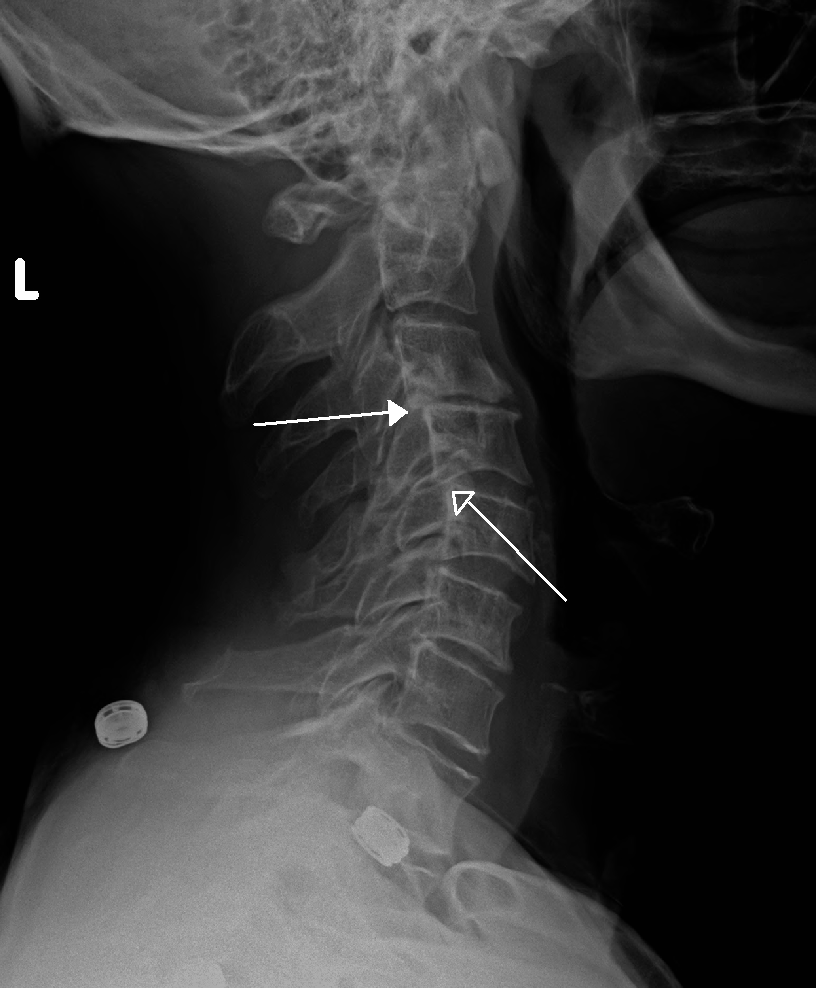
    MSMO1
    • Microcephaly-Congenital Cataract-Psoriasiform Dermatitis Syndrome Orphanet
      A rare sterol biosynthesis disorder characterized by microcephaly, bilateral congenital cataract, mild developmental delay, growth delay with short stature, psoriasiform dermatitis of variable severity, and immune dysregulation. Behavioral disorder, joint contractures, and arthralgia have also been described.
  • Retrolisthesis Wikipedia
    Retrolisthesis Grade 1 retrolistheses of C3 on C4 and C4 on C5 Specialty Orthopedics A retrolisthesis is a posterior displacement of one vertebral body with respect to the subjacent vertebra to a degree less than a luxation (dislocation) . ... British Volume, Vol 88-B, Issue SUPP_III, 450. v t e Spinal disease Deforming Spinal curvature Kyphosis Lordosis Scoliosis Other Scheuermann's disease Torticollis Spondylopathy inflammatory Spondylitis Ankylosing spondylitis Sacroiliitis Discitis Spondylodiscitis Pott disease non inflammatory Spondylosis Spondylolysis Spondylolisthesis Retrolisthesis Spinal stenosis Facet syndrome Back pain Neck pain Upper back pain Low back pain Coccydynia Sciatica Radiculopathy Intervertebral disc disorder Schmorl's nodes Degenerative disc disease Spinal disc herniation Facet joint arthrosis
  • Unilateral Nevoid Telangiectasia Wikipedia
    Please help improve it to make it understandable to non-experts , without removing the technical details. ( November 2018 ) ( Learn how and when to remove this template message ) Unilateral nevoid telangiectasia Specialty Dermatology Unilateral nevoid telangiectasia presents with fine, threadlike telangiectases , developing in a unilateral, sometimes dermatomal, distribution, with the areas most often involved being the trigeminal and C3 and C4 or adjacent areas. [1] : 589,844 [2] See also [ edit ] Skin lesion List of cutaneous conditions References [ edit ] ^ James, William; Berger, Timothy; Elston, Dirk (2005).
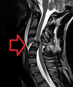
  • Neurogenic Shock Wikipedia
    Neurogenic shock Cervical spine MRI of a patient with SCI : C4 fracture and dislocation, spinal cord compression Specialty Neurology Neurogenic shock is a distributive type of shock resulting in low blood pressure , occasionally with a slowed heart rate , that is attributed to the disruption of the autonomic pathways within the spinal cord. It can occur after damage to the central nervous system , such as spinal cord injury and traumatic brain injury . Low blood pressure occurs due to decreased systemic vascular resistance as a result of lacking sympathetic tone which in turn causes pools of blood staying within the extremities and not being redirected to the core body. ... Neurogenic shock's presentation includes: [5] [6] - warm and pink skin - labored breathing - low blood pressure - dizziness - anxiety - history of trauma to head or upper spine. - if the injury is to the head or neck, hoarseness or difficulty swallowing may occur. ... External links [ edit ] Classification D ICD - 10 : R57.8 ICD - 9-CM : 785 MeSH : D012769 v t e Shock Distributive Septic shock Neurogenic shock Anaphylactic shock Toxic shock syndrome Obstructive Abdominal compartment syndrome Low volume Hemorrhage Hypovolemia Osmotic shock Other Spinal shock Cryptic shock Vasodilatory shock
  • Multiple Acyl-Coa Dehydrogenase Deficiency Orphanet
    They usually have dysplastic kidneys with multiple cysts and may also have facial dysmorphism (low-set ears, high forehead, hypertelorism and hypoplastic midface), rocker-bottom feet and anomalies of external genitalia. ... Blood acylcarnitines show increased C4-C18 species although patients may be severely carnitine depleted, which may limit the degree of these abnormalities.
    ETFDH, ETFA, ETFB, FLAD1, MUC1, CPT2, MADD, SOD2, UVRAG, VIM, TEAD2, TP63, ACTB, SMN1, SYNGAP1, OGA, PPARGC1A, CKAP4, SLC52A1, CHPT1, SLC52A3, SMN2, S100A1, SLPI, S100B, ACVRL1, RPE65, CYTB, MFAP1, GSR, DES, CSF3, CPT1B, CPT1A, CD34, SLC25A20, APRT, AMPD1, CHKB-CPT1B
    • Multiple Acyl-Coa Dehydrogenase Deficiency GeneReviews
      Pregnancy management : Successful pregnancy with low-fat, high-carbohydrate diet in late-onset MADD has been published. ... Plasma acylcarnitine profile typically shows elevations of C4, C5, C5DC, C6, C8, C10, C12, C14:1, C16, and C18:1. ... Plasma carnitine assay may show a very low free carnitine. In this setting the acylcarnitine profile may be falsely normal. ... Acylcarnitine profile may be normal in the setting of low plasma free carnitine and should be repeated after carnitine supplementation in such cases. ... Pregnancy Management Successful pregnancy with a low-fat, high-carbohydrate diet in late-onset MADD has been published [Creanza et al 2018].
    • Glutaric Acidemia Type Ii MedlinePlus
      Glutaric acidemia type II usually appears in infancy or early childhood as a sudden episode called a metabolic crisis, in which acidosis and low blood sugar (hypoglycemia) cause weakness, behavior changes such as poor feeding and decreased activity, and vomiting.
    • Glutaric Acidemia Type Ii GARD
      Treatment varies depending on the severity and symptoms but often includes a low fat, low protein, and high carbohydrate diet.
    • Multiple Acyl-Coa Dehydrogenase Deficiency OMIM
      Muscle biopsy showed free fatty acid accumulation. Low-fat diet reduced the episodes of muscle weakness. ... She never had metabolic crisis, hypoketotic hypoglycemia, or respiratory failure. Laboratory studies showed low serum carnitine, increased serum acylcarnitine levels, and elevated glutaric, ethylmalonic, 2-hydroxylglutaric, 3-methylglutaconic, and lactic acids in urine. ... Studies of an asp128-to-asn mutation of the ETFB gene (D128N; 130410.0003), identified in a patient with type III disease, showed that the residual activity of the enzyme could be rescued up to 59% of that of wildtype activity when ETFB(D128N)-transformed E. coli cells were grown at low temperature. This suggested that the effect of the ETF/ETFDH genotype in patients with milder forms of MADD, in whom residual enzyme activity allows modulation of the enzymatic phenotype, may be influenced by environmental factors such as cellular temperature.
  • Glutathionuria Wikipedia
    External links [ edit ] Classification D OMIM : 231950 MeSH : C536836 v t e Eicosanoid metabolism disorders Prostanoids PTGIS ( Essential hypertension ) TBSAX1 ( Ghosal hematodiaphyseal dysplasia ) Leukotrienes LTC4s ( Leukotriene C4 synthase deficiency ) GGT1 ( Glutathionuria ) Other/ungrouped HPGD ( Primary hypertrophic osteoathropathy ) This medical sign article is a stub .
    GGT1, RAC1, PLOD3
    • Glutathionuria OMIM
      Schulman et al. (1975) described a mildly retarded adult male with glutathionemia and marked glutathionuria, whose cultured skin fibroblasts showed very low activity of the transpeptidase. Since several studies have suggested that the transpeptidase may play a role in cellular amino acid transport, the lack of amino aciduria and amino acidemia was noteworthy. ... In addition to the previously reported features, he had hypertelorism and low-set ears. The sister was diagnosed at 25 years of age with mild mental retardation, a mild tremor, and mild hyperreflexia.
    • Gamma-Glutamyl Transpeptidase Deficiency Orphanet
      A disorder that is characterized by increased glutathione concentration in the plasma and urine. Epidemiology Gamma-glutamyl transpeptidase deficiency has been detected in seven patients in five families worldwide. Clinical description Five of the patients also had central nervous system involvement. Etiology Gamma-glutamyl transpeptidase catalyses the first step in the degradation of glutathione. No mutations have been identified in patients with gamma-glutamyl transpeptidase deficiency.
  • Complement Component 2 Deficiency OMIM
    Since two were homozygous HLA-A10, Bw18 and the third was a (A10, B11) (A2, B12.2) heterozygote, the authors suggested linkage disequilibrium between C2 deficiency, A10 and BW18. Awdeh et al. (1981) did C4 allotyping of 13 homozygous C2-deficient persons and found that 23 of 25 haplotypes were of the relatively rare type C4A*4B*2. ... Animal Model Although guinea pigs with C2 and C4 deficiency appeared healthy, Bottger et al. (1986) found that they had serologic characteristics of immune complex disease.
    C2, CYP21A2, MBL2, ACADVL, C4A, C4B, CFH, HLA-A, HLA-DRB5, OTOA, C4B_2
    • Complement 2 Deficiency Wikipedia
      Complement 2 deficiency Specialty Hematology Complement 2 deficiency is a type of complement deficiency caused by any one of several different alterations in the structure of complement component 2 . It has been associated with an increase in infections . [1] [2] It can present similarly to systemic lupus erythematosus (SLE). [3] References [ edit ] ^ Alper CA, Xu J, Cosmopoulos K, et al. (July 2003). "Immunoglobulin deficiencies and susceptibility to infection among homozygotes and heterozygotes for C2 deficiency" (PDF) . J. Clin. Immunol . 23 (4): 297–305. doi : 10.1023/A:1024540917593 . PMID 12959222 . ^ Sherwood L. Gorbach; John G. Bartlett; Neil R. Blacklow (2004). Infectious diseases .
    • Complement Component 2 Deficiency MedlinePlus
      Complement component 2 deficiency is a disorder that causes the immune system to malfunction, resulting in a form of immunodeficiency. Immunodeficiencies are conditions in which the immune system is not able to protect the body effectively from foreign invaders such as bacteria and viruses. People with complement component 2 deficiency have a significantly increased risk of recurrent bacterial infections, specifically of the lungs (pneumonia), the membrane covering the brain and spinal cord (meningitis), and the blood (sepsis ), which may be life-threatening. These infections most commonly occur in infancy and childhood and become less frequent in adolescence and adulthood. Complement component 2 deficiency is also associated with an increased risk of developing autoimmune disorders such as systemic lupus erythematosus (SLE) or vasculitis.
    • Complement Component 2 Deficiency GARD
      Complement component 2 deficiency (C2D) is a genetic condition that affects the immune system. Signs and symptoms include recurrent bacterial infections and risk for a variety of autoimmune conditions . Infections can be very serious and are common in early life. They become less frequent during the teen and adult years. The most frequent autoimmune conditions associated with C2D are lupus (10-20%) and vasculitis. C2D is caused by mutations in the C2 gene and is inherited in an autosomal recessive fashion.
  • Cryoglobulinemia Wikipedia
    It has been proposed that these cases be termed an intermediate type II-III variant of cryoglobulinemic disease and that some of the type III cases associated with the expression of low levels of a one or more isotypes of circulating monoclonal immunoglobulin(s) are in transition to type II disease. [7] [10] Signs and symptoms [ edit ] The clinical features of cryoglobulinemic disease can reflect those due not only to the circulation of cryoglobulins but also to any underlying hematological premalignant or malignant disorder, infectious disease, or autoimmune syndrome. ... Mechanism [ edit ] Cryoglobulins [ edit ] Cryoglobulins consists of one or more of the following components: monoclonal or polyclonal IgM , IgG , IgA antibodies, monoclonal κ , or λ free light chain portions of these antibodies, and proteins of the blood complement system , particularly complement component 4 (C4). The particular components involved are a reflection of the disorders which are associated with, and considered to be the cause of, the cryoglobulinemic disease. ... Non-IgM monoclonal immunoglobulin-based cases of cryoglobulinemic disease are less commonly associated with other B-cell lymphocytic diseases viz., Non-Hodgkin lymphoma , Hodgkin lymphoma , B-cell chronic lymphocytic leukemia , and Castleman disease ; they occur rarely in non-B cell hematological disorders such as myelodysplastic syndromes and chronic myelogenous leukemia . [7] Among these purely monoclonal immunoglobulin causes of cryoglobulinemic disease, Waldenström macroglobulinemia and multiple myeloma together account for ≈40% of cases; their pre-malignant precursors account for ≈44% of cases; and the other cited hematological diseases account for ≈16% of cases. [2] [3] Mixtures of monoclonal or polyclonal IgM, IgG, and/or IgA along with blood complement proteins such as C4 are the cryoglobulins associated with cases of infectious diseases, particularly hepatitis C infection, HIV infection, and Hepatitis C and HIV coinfection , and, less commonly or rarely, with cases of other infectious diseases such as hepatitis B infection, hepatitis A infection, cytomegalovirus infection, Epstein–Barr virus infection , Lyme disease , syphilis , lepromatous leprosy , Q fever , poststreptococcal nephritis , subacute bacterial endocarditis , coccidioidomycosis , malaria , schistosomiasis , echinococcosis , toxoplasmosis , and Kala-azar . These mixed-protein cryoglobulins are also associated with autoimmune diseases , particularly Sjögren syndrome , less commonly systemic lupus erythematosus and rheumatoid arthritis , and rarely polyarteritis nodosa , systemic sclerosis , temporal arteritis , polymyositis , Henoch–Schönlein purpura , pemphigus vulgaris , sarcoidosis , inflammatory bowel diseases , and others. [7] In these mixed-protein depositions, the monoclonal or polyclonal IgM typically possesses rheumatoid factor activity and therefore binds to the Fc region of polyclonal IgG antibodies, activates the blood complement system, and complexes with complement components to form precipitates composed of IgM, IgG or IgG, and complement components, particularly complement component 4 (C4). [3] Diagnosis [ edit ] Cryoglobulinemia and cryoglobulinemic disease must be distinguished from cryofibrinogenemia or cryofibrinogenemic disease, conditions which involve the cold-induced intravascular deposition of circulating native fibrinogens. [13] [14] These molecules precipitate at lower temperatures (e.g., 4 °C). ... Other routine tests include measuring blood levels of rheumatoid factor activity, complement C4, other complement components, and hepatitic C antigen .
    TSLP, NOTCH4, MYD88, HLA-DRB1, CXCL10, TNF, GPT, IFNA13, IFNL3, IFNA1, IFNG, TLR2, NLRP3, CCR5, BTG3, TNFSF13B, IVNS1ABP, CXCL13, NR1D2, IL1R2, VIPR1, VCAM1, CRYGD, SMN2, IGHG3, SMN1, CCL2, PTPN11, PAEP, BCL2, HLA-A, MYC, LAG3, KIR3DL1, ITGAM, IL1RN, ADK
    • Cryoglobulinemia Mayo Clinic
      Overview Cryoglobulins are abnormal proteins in the blood. If you have cryoglobulinemia (kry-o-glob-u-lih-NEE-me-uh), these proteins may clump together at temperatures below 98.6 F (37 C). These gelatinous protein clumps can impede your blood circulation, which can damage your skin, joints, nerves and organs — particularly your kidneys and liver. Cryoglobulinemia care at Mayo Clinic Symptoms Symptoms usually come and go, and may include: Skin lesions. Most people with cryoglobulinemia develop purplish skin lesions on their legs. In some people, leg ulcers also occur. Joint pain. Symptoms resembling rheumatoid arthritis are common in cryoglobulinemia.
    • Simple Cryoglobulinemia GARD
      Simple cryoglobulinemia occurs when the body makes an abnormal immune system protein called a cryoglobulin. At temperatures less than 98.6 degrees Fahrenheit (normal body temperature), cryoglobulins become solid or gel-like and can block blood vessels. This causes a variety of health problems. Many people with cryoglobulins will not experience any symptoms. If symptoms occur, they may include skin ulcers, purple skin spots ( purpura ), numbness in the fingers and toes ( Raynaud's phenomenon ), joint pain, and kidney problems. The underlying cause is unknown. Simple cryoglobulinemia is typically associated with immune system cancers, such as multiple myeloma or non-Hodgkin lymphoma.
  • Hemolytic Uremic Syndrome, Atypical, Susceptibility To, 6 OMIM
    Four patients had decreased serum C3 (120700), consistent with activation of the alternative complement pathway. C4 (120810) levels were normal. Molecular Genetics In 7 (4.6%) of 153 patients with aHUS, Delvaeye et al. (2009) identified 6 different heterozygous mutations in the THBD gene (see, e.g., 188040.0005-188040.0008).
    CFH, DGKE, CD46, C3, CFB, THBD, CFHR3, CFHR1, CFI, VTN, BAAT, ADAMTS13, TRIM25, C17orf67, CAPG, C5, CFHR5, VWF, C4BPA, C5AR1, IGAN1, PLG, C4BPB, CRP, GRHPR, CFHR4, C3AR1, TNF, GPR182, CABIN1, HPLH1, SMARCAL1, KRT20, SPZ1, PRSS55, CRISP2, THBS1, CRYGD, HP, ETFA, FHL1, MTOR, G6PD, GPI, CR1, CPB1, CD40LG, SPTA1, IL5, CD36, MUC1, MS4A1, NHS, PIGA, MASP1, NFE2L2
    • Hemolytic Uremic Syndrome, Atypical, Susceptibility To, 5 OMIM
      A number sign (#) is used with this entry because susceptibility to the development of atypical hemolytic uremic syndrome-5 (AHUS5) can be conferred by mutation in the gene encoding complement component-3 (C3; 120700). For a general phenotypic description and a discussion of genetic heterogeneity of aHUS, see AHUS1 (235400). Clinical Features Fremeaux-Bacchi et al. (2008) reported 11 probands with atypical HUS. Further pedigree analysis showed that 1 proband had 2 additional affected family members and another had 1 additional affected family member. Age at onset ranged from 8 months to 40 years. Most developed end-stage renal disease, and all had decreased serum C3.
    • Hemolytic Uremic Syndrome, Atypical, Susceptibility To, 2 OMIM
      A number sign (#) is used with this entry because susceptibility to the development of atypical hemolytic uremic syndrome-2 (AHUS2) can be conferred by variation in the gene encoding membrane cofactor protein (CD46, MCP; 120920) on chromosome 1q32. For a general phenotypic description and a discussion of genetic heterogeneity of aHUS, see AHUS1 (235400), which is caused by mutation in the CFH gene (134370). Some patients with aHUS may have mutations in multiple genes involved in the complement pathway (Esparza-Gordillo et al., 2006). Clinical Features Pirson et al. (1987) described 3 Belgian brothers who developed atypical HUS at ages 27, 31, and 35 years. C3 levels at presentation were normal and there was no recovery of renal function.
    • Hemolytic Uremic Syndrome, Atypical, Susceptibility To, 3 OMIM
      A number sign (#) is used with this entry because susceptibility to the development of atypical hemolytic uremic syndrome-3 (AHUS3) can be conferred by heterozygous mutation in the gene encoding complement factor I (CFI; 217030) on chromosome 4q25. For a general phenotypic description and a discussion of genetic heterogeneity of aHUS, see AHUS1 (235400). Clinical Features Fremeaux-Bacchi et al. (2004) reported 3 unrelated patients with aHUS. The first patient was a 32-year-old woman who developed aHUS after pregnancy. Renal biopsy showed thrombotic microangiopathy. She had decreased serum factor I, factor B (CFB; 138470), and C3, indicating consumptive depletion of these complement proteins.
    • Atypical Hemolytic Uremic Syndrome Orphanet
      A rare thrombotic microangiopathy disorder characterized by mechanical hemolytic anemia, thrombocytopenia, and renal dysfunction.
    • Atypical Hemolytic Uremic Syndrome Wikipedia
      Including subsequent relapses, a total of approximately two-thirds (65%) of patients died, required dialysis, or had permanent renal damage within the first year after diagnosis despite plasma exchange or plasma infusion (PE/PI). [6] Contents 1 Signs and symptoms 1.1 Comorbidities 2 Mechanisms 3 Diagnosis 4 Treatment 4.1 Plasma exchange/infusion 4.2 Monoclonal antibody therapy 4.3 Dialysis 4.4 Kidney transplantation 5 Prognosis 6 Epidemiology 7 Society and culture 7.1 Naming 7.2 Research directions 8 References 9 External links Signs and symptoms [ edit ] Clinical signs and symptoms of complement-mediated TMA can include abdominal pain, [8] confusion, [8] fatigue, [5] edema (swelling), [9] nausea/vomiting [10] and diarrhea. [11] aHUS often presents with malaise and fatigue, as well as microangiopathic anemia. [7] : 1931 However, severe abdominal pain and bloody diarrhea are unusual. [7] : 1931 Laboratory tests may also reveal low levels of platelets (cells in the blood that aid in clotting), [1] elevated lactate dehydrogenase (LDH, a chemical released from damaged cells, and which is therefore a marker of cellular damage), [6] decreased haptoglobin (indicative of the breakdown of red blood cells), [6] anemia (low red blood cell count)/schistocytes (damaged red blood cells), [1] [6] elevated creatinine (indicative of kidney dysfunction), [12] and proteinuria (indicative of kidney injury). [13] Patients with aHUS often present with an abrupt onset of systemic signs and symptoms such as acute kidney failure, [1] hypertension (high blood pressure), [5] myocardial infarction (heart attack), [14] stroke, [8] lung complications, [14] pancreatitis (inflammation of the pancreas), [10] liver necrosis (death of liver cells or tissue), [5] encephalopathy (brain dysfunction), [5] seizure, [15] or coma. [16] Failure of neurologic, cardiac, kidney, and gastrointestinal (GI) organs, as well as death, can occur unpredictably at any time, either very quickly or following prolonged symptomatic or asymptomatic disease progression. [1] [13] [17] [18] [19] For example, approximately 1 in 6 patients with aHUS initially will present with proteinuria or hematuria without acute kidney failure. [13] Patients who survive the presenting signs and symptoms endure a chronic thrombotic and inflammatory state, which puts many of them at lifelong elevated risk of sudden blood clotting, kidney failure, other severe complications and premature death. [9] [20] Comorbidities [ edit ] Although many patients experience aHUS as a single disease, comorbidities are common. ... In addition, there are other conditions that can cause TMA as a secondary manifestation; these entities include systemic lupus erythematosus (SLE), malignant hypertension, progressive systemic sclerosis (PSS, also known as scleroderma), the pregnancy-associated HELLP (hemolysis, liver dysfunction, and low platelets) syndrome, and toxic drug reaction (e.g., to cocaine, cyclosporine, or tacrolimus). ... PE/PI is associated with significant safety risks, including risk of infection, allergic reactions, thrombosis, loss of vascular access, and poor quality of life. [22] [26] Importantly, terminal complement activation has been shown to be chronically present on the surface of platelets in patients with aHUS who appear to be clinically well while receiving chronic PE/PI. [9] [27] Guidelines issued by the European Paediatric Study Group for HUS recommend rapid administration of plasma exchange or plasma infusion (PE/PI), intensively administered daily for 5 days and then with reducing frequency. [22] However, the American Society for Apheresis offers a "weak" recommendation for plasma exchange to treat aHUS, due to the "low" or "very low" quality of evidence supporting its use.
    • Atypical Hemolytic Uremic Syndrome GARD
      Atypical hemolytic uremic syndrome (aHUS) is a disease that causes abnormal blood clots to form in small blood vessels in the kidneys. These clots can cause serious medical problems if they restrict or block blood flow, including hemolytic anemia, thrombocytopenia, and kidney failure. It can occur at any age and is often caused by a combination of environmental and genetic factors. Genetic factors involve genes that code for proteins that help control the complement system (part of your body’s immune system). Environmental factors include certain medications (such as anticancer drugs), chronic diseases (e.g., systemic sclerosis and malignant hypertension), viral or bacterial infections, cancers, organ transplantation, and pregnancy.
    • Hemolytic Uremic Syndrome, Atypical, Susceptibility To, 1 OMIM
      Thompson and Winterborn (1981) reported an 8-month-old Asian boy with very low levels of plasma factor H who presented with the hemolytic uremic syndrome. ... In total, 8 patients died at ages 3 weeks to 10 months. Factor H levels were low to undetectable in all 4 patients studied, and C3 levels were decreased in 9 of 10 infants tested. ... Pathogenesis Atypical Hemolytic Uremic Syndrome Low levels of factor H in patients with aHUS were reported by Roodhooft et al. (1990), Pichette et al. (1994), and others. ... All cases with factor H abnormalities had low C3 serum concentrations. Noris et al. (1999) concluded that reduced C3 in familial HUS is likely related to a genetically determined deficiency of factor H. ... Plasma from a 54-year-old woman with HUS had a low capacity to stimulate PGI2 production by rat aortic rings.
    • Hemolytic Uremic Syndrome, Atypical, Susceptibility To, 4 OMIM
      ., 1981), Goicoechea de Jorge et al. (2007) identified a heterozygous mutation in the CFB gene (F286L; 138470.0005) that segregated with low levels of C3. Functional expression studies showed that the mutant CFB resulted in increased formation of the C3bB complex, indicating a gain-of-function effect that enhanced the generation of C3b.
  • Meier-Gorlin Syndrome 7 OMIM
    INHERITANCE - Autosomal recessive GROWTH Height - Short stature Weight - Low weight Other - Growth failure, progressive HEAD & NECK Head - Craniosynostosis - Microcephaly, progressive Ears - Microtia - Hearing loss Eyes - Thin eyebrows - Proptosis - Strabismus - Myopia Nose - Choanal atresia Mouth - Small mouth - High palate - Cleft palate CARDIOVASCULAR Heart - Atrial septal defect - Ventricular septal defect - Atrioventricular canal - Atrioventricular conduction block RESPIRATORY Lung - Pulmonary hypoplasia (in 1 patient) CHEST Breasts - Breast agenesis ABDOMEN Gastrointestinal - Anterior anus - Anal stenosis - Imperforate anus - Anorectal malformation - Duodenal stenosis GENITOURINARY External Genitalia (Male) - Hypospadias - Micropenis - Urethral stricture External Genitalia (Female) - Clitoromegaly Internal Genitalia (Male) - Undescended testes Ureters - Vesicoureteral reflux SKELETAL Skull - Microcephaly, progressive - Unicoronal or bicoronal craniosynostosis - Lambdoid or bilateral lambdoid craniosynostosis - Sagittal craniosynostosis - Large anterior fontanel - Copper-beaten appearance of skull Spine - Scoliosis (in 1 patient) - C1-C3 fusion (in 1 patient) - C4-C7 fusion (in 1 patient) - Thoracic vertebral segmentation defects (in 1 patient) Limbs - Patellar aplasia/hypoplasia - Bilateral radial head dislocation - Bowed legs (in 1 patient) - Joint laxity (in 1 patient) Hands - Digital clubbing (in 1 patient) - Syndactyly of second, third, and fourth fingers, mild (in 1 patient) - Preaxial polydactyly, bilateral (in 1 patient) Feet - Syndactyly of second and third toes NEUROLOGIC Central Nervous System - Developmental delay, mild to severe - Chiari I malformation (in 1 patient) MOLECULAR BASIS - Caused by mutation in the cell division cycle 45, S. cerevisiae, homolog-like gene (CDC45L, 603465.0001 ) ▲ Close
    ORC1, CDT1, GMNN, ORC6, CDC6, ORC4, CDC45, BMP5, DRD2, SLC6A3, CYP2D6, MAPK1, MYH9, TLR4, MTOR, RBP4, MCM5, PSEN1, COMT, PTH, MAPK3, SOD1, RELA, PRKCA, PRKACG, RGS2, ATXN2, SLC18A2, VEGFA, SYT1, TGFB1, TNF, PIK3CG, PDPN, POSTN, CHEK2, PLCB1, SIRT1, ORC3, DISC1, CLEC7A, GORASP1, WNK1, PLA2G4A, ACTG1, PIK3CA, HTR2A, ASPH, BRCA2, BSG, CD2, COL1A1, CRP, CCN2, DRD3, EPHB2, FANCD2, FASN, FOXM1, GNAS, GRIK3, IL6, SERPINE1, IL10, INSR, ITPR1, LEP, CYP4F3, LY6E, LYZ, MAOB, MCM2, ATXN3, NOS2, ACTB, APOE, PAEP, ACHE
    • Meier-Gorlin Syndrome GARD
      Meier-Gorlin syndrome (MGS) is a very rare inherited condition characterized by very small ears and ear canals, short stature, and absent or very small kneecaps (patellae). Other signs and symptoms can include hearing loss, feeding problems, respiratory tract abnormalities, small chin, and small head size. People with MGS may also have characteristic facial features including a small mouth, full lips and a narrow nose. Females with MGS may have underdeveloped breasts. Most people with this syndrome have normal intelligence. There are many forms of MGS and it can be caused by mutations in any of eight different genes: ORC1 , ORC4 , ORC6 , CDT1 , CDC6 , CDC45L , MCM5 and GMNN .
    • Meier-Gorlin Syndrome 4 OMIM
      INHERITANCE - Autosomal recessive GROWTH Height - Short stature - Birth length less than 3rd percentile Weight - Birth weight less than 3rd percentile Other - Intrauterine growth retardation (IUGR) - Failure to thrive HEAD & NECK Head - Microcephaly Ears - Microtia - Low-set ears - Abnormally formed ears Mouth - Small mouth - Full lips - Maxillary hypoplasia - Mandibular hypoplasia RESPIRATORY - Respiratory problems Lung - Emphysema, congenital CHEST Ribs Sternum Clavicles & Scapulae - Hook-shaped clavicles - Abnormal glenoid fossa Breasts - Breast hypoplasia ABDOMEN Gastrointestinal - Feeding difficulties in early infancy GENITOURINARY External Genitalia (Male) - Cryptorchidism SKELETAL - Delayed bone age Limbs - Slender long bones - Absent patellae - Genu recurvatum NEUROLOGIC Central Nervous System - No mental retardation - Intellect high (in some patients) MOLECULAR BASIS - Caused by mutation in the chromatin licensing and DNA replication factor 1 gene (CDT1, 605525.0001 ) ▲ Close
    • Meier-Gorlin Syndrome 6 OMIM
      She had delayed bone age (-3.5 SD from mean for age) with low IGF1 (147440) and normal IGFBP3 (146732). ... INHERITANCE - Autosomal dominant GROWTH Height - Short stature - Small for gestational age Weight - Low birthweight for gestational age Other - Failure to thrive HEAD & NECK Head - Microcephaly Face - Frontal bossing - High forehead - Midface hypoplasia - Micrognathia - Retrognathia Ears - Microtia - Posteriorly rotated ears - Hearing loss, conductive Eyes - Strabismus - Downslanting palpebral fissures - Entropion Nose - Short nose - Upturned nose - Hypoplastic nares - Anteverted nares - Depressed nasal bridge Mouth - Full lips - Cleft palate RESPIRATORY - Recurrent respiratory infections Larynx - Laryngomalacia Airways - Bronchomalacia - Tracheomalacia Lung - Emphysema ABDOMEN External Features - Umbilical hernia Gastrointestinal - Gastroesophageal reflux - Feeding problems in infancy GENITOURINARY External Genitalia (Female) - Hypoplastic labia majora Internal Genitalia (Male) - Cryptorchidism SKELETAL - Delayed bone age Spine - Lumbar lordosis, excessive Pelvis - Hip dysplasia Limbs - Aplastic patellae Hands - Midphalangeal hypoplasia of the 5th fingers (bilateral) - Palmar crease Feet - Planovalgus - Sandal gap NEUROLOGIC Central Nervous System - Speech delay - Motor delay - Intellectual disability - Delayed myelination - Simplified gyral pattern, mild - Speech delay Peripheral Nervous System - 'Onion bulb' formations seen on nerve biopsy ENDOCRINE FEATURES - Delayed puberty - Growth hormone deficiency MOLECULAR BASIS - Caused by mutation in the geminin gene (GMNN, 602842.0001 ) ▲ Close
    • Meier-Gorlin Syndrome 2 OMIM
      A number sign (#) is used with this entry because of evidence that Meier-Gorlin syndrome-2 (MGORS2) is caused by homozygous or compound heterozygous mutation in the ORC4 gene (603056) on chromosome 2q23. For a general phenotypic description and a discussion of genetic heterogeneity of Meier-Gorlin syndrome, see 224690. Clinical Features Bongers et al. (2001) reported 5.25-year-old monozygotic twin sisters, born of consanguineous American parents of Acadian/French Creole ancestry, who had intrauterine growth retardation and were noted to be microcephalic at birth. Examination at 5 years of age showed height and weight below the 3rd centile for both girls, with head circumferences at the 3rd centile. Both had dolichocephaly, mild micrognathia, flat philtrum with small mouth, and slight hypoplasia of the nasal alae.
    • Meier-Gorlin Syndrome 3 OMIM
      INHERITANCE - Autosomal recessive GROWTH Height - Short stature - Birth length less than 3rd percentile Weight - Birth weight less than 3rd percentile Other - Failure to thrive - Intrauterine growth retardation (IUGR) HEAD & NECK Head - Microcephaly Face - Triangular face - Maxillary hypoplasia - Mandibular hypoplasia - Microretrognathia Ears - Microtia, bilateral - Ears abnormally formed - Low-set ears - Posteriorly rotated ears Eyes - Downslanting palpebral fissures Nose - Convex nasal profile - High nasal bridge Mouth - Full lips - Small mouth RESPIRATORY Larynx - Laryngomalacia Airways - Tracheomalacia - Bronchomalacia Lung - Pulmonary infections, recurrent - Dyspnea secondary to thorax morphology CHEST Ribs Sternum Clavicles & Scapulae - Narrow chest - Short thorax - Short ribs - Lack of sternal ossification Breasts - Breast hypoplasia ABDOMEN Gastrointestinal - Gastroesophageal reflux - Feeding problems in early infancy GENITOURINARY External Genitalia (Male) - Cryptorchidism - Hypoplastic scrotum - Micropenis - Hypospadias External Genitalia (Female) - Hypertrophic clitoris - Hypoplastic labia minora/majora SKELETAL - Delayed bone age Skull - Frontal circular lacuna Limbs - Slender long bones - Abnormal humeral epiphyses with flat metaphyses - Abnormal femoral epiphyses with flat metaphyses - Abnormal tibial epiphyses - Coxa vara - Absent or hypoplastic patellae - Genu varum Hands - Clinodactyly, fifth finger Feet - Club feet SKIN, NAILS, & HAIR Hair - Absent or sparse axillary hair - Absent or sparse pubic hair NEUROLOGIC Central Nervous System - Delayed motor development (in some patients) - Delayed speech development (in some patients) MOLECULAR BASIS - Caused by mutation in origin recognition complex, subunit 6, gene (ORC6, 607213.0001 ) ▲ Close
    • Meier-Gorlin Syndrome 1 OMIM
      INHERITANCE - Autosomal recessive GROWTH Height - Short stature - Birth length less than 3rd percentile Weight - Birth weight less than 3rd percentile Other - Failure to thrive - Intrauterine growth retardation HEAD & NECK Head - Microcephaly - Small anterior fontanel Face - Micrognathia - Maxillary hypoplasia - Mandibular hypoplasia - Frontal bossing Ears - Bilateral microtia - Hearing loss - Mondini malformation - Low-set ears - Atretic auditory canal Eyes - Strabismus - Long eyelashes - Short palpebral fissures Mouth - Small mouth - Full lips - Cleft palate - High-arched palate Teeth - Small teeth RESPIRATORY - Respiratory distress (neonate) Lung - Emphysema, congenital (in some patients) CHEST External Features - Chest asymmetry Ribs Sternum Clavicles & Scapulae - Pectus carinatum - Lack of sternal ossification - Slender ribs - Flat or absent glenoid fossae - Hooked clavicles - Short ribs Breasts - Breast hypoplasia ABDOMEN Gastrointestinal - Feeding problems - Gastroesophageal reflux GENITOURINARY External Genitalia (Male) - Shawl scrotum - Micropenis External Genitalia (Female) - Clitoromegaly - Hypoplastic labia minora - Hypoplastic labia majora Internal Genitalia (Male) - Cryptorchidism SKELETAL - Delayed bone age - Joint laxity - Joint contractures Spine - Hemivertebrae Pelvis - Blount osteochondritis dissecans - Aseptic femoral necrosis - Coxa valga/vara Limbs - Aplastic or hypoplastic patellae - Elbow dislocation - Epiphyseal flattening - Slender long bones - Genu valgum - Genu varum Hands - Fifth finger clinodactyly - Small hands - Camptodactyly Feet - Cutaneous syndactyly (2nd-3rd, 4th-5th) - Talipes equinovarus SKIN, NAILS, & HAIR Skin - Thin skin - Prominent vasculature (nose and forehead) Nails - Hyperconvex nails Hair - Long eyelashes NEUROLOGIC Central Nervous System - Mental retardation PRENATAL MANIFESTATIONS Delivery - Breech presentation MISCELLANEOUS - Genetic heterogeneity MOLECULAR BASIS - Caused by mutation in the origin recognition complex, subunit 1, gene (ORC1, 601902.0001 ) ▲ Close
    • Ear-Patella-Short Stature Syndrome Orphanet
      Ear-patella-short stature syndrome is an association of malformations including bilateral microtia (severe hypoplasia of ear pinnae), absent patellae, short stature, poor weight gain, and characteristic facial features such as high forehead, micrognathism with full lips and small mouth, and accentuated nasolabial folds (smile wrinkles linking the nostrils to the labial commissure). Epidemiology Ear-patella-short stature syndrome is a rare condition, with less than 50 cases reported in the literature. Clinical description Other skeletal anomalies include dislocation of the elbow, slender ribs and long bones, abnormal modelling of the glenoid fossas with hooked clavicles, and clinodactyly. Bone age is significantly delayed and long bone epiphyses are flattened. Hypoplastic genitalia have been reported in affected boys and girls. Some patients have severe deafness, which may impair neuromotor and mental development, and cognitive ability may be subnormal.
    • Meier-Gorlin Syndrome 5 OMIM
      INHERITANCE - Autosomal recessive GROWTH Height - Short stature - Birth length less than 3rd percentile Weight - Birth weight less than 3rd percentile Other - Intrauterine growth retardation (IUGR) - Failure to thrive HEAD & NECK Head - Microcephaly Face - Triangular face - Long philtrum - Maxillary hypoplasia - Mandibular hypoplasia - Micrognathia Ears - Microtia, bilateral - Low-set ears - Absent helices, bilaterally - Hypoplastic lobules - Small external auditory meatus Mouth - Lips full - Palate cleft, submucous Teeth - Teeth small CHEST Ribs Sternum Clavicles & Scapulae - Shoulder hypermobility ABDOMEN Gastrointestinal - Gastroesophageal reflux in early infancy - Feeding problems in early infancy GENITOURINARY External Genitalia (Male) - Micropenis - Cryptorchidism SKELETAL - Delayed bone age Skull - Prominent metopic suture Limbs - Slender long bones - Absent patellae - Elbow hypermobility - Elbow dislocation - Knee hypermobility - Hypoplastic and irregular femoral epiphyses - Hypoplastic and irregular tibial epiphyses Hands - Clinodactyly, fifth fingers - Finger hypermobility Feet - Clinodactyly, fifth toes NEUROLOGIC Central Nervous System - Psychomotor retardation, mild MOLECULAR BASIS - Caused by mutation in the cell division cycle 6 gene (CDC6, 602627.0001 ) ▲ Close
  • Human Papillomavirus Type 18 Integration Site 2 OMIM
    In 2 cervical carcinoma cell lines, HeLa and C4-I, HPV18 DNA was integrated in chromosome 8, 5-prime to the MYC gene (190080).
    HPV18I2
  • ←
  • 1
  • 2
  • 3
  • 4
  • 5
  • 6
  • ...
  • 319
  • 320
  • →

FindZebra

contact@findzebra.com