Retinitis Pigmentosa 51

A number sign (#) is used with this entry because of evidence that retinitis pigmentosa-51 (RP51) is caused by homozygous mutation in the TTC8 gene (608132) on chromosome 14q31.
Mutation in the TTC8 gene can also cause Bardet-Biedl syndrome-8 (BBS8; 615985), in which retinitis pigmentosa is one of the primary features.
For a general phenotypic description and a discussion of genetic heterogeneity of retinitis pigmentosa, see 268000.
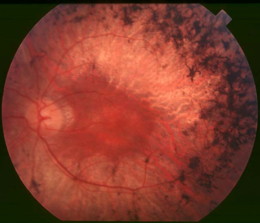
Clinical FeaturesRiazuddin et al. (2010) studied 4 affected and 7 unaffected members of a large consanguineous Pakistani family segregating autosomal recessive retinitis pigmentosa (RP). Medical records of the 4 affected individuals were suggestive of early-onset RP, with diagnoses made between 2 years and 4 years of age. Fundus photographs showed typical changes of RP, including attenuation of retinal arteries and bone spicule pigment deposits in the midperiphery of the retina. The diagnosis was confirmed by electroretinography (ERG), which showed typical RP changes with loss of both rod and cone responses in affected individuals but not in unaffected sibs or in the unaffected mother. None of the affected individuals had evidence of syndromic disease; specifically, there were no features consistent with Bardet-Biedl syndrome (BBS8; see 209900), which is also caused by TTC8 mutation. None had renal problems, and their body mass indices were within the normal range. There were no signs of polydactyly or midline defects or facies reminiscent of BBS, and no evidence of developmental delay, appreciable cognitive impairment, or inappropriate social behavior.
Goyal et al. (2016) studied a 4-generation consanguineous family of North Indian origin in which 2 brothers and their male cousin had RP. The proband was a 22-year-old man who had noticed night blindness and photophobia since age 17 years, with loss of central vision at age 18. His affected brother lost central vision at age 16 years, and their cousin had onset of RP at age 2 years with loss of central vision at age 5. Funduscopy in all 3 showed attenuated retinal vessels, bone spicule-like pigmentation in the midperiphery of the retina, macular degeneration, retinal pigment epithelium (RPE) degeneration, and waxy pallor of the optic disc. Visual acuity was reduced, and all 3 had high myopia. OCT in the proband's affected brother showed fraying of the rod and cone layers, hyperflexed areas in the RPE layer due to accumulation of pigments, and macular degeneration; ERG testing showed extinguished rod and cone responses. None of the affected individuals exhibited any extraocular characteristics of BBS, and thus were considered to represent nonsyndromic autosomal recessive RP with macular degeneration.
MappingIn a large consanguineous Pakistani family segregating autosomal recessive retinitis pigmentosa, Riazuddin et al. (2010) performed a genomewide scan and obtained a 2-point lod score of 3.12 at marker D14S256 (theta = 0.0). Recombination events defined a 13.4-cM (5.6-Mb) critical interval between D14S612 and D14S1050 on chromosome 14q, harboring more than 100 annotated genes.
Molecular GeneticsIn a large consanguineous Pakistani family segregating autosomal recessive retinitis pigmentosa (RP) mapping to chromosome 14q, Riazuddin et al. (2010) sequenced candidate genes and identified a homozygous splice site mutation in the TTC8 gene (608132.0005) that segregated with disease and was not found in 384 Pakistani control chromosomes or 384 chromosomes of northern European descent.
In the proband from a consanguineous North Indian family with RP and macular degeneration mapping to chromosome 14q, Goyal et al. (2016) performed whole-exome sequencing and identified homozygosity for a missense mutation in the TTC8 gene (Q449H; 608132.0006). The mutation segregated with disease in the family and was not found in 100 ethnically matched controls.