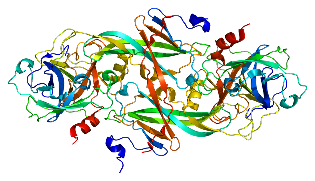
Factor Xi Deficiency

A number sign (#) is used with this entry because factor XI deficiency is caused by homozygous, compound heterozygous, or heterozygous mutation in the F11 gene (264900) on chromosome 4q35.
DescriptionFactor XI deficiency is an autosomal bleeding disorder characterized by reduced levels of factor XI in plasma (less than 15 IU/dL). Bleeding occurs mainly after trauma or surgery. On the basis of the concordance or discordance of F11 antigen and activity, the disorder is classified into the more frequent cross-reactive negative (CRM-) and the rarer CRM positive (CRM+) (summary by Duga and Salomon, 2009).
Clinical FeaturesMannhalter et al. (1987) demonstrated a deficiency of factor XI in a homozygous girl who was positive for crossreacting material. The ratio of factor XI coagulant activity to factor XI antigen was 0.04 for the proposita as compared with 0.7 to 0.74 in other family members and 1.04 +/- 0.15 in 12 normal individuals.
Dzik et al. (1987) reported the development of factor XI deficiency in a recipient of liver transplantation from a patient with said deficiency.
Clarkson et al. (1991) described a 54-year-old white man who acquired factor XI deficiency from a liver transplant performed for chronic active hepatitis secondary to hepatitis B infection. The donor of the liver was of Ashkenazi Jewish descent and had a history of bleeding after dental procedures. Before his death from intracerebral bleeding, he had been shown to have an isolated prolonged PTT value. The experience demonstrated that the liver is the only site of factor XI production.
Litz et al. (1988) studied a kindred in which 8 individuals by measurement and a ninth individual by presumption from the position in the pedigree were heterozygous. The family was detected through a propositus investigated for a spontaneous subcutaneous hematoma. Several of the 8 deficient individuals had recurrent nosebleeds, easy bruisability, recurrent gumbleeds, and/or postsurgical oozing for greater than 72 hours.
Although a homozygous or compound heterozygous deficiency of factor XI results in a variable bleeding phenotype, the clinical presentation in heterozygotes is less predictable. As reviewed by Bolton-Maggs (1996), however, most studies of heterozygotes suggest that 20 to 50% bleed excessively. The discrepancies in several studies may be related in part to differences in the definitions of 'excessive bleeding,' to the variability of the injuries, and to the different proportions of unchallenged patients. The variable manifestations of heterozygous factor XI deficiency may also be related to different genotypic forms of the heterozygotes or to other as yet uncharacterized imbalances in coagulation.
Heterozygotes for factor XI deficiency can have major bleeding problems. Indeed, Ragni et al. (1985) found no difference in the frequency of bleeders and nonbleeders between homozygotes and heterozygotes.
InheritanceFactor XI deficiency is generally inherited as a recessive trait; however, the dimeric structure of circulating F11 might result in a dominant-negative effect through intracellular heterodimer formation, eventually leading to a dominant pattern of inheritance, as clearly demonstrated by heterozygous mutation in the F11 gene (summary by Duga and Salomon, 2009).
Population GeneticsBiggs and MacFarlane (1962) noted that almost all reported patients with F11 deficiency were of Jewish extraction. Rosenthal (1964) collected 72 cases from 46 Jewish families. According to Seligsohn (1979), the homozygote frequency in Ashkenazi Jews in Israel is about 1 in 190, and the heterozygote frequency is about 1 in 8. Accordingly, factor XI deficiency is one of the most frequent genetic disorders in this ethnic group.
Factor XI deficiency is also common in the French Basque population (Bauduer et al., 1999; Zivelin et al., 2002).
Among 1,501 individuals of Ashkenazi Jewish origin screened for factor XI deficiency carrier status, Lazarin et al. (2013) found a carrier frequency of approximately 1 in 13. Among 15,724 ethnically diverse individuals the carrier frequency was 1 in 92.
Molecular GeneticsAsakai et al. (1989) identified 3 independent point mutations in the F11 gene of 6 unrelated Ashkenazi patients. One mutation disrupted normal mRNA splicing (264900.0001), another caused premature polypeptide termination (264900.0002), and the last resulted in a missense mutation (264900.0003). No correlation could be demonstrated between the specific genotype and the bleeding tendency in the subjects studied. In a survey of 53 apparently normal, unrelated Ashkenazi Jews, Asakai et al. (1989) found 2 who were heterozygous for the missense mutation, but none with the splicing or nonsense mutations. This situation is similar to that in Tay-Sachs disease (272800) and Gaucher disease (230800), in which several mutations have high frequency in Ashkenazi Jews.
Peretz et al. (1993) described a mutation in the F11 gene in an Ashkenazi Jewish patient. Pugh et al. (1995) reported 6 additional F11 mutations in patients with factor 11 deficiency (see, e.g., 264900.0004 and 264900.0005) and noted that 5 mutations had been identified in non-Ashkenazi patients, 2 in Japanese patients and 3 in English patients.
Mitchell et al. (1999) performed SSCP analysis of the F11 gene in 3 patients with heterozygous factor XI deficiency. Three missense mutations were found: arg308 to cys (264900.0009), ala412 to val (264900.0010), and ser576 to arg (264900.0011). In these 3 patients the factor XI antigen level varied from 22.9 to 38.6 and factor XI activity varied from 27 to 50%. They speculated about the mechanism by which these mutations in heterozygous state lead to clinical manifestations.
Salomon et al. (2003) studied the prevalence of acquired inhibitors against factor XI in patients with severe factor XI deficiency, discerned whether these inhibitors are related to specific mutations, and characterized their activity. Of 118 Israeli patients, 7 had an inhibitor; all belonged to a subgroup of 21 homozygotes for glu117-to-ter (264900.0002) who had a history of plasma replacement therapy. Three additional patients with inhibitors from the UK and the US also had this genotype and were exposed to plasma. The inhibitors affected factor XI activation by thrombin or factor XIIa and activation of factor IX by factor XIa.
Animal ModelPTA, or factor XI, deficiency, analogous to that in man and cattle, has been found in a family of Springer Spaniel dogs (Dodds and Kull, 1971).
HistoryFactor XI deficiency was first described by Rosenthal et al. (1953).
In a chromosomally normal, Ashkenazi Jewish boy with Prader-Willi syndrome (176270), Futterweit et al. (1986) found factor XI deficiency (with factor XI levels in the homozygous range). Although no deletion of chromosome 15 was visualized, submicroscopic deletion is possible in such cases. There was no family history of bleeding, but tests for heterozygosity in the parents were apparently not done.