Retinitis Pigmentosa 36

A number sign (#) is used with this entry because of evidence that retinitis pigmentosa-36 (RP36) is caused by homozygous mutation in the PRCD gene (610598) on chromosome 17q25.
For a phenotypic description and a discussion of genetic heterogeneity of retinitis pigmentosa, see 268000.
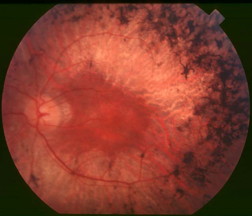
Clinical FeaturesZangerl et al. (2006) examined a 32-year-old woman from Bangladesh who first noticed difficulty seeing at night as a child. Funduscopic examination revealed optic discs with fairly normal color but markedly attenuated arterioles. Extensive bone-spicule-like pigmentation was present in all 4 quadrants of both eyes, with highest density at the equator, and was admixed with lighter colored deposits at the level of the retinal pigment epithelium. Small patches of geographic atrophy near fixation in both eyes were also observed. ERGs showed no response from either eye under any stimulus condition. The patient was born of first-cousin parents with normal vision and had 4 unaffected sibs, 1 brother who became blind at a young age, and 2 other sibs with uncharacterized vision problems.
Nevet et al. (2010) studied 24 individuals with retinitis pigmentosa from a Muslim Arab village with a high degree of inbreeding. Both scotopic and photopic electroretinograms (ERGs) were nonrecordable in all tested individuals, as early as 6 years of age. Typical bone spicule-type pigment deposits were noted in most patients. Additional findings included signs of macular degeneration (macular edema and puckering, macular scarring, and bull's eye maculopathy), optic disc pallor, and cataract. Most patients were also myopic.
MappingNevet et al. (2010) performed haplotype analysis in a consanguineous family with retinitis pigmentosa from a Muslim Arab village and identified a haplotype of 2 polymorphic marker alleles linked to the PRCD gene on chromosome 17q25.1 that cosegregated with RP in the family. Genetic analysis in 5 additional RP families from the same village showed cosegregation of the PRCD-linked haplotype with the disease in all of them.
Molecular GeneticsZangerl et al. (2006) analyzed the PRCD gene in 1,836 patients with RP and other inherited retinal disorders. In a 32-year-old woman from Bangladesh with autosomal recessive RP, they identified homozygosity for a missense mutation in the PRCD gene (C2Y; 610598.0001) that was identical to a mutation observed in dogs with progressive rod-cone degeneration.
In an individual from a Muslim Arab village with a high degree of inbreeding, who had RP mapping to the PRCD gene, Nevet et al. (2010) identified homozygosity for a nonsense mutation in the PRCD gene (R22X; 610598.0002). Analysis of 17 additional affected individuals and 28 unaffected relatives from 9 families revealed homozygosity for R22X in all affected individuals but in none of the unaffected family members. The mutation was also found in heterozygosity in 16 of 159 randomly selected adults from the village, indicating a carrier frequency of 10%. No carriers of R22X were detected among 130 unrelated Israeli Muslim Arab controls from Northern Israel, showing that this founder mutation is rare in the surrounding Muslim Arab population.