Silicosis

Silicosis is a form of occupational lung disease caused by inhalation of crystalline silica dust. It is marked by inflammation and scarring in the form of nodular lesions in the upper lobes of the lungs. It is a type of pneumoconiosis. Silicosis (particularly the acute form) is characterized by shortness of breath, cough, fever, and cyanosis (bluish skin). It may often be misdiagnosed as pulmonary edema (fluid in the lungs), pneumonia, or tuberculosis.
Silicosis resulted in at least 43,000 deaths globally in 2013, down from at least 50,000 deaths in 1990.
The name silicosis (from the Latin silex, or flint) was originally used in 1870 by Achille Visconti (1836–1911), prosector in the Ospedale Maggiore of Milan. The recognition of respiratory problems from breathing in dust dates to ancient Greeks and Romans. Agricola, in the mid-16th century, wrote about lung problems from dust inhalation in miners. In 1713, Bernardino Ramazzini noted asthmatic symptoms and sand-like substances in the lungs of stone cutters. With industrialization, as opposed to hand tools, came increased production of dust. The pneumatic hammer drill was introduced in 1897 and sandblasting was introduced in about 1904, both significantly contributing to the increased prevalence of silicosis.
Signs and symptoms
Because chronic silicosis is slow to develop, signs and symptoms may not appear until years after exposure. Signs and symptoms include:
- Dyspnea (shortness of breath) exacerbated by exertion
- Cough, often persistent and sometimes severe
- Fatigue
- Tachypnea (rapid breathing) which is often labored,
- Loss of appetite and weight loss
- Chest pain
- Fever
- Gradual darkening of skin (blue skin)
- Gradual dark shallow rifts in nails eventually leading to cracks as protein fibers within nail beds are destroyed.
In advanced cases, the following may also occur:
- Cyanosis, pallor along upper parts of body (blue skin)
- Cor pulmonale (right ventricle heart disease)
- Respiratory insufficiency
Patients with silicosis are particularly susceptible to tuberculosis (TB) infection—known as silicotuberculosis. The reason for the increased risk—3 fold increased incidence—is not well understood. It is thought that silica damages pulmonary macrophages, inhibiting their ability to kill mycobacteria. Even workers with prolonged silica exposure, but without silicosis, are at a similarly increased risk for TB.
Pulmonary complications of silicosis also include chronic bronchitis and airflow limitation (indistinguishable from that caused by smoking), non-tuberculous Mycobacterium infection, fungal lung infection, compensatory emphysema, and pneumothorax. There are some data revealing an association between silicosis and certain autoimmune diseases, including nephritis, scleroderma, and systemic lupus erythematosus, especially in acute or accelerated silicosis.
In 1996, the International Agency for Research on Cancer (IARC) reviewed the medical data and classified crystalline silica as "carcinogenic to humans." The risk was best seen in cases with underlying silicosis, with relative risks for lung cancer of 2–4. Numerous subsequent studies have been published confirming this risk. In 2006, Pelucchi et al. concluded, "The silicosis-cancer association is now established, in agreement with other studies and meta-analysis."
Pathophysiology

When small silica dust particles are inhaled, they can embed themselves deeply into the tiny alveolar sacs and ducts in the lungs, where oxygen and carbon dioxide gases are exchanged. There, the lungs cannot clear out the dust by mucous or coughing.
When fine particles of crystalline silica dust are deposited in the lungs, macrophages that ingest the dust particles will set off an inflammatory response by releasing tumor necrosis factors, interleukin-1, leukotriene B4 and other cytokines. In turn, these stimulate fibroblasts to proliferate and produce collagen around the silica particle, thus resulting in fibrosis and the formation of the nodular lesions. The inflammatory effects of crystalline silica are apparently mediated by the NALP3 inflammasome.
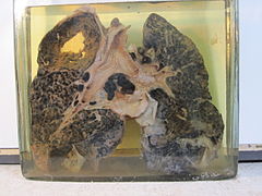
Characteristic lung tissue pathology in nodular silicosis consists of fibrotic nodules with concentric "onion-skinned" arrangement of collagen fibers, central hyalinization, and a cellular peripheral zone, with lightly birefringent particles seen under polarized light. The silicotic nodule represents a specific tissue response to crystalline silica. In acute silicosis, microscopic pathology shows a periodic acid-Schiff positive alveolar exudate (alveolar lipoproteinosis) and a cellular infiltrate of the alveolar walls.
Silica
Silicon (Si) is the second most common element in the Earth's crust (oxygen is the most common). The compound silica, also known as silicon dioxide (SiO2), is formed from silicon and oxygen atoms. Since oxygen and silicon make up about 75% of the Earth's crust, the compound silica is quite common. It is found in many rocks, such as granite, sandstone, gneiss and slate, and in some metallic ores. Silica can be a main component of sand. It can also be in soil, mortar, plaster, and shingles. The cutting, breaking, crushing, drilling, grinding, or abrasive blasting of these materials may produce fine to ultra fine airborne silica dust.
Silica occurs in 3 forms: crystalline, microcrystalline (or cryptocrystalline) and amorphous (non-crystalline). "Free" silica is composed of pure silicon dioxide, not combined with other elements, whereas silicates (e.g., talc, asbestos, and mica) are SiO2 combined with an appreciable portion of cations.
- Crystalline silica exists in 7 different forms (polymorphs), depending upon the temperature of formation. The main 3 polymorphs are quartz, cristobalite, and tridymite. Quartz is the second most common mineral in the world (next to feldspar).
- Microcrystalline silica consists of minute quartz crystals bonded together with amorphous silica. Examples include flint and chert.
- Amorphous silica consists of kieselgur (diatomite), from the skeletons of diatoms, and vitreous silica, produced by heating and then rapid cooling of crystalline silica. Amorphous silica is less toxic than crystalline, but not biologically inert, and diatomite, when heated, can convert to tridymite or cristobalite.
Silica flour is nearly pure SiO2 finely ground. Silica flour has been used as a polisher or buffer, as well as paint extender, abrasive, and filler for cosmetics. Silica flour has been associated with all types of silicosis, including acute silicosis.
Silicosis is due to deposition of fine respirable dust (less than 10 micrometers in diameter) containing crystalline silicon dioxide in the form of alpha-quartz, cristobalite, or tridymite.
Diagnosis
There are three key elements to the diagnosis of silicosis. First, the patient history should reveal exposure to sufficient silica dust to cause this illness. Second, chest imaging (usually chest x-ray) that reveals findings consistent with silicosis. Third, there are no underlying illnesses that are more likely to be causing the abnormalities. Physical examination is usually unremarkable unless there is complicated disease. Also, the examination findings are not specific for silicosis. Pulmonary function testing may reveal airflow limitation, restrictive defects, reduced diffusion capacity, mixed defects, or may be normal (especially without complicated disease). Most cases of silicosis do not require tissue biopsy for diagnosis, but this may be necessary in some cases, primarily to exclude other conditions.
For uncomplicated silicosis, chest x-ray will confirm the presence of small (< 10 mm) nodules in the lungs, especially in the upper lung zones. Using the ILO classification system, these are of profusion 1/0 or greater and shape/size "p", "q", or "r". Lung zone involvement and profusion increases with disease progression. In advanced cases of silicosis, large opacity (> 1 cm) occurs from coalescence of small opacities, particularly in the upper lung zones. With retraction of the lung tissue, there is compensatory emphysema. Enlargement of the hilum is common with chronic and accelerated silicosis. In about 5–10% of cases, the nodes will calcify circumferentially, producing so-called "eggshell" calcification. This finding is not pathognomonic (diagnostic) of silicosis. In some cases, the pulmonary nodules may also become calcified.
A computed tomography or CT scan can also provide a mode detailed analysis of the lungs, and can reveal cavitation due to concomitant mycobacterial infection.

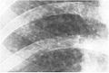
Chest X-ray showing uncomplicated silicosis

Complicated silicosis
Silicosis ILO Classification 2-2 R-R

Fibrothorax and pleural effusion caused by silicosis


Classification
Classification of silicosis is made according to the disease's severity (including radiographic pattern), onset, and rapidity of progression. These include:
- Chronic simple silicosis
- Usually resulting from long-term exposure (10 years or more) to relatively low concentrations of silica dust and usually appearing 10–30 years after first exposure. This is the most common type of silicosis. Patients with this type of silicosis, especially early on, may not have obvious signs or symptoms of disease, but abnormalities may be detected by x-ray. Chronic cough and exertional dyspnea (shortness of breath) are common findings. Radiographically, chronic simple silicosis reveals a profusion of small (<10 mm in diameter) opacities, typically rounded, and predominating in the upper lung zones.
- Accelerated silicosis
- Silicosis that develops 5–10 years after first exposure to higher concentrations of silica dust. Symptoms and x-ray findings are similar to chronic simple silicosis, but occur earlier and tend to progress more rapidly. Patients with accelerated silicosis are at greater risk for complicated disease, including progressive massive fibrosis (PMF).
- Complicated silicosis
- Silicosis can become "complicated" by the development of severe scarring (progressive massive fibrosis, or also known as conglomerate silicosis), where the small nodules gradually become confluent, reaching a size of 1 cm or greater. PMF is associated with more severe symptoms and respiratory impairment than simple disease. Silicosis can also be complicated by other lung disease, such as tuberculosis, non-tuberculous mycobacterial infection, and fungal infection, certain autoimmune diseases, and lung cancer. Complicated silicosis is more common with accelerated silicosis than with the chronic variety.
- Acute silicosis
- Silicosis that develops a few weeks to 5 years after exposure to high concentrations of respirable silica dust. This is also known as silicoproteinosis. Symptoms of acute silicosis include more rapid onset of severe disabling shortness of breath, cough, weakness, and weight loss, often leading to death. The x-ray usually reveals a diffuse alveolar filling with air bronchograms, described as a ground-glass appearance, and similar to pneumonia, pulmonary edema, alveolar hemorrhage, and alveolar cell lung cancer.
Prevention
 Play media
The best way to prevent silicosis is to avoid worker exposure to dust. The next best preventive measure is to control the dust. Water spray is often used where dust emanates to control the kick up of silica dust. To avoid dust accumulating on clothing and skin, place clothes in a seal-able bag and, if possible, shower once returning home. When dust starts accumulating around a workplace, utilize an industrial vacuum to contain and transport dust to a safe location. Dust can also be controlled through personal dry air filtering.
Preventing silicosis may require specific measures. One example is during tunnel construction where purpose-designed cabins are used in addition to air scrubbers to filter the air during construction. Items to be considered when selecting respiratory protection include whether it provides the correct level of protection, if facial fit testing has been provided, if the wearer is absent of facial hair, and how filters will be replaced.
Treatment
Silicosis is a permanent disease with no cure. Treatment options currently available focus on alleviating the symptoms and preventing any further progress of the condition. These include:
- Stopping further exposure to airborne silica, silica dust and other lung irritants, including tobacco smoking.
- Cough suppressants.
- Antibiotics for bacterial lung infection.
- Tuberculosis (TB) prophylaxis for those with positive tuberculin skin test or IGRA blood test.
- Prolonged anti-tuberculosis (multi-drug regimen) for those with active TB.
- Chest physiotherapy to help the bronchial drainage of mucus.
- Oxygen administration to treat hypoxemia, if present.
- Bronchodilators to facilitate breathing.
- Lung transplantation to replace the damaged lung tissue is the most effective treatment, but is associated with severe risks of its own from the lung transplant surgery as well as from consequences of long-term immunosuppression (e.g., opportunistic infections).
- For acute silicosis, bronchoalveolar lavage may alleviate symptoms, but does not decrease overall mortality.
Epidemiology
Silicosis resulted in 46,000 deaths in 2013 down from 55,000 deaths in 1990.
Occupational silicosis
Silicosis is the most common occupational lung disease worldwide. It occurs everywhere, but is especially common in developing countries. From 1991 to 1995, China reported more than 24,000 deaths due to silicosis each year. It also affects developed nations. In the United States, it is estimated that between one and two million workers have had occupational exposure to crystalline silica dust and 59,000 of these workers will develop silicosis sometime in the course of their lives.
According to CDC data, silicosis in the United States is relatively rare. The incidence of deaths due to silicosis declined by 84% between 1968 and 1999, and only 187 deaths in 1999 had silicosis as the underlying or contributing cause. Additionally, cases of silicosis in Michigan, New Jersey, and Ohio are highly correlated to industry and occupation.
Although silicosis has been known for centuries, the industrialization of mining has led to an increase in silicosis cases. Pneumatic drilling in mines and less commonly, mining using explosives, would raise fine-ultra fine crystalline silica dust (rock dust). In the United States, a 1930 epidemic of silicosis due to the construction of the Hawk's Nest Tunnel near Gauley Bridge, West Virginia caused the death of at least 400 workers. Other accounts place the mortality figure at well over 1000 workers, primarily African American transient workers from the southern United States. Workers who became ill were fired and left the region, making an exact mortality account difficult. The Hawks Nest Tunnel Disaster is known as "America's worst industrial disaster. The prevalence of silicosis led some men to grow what is called a miner's mustache, in an attempt to intercept as much dust as possible.
Chronic simple silicosis has been reported to occur from environmental exposures to silica in regions with high silica soil content and frequent dust storms.
Also, the mining establishment of Delamar Ghost Town, Nevada was ruined by a dry-mining process that produced a silicosis-causing dust. After hundreds of deaths from silicosis, the town was nicknamed The Widowmaker. The problem in those days was somewhat resolved with an addition of a nozzle to the drill which sprayed a mist of water, turning dust raised by drilling into mud, but this inhibited mining work.
Because of work-exposure to silica dust, silicosis is an occupational hazard to construction, demolition, mining, sandblasting, quarry, tunnelling, ceramics and foundry workers, as well as grinders, stone cutters, stone countertops, refractory brick workers, tombstone workers, workers in the oil and gas industry, pottery workers, fiberglass manufacturing, glass manufacturing, flint knappers and others. Brief or casual exposure to low levels of crystalline silica dust are said to not produce clinically significant lung disease.
In less developed countries where work conditions are poor and respiratory equipment is seldom used, the life expectancy is low (e.g. for silver miners in Potosí, Bolivia is around 40 years due to silicosis).
Recently, silicosis in Turkish denim sandblasters was detected as a new cause of silicosis due to recurring, poor working conditions.
Silicosis is seen in horses associated with inhalation of dust from certain cristobalite-containing soils in California.
Social realist artist Noel Counihan depicted men who worked in industrial mines in Australia in the 1940s dying of silicosis in his series of six prints, 'The miners' (1947 linocuts). While it has been long-thought that cases of silicosis in Australia were no longer possible, the recent reported epidemic in 2018 demonstrated that additional protections for workers were needed. Some have spoken publicly on the need to re-learn lessons from past experiences to prevent further disease. and 2019 The Australian Government Department of Health established a National Dust Disease Taskforce in response to the number of reported cases of silicosis in 2018.
Desert lung disease
A non-occupational form of silicosis has been described that is caused by long-term exposure to sand dust in desert areas, with cases reported from the Sahara, Libyan desert and the Negev. The disease is caused by deposition of this dust in the lung. Desert lung disease may be related to Al Eskan disease, a lung disorder thought to be caused by exposure to sand dust containing organic antigens, which was first diagnosed after the 1990 Gulf war. The relative importance of the silica particles themselves and the microorganisms that they carry in these health effects remains unclear.
Regulation
In March 2016, OSHA officially mandated that companies must provide certain safety measures for employees who work with or around silica, in order to prevent silicosis, lung cancer, and other silica-related diseases.
Key provisions
- Reduce the permissible exposure limit (PEL) for respirable crystalline silica to 50 micrograms per cubic meter of air, averaged over an 8-hour shift.
- Use engineering controls (such as water or ventilation) to limit worker exposure to silica dust; provide respirators when engineering controls cannot adequately limit exposure; limit worker access to high exposure areas; develop a written exposure control plan, offer medical exams to highly exposed workers, and train workers on silica risks and how to limit exposures. However, recent studies have found workers who grind granite counters and use water controls to eliminate dust are becoming ill from exposure to dust after water has evaporated the next day.
- Provide medical exams to monitor highly exposed workers and gives them information about their lung health.
An additional provision exists for small business who are provided flexibility.
Compliance schedule
Both standards contained in the final rule take effect on June 23, 2016, after which industries have one to five years to comply with most requirements, based on the following schedule:
- Construction – June 23, 2017, one year after the effective date.
- General Industry and Maritime – June 23, 2018, two years after the effective date.
- Hydraulic Fracturing – June 23, 2018, two years after the effective date for all provisions except Engineering Controls, which have a compliance date of June 23, 2021.
See also
- Pneumoconiosis
- Asbestosis – Pneumoconiosis caused by inhalation and retention of asbestos fibers
- Health effects arising from the September 11 attacks
- Hawks Nest Tunnel disaster – Tunnel in West Virginia where hundreds of workers contracted silicosis
- Dust pneumonia