Myocarditis

Myocarditis, also known as inflammatory cardiomyopathy, is inflammation of the heart muscle. Symptoms can include shortness of breath, chest pain, decreased ability to exercise, and an irregular heartbeat. The duration of problems can vary from hours to months. Complications may include heart failure due to dilated cardiomyopathy or cardiac arrest.
Myocarditis is most often due to a viral infection. Other causes include bacterial infections, certain medications, toxins, and autoimmune disorders. A diagnosis may be supported by an electrocardiogram (ECG), increased troponin, heart MRI, and occasionally a heart biopsy. An ultrasound of the heart is important to rule out other potential causes such as heart valve problems.
Treatment depends on both the severity and the cause. Medications such as ACE inhibitors, beta blockers, and diuretics are often used. A period of no exercise is typically recommended during recovery. Corticosteroids or intravenous immunoglobulin (IVIG) may be useful in certain cases. In severe cases an implantable cardiac defibrillator or heart transplant may be recommended.
In 2013, about 1.5 million cases of acute myocarditis occurred. While people of all ages are affected, the young are most often affected. It is slightly more common in males than females. Most cases are mild. In 2015 cardiomyopathy, including myocarditis, resulted in 354,000 deaths up from 294,000 in 1990. The initial descriptions of the condition are from the mid-1800s.
Signs and symptoms
The signs and symptoms associated with myocarditis are varied, and relate either to the actual inflammation of the myocardium or to the weakness of the heart muscle that is secondary to the inflammation. Signs and symptoms of myocarditis include the following:
- Chest pain (often described as "stabbing" in character)
- Congestive heart failure (leading to swelling, shortness of breath and liver congestion)
- Palpitations (due to abnormal heart rhythms)
- Dullness of heart sounds
- Sudden death (in young adults, myocarditis causes up to 20% of all cases of sudden death)
- Fever (especially when infectious, e.g., in rheumatic fever)
- Symptoms in young children tend to be more nonspecific, with generalized malaise, poor appetite, abdominal pain, and chronic cough. Later stages of the illness will present with respiratory symptoms with increased work of breathing, and is often mistaken for asthma.
Since myocarditis is often due to a viral illness, many patients give a history of symptoms consistent with a recent viral infection, including fever, rash, diarrhea, joint pains, and easily becoming tired.
Myocarditis is often associated with pericarditis, and many people with myocarditis present with signs and symptoms that suggest myocarditis and pericarditis at the same time.
Causes
A large number of causes of myocarditis have been identified, but often a cause cannot be found. In Europe and North America, viruses are common culprits. Worldwide, however, the most common cause is Chagas disease, an illness endemic to Central and South America that is due to infection by the protozoan Trypanosoma cruzi. In viral myocarditis, the Coxsackie B family of the single-stranded RNA viruses, in particular the plus-strand RNA virus Coxsackievirus B3 and Coxsackievirus B5 are the most frequent cause. Many of the causes listed below, particularly those involving protozoa, fungi, parasites, allergy, autoimmune disorders, and drugs are also causes of eosinophilic myocarditis.
Infections
- Viral: adenovirus, parvovirus B19, coxsackie virus, rubella virus, polio virus, Epstein-Barr virus, hepatitis C, severe acute respiratory syndrome coronavirus 2
- Protozoan: Trypanosoma cruzi (causing Chagas disease) and Toxoplasma gondii
- Bacterial: Brucella, Corynebacterium diphtheriae, gonococcus, Haemophilus influenzae, Actinomyces, Tropheryma whipplei, Vibrio cholerae, Borrelia burgdorferi, leptospirosis, Rickettsia, Mycoplasma pneumoniae
- Fungal: Aspergillus
- Parasitic: ascaris, Echinococcus granulosus, Paragonimus westermani, schistosoma, Taenia solium, Trichinella spiralis, visceral larva migrans, Wuchereria bancrofti
Bacterial myocarditis is rare in patients without immunodeficiency.
Toxins
- Drugs, including alcohol, anthracyclines and some other forms of chemotherapy, and antipsychotics, e.g., clozapine, also some designer drugs such as mephedrone
Immunologic
- Allergic (acetazolamide, amitriptyline)
- Rejection after a heart transplant
- Autoantigens (scleroderma, systemic lupus erythematosus, sarcoidosis, systemic vasculitis such as eosinophilic granulomatosis with polyangiitis, and granulomatosis with polyangiitis, Kawasaki disease, idiopathic hypereosinophilic syndrome)
- Toxins (arsenic, toxic shock syndrome toxin, carbon monoxide, or snake venom)
- Heavy metals (copper or iron)
Physical agents
- Electric shock, hyperpyrexia, and radiation
Mechanism
Most forms of myocarditis involve the infiltration of heart tissues by one or two types of pro-inflammatory blood cells, lymphocytes and macrophages plus two respective descendants of these cells, NK cells and macrophages. Eosinophilic myocarditis is a subtype of myocarditis in which cardiac tissue is infiltrated by another type of pro-inflammatory blood cell, the eosinophil. Eosinophilic myocarditis is further distinguished from non-eosinophilic myocarditis by having a different set of causes and recommended treatments. Coxsackie B, specifically B3 and B5, has been found to interact with coxsackievirus-adenovirus receptor (CAR) and decay-accelerating factor (DAF). However, other proteins have also been identified that allow Coxsackieviruses to bind to cardiac cells. The natural function of CAR and mechanism that the Coxsackievirus uses to infect the cardiac muscle is still unknown. The mechanism by which coxsackie B viruses (CBVs) trigger inflammation is believed to be through the recognition of CBV virions by Toll-like receptors.
Diagnosis
 91/PericarditisMyocarditis.jpg/310px-PericarditisMyocarditis.jpg" decoding="async" width="310" height="148" class="thumbimage" srcset="//upload.wikimedia.org/wikipedia/commons/thumb/9/91/PericarditisMyocarditis.jpg/465px-PericarditisMyocarditis.jpg 1.5x, //upload.wikimedia.org/wikipedia/commons/thumb/9/91/PericarditisMyocarditis.jpg/620px-PericarditisMyocarditis.jpg 2x" data-file-width="4204" data-file-height="2001">
Myocarditis refers to an underlying process that causes inflammation and injury of the heart. It does not refer to inflammation of the heart as a consequence of some other insult. Many secondary causes, such as a heart attack, can lead to inflammation of the myocardium and therefore the diagnosis of myocarditis cannot be made by evidence of inflammation of the myocardium alone.
Myocardial inflammation can be suspected on the basis of electrocardiographic (ECG) results, elevated C-reactive protein (CRP) and/or erythrocyte sedimentation rate (ESR), and increased IgM (serology) against viruses known to affect the myocardium. Markers of myocardial damage (troponin or creatine kinase cardiac isoenzymes) are elevated.
The ECG findings most commonly seen in myocarditis are diffuse T wave inversions; saddle-shaped ST-segment elevations may be present (these are also seen in pericarditis).
The gold standard is the biopsy of the myocardium, in general done in the setting of angiography. A small tissue sample of the endocardium and myocardium is taken and investigated. The cause for the myocarditis can be only diagnosed by a biopsy. Endomyocardial biopsy samples are assessed for histopathology (how the tissue looks like under the microscope: myocardial interstitium may show abundant edema and inflammatory infiltrate, rich in lymphocytes and macrophages. Focal destruction of myocytes explains the myocardial pump failure. In addition samples may be assessed with immunohistochemistry to determine which types of immune cells are involved in the reaction and how they are distributed. Furthermore, PCR and/or RT-PCR may be performed to identify particular viruses. Finally, further diagnostic methods like microRNA assays and gene-expression profile may be performed.
Cardiac magnetic resonance imaging (cMRI or CMR) has been shown to be very useful in diagnosing myocarditis by visualizing markers for inflammation of the myocardium. Recently, consensus criteria for the diagnosis of myocarditis by CMR have been published.
 Play media
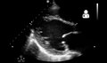
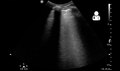
Ultrasound showing cardiogenic shock due to myocarditis
 Play media
Ultrasound showing cardiogenic shock due to myocarditis
 Play media
Ultrasound showing cardiogenic shock due to myocarditis


Treatment
As with most viral infections, symptomatic treatment is the only form of therapy for most forms of myocarditis. In the acute phase, supportive therapy, including bed rest, is indicated.
Medication
In people with symptoms, digoxin and diuretics may help. For people with moderate to severe dysfunction, cardiac function can be supported by use of inotropes such as milrinone in the acute phase, followed by oral therapy with ACE inhibitors when tolerated.
Systemic corticosteroids may have beneficial effects in people with proven myocarditis. However, data on the usefulness of corticosteroids should be interpreted with caution, since 58% of adults recover spontaneously, while most studies on children lack control groups.
A 2015 Cochrane review found no evidence of benefit of using intravenous immunoglobulin (IVIG) in adults and tentative benefit in certain children. It is not recommended routinely until there is better evidence.
Surgery
People who do not respond to conventional therapy may be candidates for bridge therapy with left ventricular assist devices. Heart transplantation is reserved for people who fail to improve with conventional therapy.
Extracorporeal membrane oxygenation may be used in those who are about to go into cardiac arrest.
Alternative medicine
Studies have shown no benefit for the use of herbal medicine on all cause mortality in viral myocarditis.
Epidemiology
The exact incidence of myocarditis is unknown. However, in series of routine autopsies, 1–9% of all patients had evidence of myocardial inflammation. In young adults, up to 20% of all cases of sudden death are due to myocarditis.
Among patients with HIV, myocarditis is the most common cardiac pathological finding at autopsy, with a prevalence of 50% or more.
Myocarditis is the third most common cause of death among young adults with a cumulative incidence rate globally of 1.5 cases per 100,000 persons annually. Myocarditis accounts for approximately 20% of sudden cardiac death in a variety of populations. Populations that experience this increased mortality rate include: adults under 40, young athletes, U.S. Air Force recruits, and elite Swedish orienteers. The prevalence rate of myocarditis is about 22 cases per 100,000 persons annually. With individuals who develop myocarditis, the first year is difficult as a collection of cases have shown there is a 20% mortality rate.
</ref> One rare instance of myocarditis is viral fulminant myocarditis; fulminant myocarditis involves rapid onset cardiac inflammation and a mortality rate of 40-70%. When looking at different causes of myocarditis, viral infection is the most prevalent, especially in children; however, the prevalence rate of myocarditis is often underestimated since the condition is easily overlooked. Viral myocarditis being an outcome of viral infection depends heavily on genetic host factors and the pathogenicity unique to the virus. One notable instance of viral myocarditis is the involvement of the SARS-CoV-2 virus; fulminant myocarditis from cardiac damage and SARS-CoV-2 has been associated with high mortality rates. Some incidences of acute myocarditis can be attributed to the exposure of drugs or toxic substances and abnormal immunoreactivity. The following agents may be other causes of myocarditis in various populations also, as previously highlighted in a prior section: protozoa, viruses, bacteria, rickettsia, and fungi. If one tests positive for an acute viral infection, clinical developments have discovered that 1% to 5% of said population may show some form of myocarditis.
When looking at the population affected, myocarditis is more common in pregnant women in addition to children and those who are immunocompromised. Myocarditis, however, has shown to be more common in the male population than in the female. Multiple studies report a ratio of 1:1.3-1.7 of female-male prevalence of myocarditis. Young males specifically have a higher incidence rate than any other population due to their testosterone levels creating a greater inflammatory response that increases chance of cardiac pathologies such as Cardiomyopathy, heart failure, or Myocarditis. While males tend to have a higher tendency of developing myocarditis, females tend to display more severe signs and symptoms such as ventricular tachycardia and ventricular fibrillation at an older age. Clinical patterns can assist in the diagnosis of myocarditis among the affected population. Due to the asymptomatic nature of Myocarditis, much information about the epidemiology of the disease is due to postmortem research. In a study of 3,055 patients with acute or chronic myocarditis, 72% presented with difficulty or labored breathing, 32% with chest pain, and another 18% with arrhythmias. Clinical observation suggests the possibility of a relationship between immunization and cardiac related symptoms; myocarditis and pericarditis have been observed to have a 200 times higher incidence rate post smallpox vaccine compared to pre smallpox vaccine.
History
Cases of myocarditis have been documented as early as the 1600s, but the term "myocarditis", implying an inflammatory process of the myocardium, was introduced by German physician Joseph Friedrich Sobernheim in 1837. However, the term has been confused with other cardiovascular conditions, such as hypertension and ischemic heart disease. Following admonition regarding the indiscriminate use of myocarditis as a diagnosis from authorities such as British cardiologist Sir Thomas Lewis and American cardiologist and a co-founder of the American Heart Association Paul White, myocarditis was under-diagnosed.
Although myocarditis is clinically and pathologically clearly defined as "inflammation of the myocardium", its definition, classification, diagnosis, and treatment are subject to continued controversy, but endomyocardial biopsy has helped define the natural history of myocarditis and clarify clinicopathological correlations.