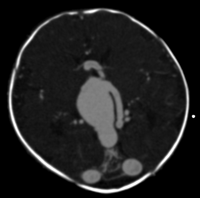
Vein Of Galen Aneurysmal Malformation

A congenital vascular malformation characterized by dilation of the embryonic precursor of the vein of Galen. It is a sporadic lesion that occurs during embryogenesis.
Epidemiology
The lesion is rare, with less than 800 cases (representing less than 10% of all cerebral arteriovenous malformations) reported so far. Vein of Galen aneurysmal malformation (VGAM) is more frequent in males than in females.
Clinical description
Cardiac insufficiency of variable severity is the principle manifestation that leads to detection of the malformation in newborns. It is sometimes associated with respiratory problems and/or hepatic, renal, and/or cerebral (encephalomalacia) manifestations, the main concern for therapeutic management. In infants, macrocrania may lead to suspicion of VGAM. Epilepsy, neuropsychological delay and neurologic deficiency are indicators of the malformation in older children. Cerebral hemorrhage only occurs in older children with progressive forms of VGAM.
Diagnostic methods
In newborns, the cardiac manifestations dominate the clinical picture. Trans-fontanel ultrasonography should be conducted before MRI.
Differential diagnosis
It is important to differentiate VGAM from congenital cardiopathy (by means of trans-fontanel ultrasonography).
Antenatal diagnosis
VGAMs are sometimes detected through ultrasound examinations during the third trimester of pregnancy. In this case, only two findings justify medical termination of the pregnancy: severe cardiac insufficiency and cerebral damage detected by fetal MRI.
Genetic counseling
In infants with macrocrania, the diagnosis relies on MRI. During the neonatal period, investigations for encephalomalacia (antenatal MRI, trans-fontanel ultrasonography, CT scans) are essential.
Management and treatment
Monitoring of a newborn with a VGAM that is either naturally well tolerated or controlled by medical treatment should consist of monthly clinical and neuropsychological examinations, and MRI or CT scans from birth to 3 months of age. During the neonatal period, early emergency treatment is only required in case of severe or poorly tolerated cardiac insufficiency. The child should be managed by a pediatric intensive care unit and an experienced team of pediatric neurologists. For asymptomatic infants, therapeutic management of the VGAM should be delayed until four or five months after birth, but may be required earlier if the VGAM is life-threatening or may affect cerebral development. Treatment is endovascular, using a femoral transarterial approach. Arteriovenous shunts are embolized with a glue, usually requiring two or three interventions. Hydrocephaly due to venous congestion may require a ventricular diversion valve or a ventriculocisternostomy. Radiosurgery is not a therapeutic option. Surgery is not indicated as a treatment for VGAM, except in very rare cases. Patients undergoing VGAM treatment should have regular programmed clinical and morphological examinations. The aim of these examinations is to detect rapid increases in cranial circumference, delays in psychomotor development, or pial or more profound venous reflux. Additional interventions involving targeted embolisation may be required, using either a venous or arterial approach depending on the architecture of the lesion.
Prognosis
The neurological prognosis for VGAM with early treatment is excellent; however, the prognosis is poorer for patients with severe neonatal malformations.