Retinitis Pigmentosa 59

A number sign (#) is used with this entry because of evidence that retinitis pigmentosa-59 (RP59) is caused by homozygous mutation in the DHDDS gene (608172) on chromosome 1p36.
Congenital disorder of glycosylation type Ibb (CDG1BB) can be caused by compound heterozygous mutation in the DHDDs gene. One such patient has been reported.
For a general phenotypic description and a discussion of genetic heterogeneity of retinitis pigmentosa, see 268000; for congenital disorder of glycosylation, see 212065.
Clinical FeaturesZuchner et al. (2011) studied an Ashkenazi Jewish family in which 3 of 4 sibs were diagnosed with retinitis pigmentosa (RP) in their teenage years. Early symptoms consisted of impaired night and peripheral vision. Clinical examination of the affected individuals revealed pigmentary retinal degeneration, and the diagnosis of RP was confirmed by rod and cone responses on electroretinograms (ERGs). The remainder of the physical examination was unremarkable, and laboratory studies, including x-ray bone body survey and bone density scan, were all normal, although 2 of the affected sibs had a history of lytic bone disease diagnosed 15 years previously.
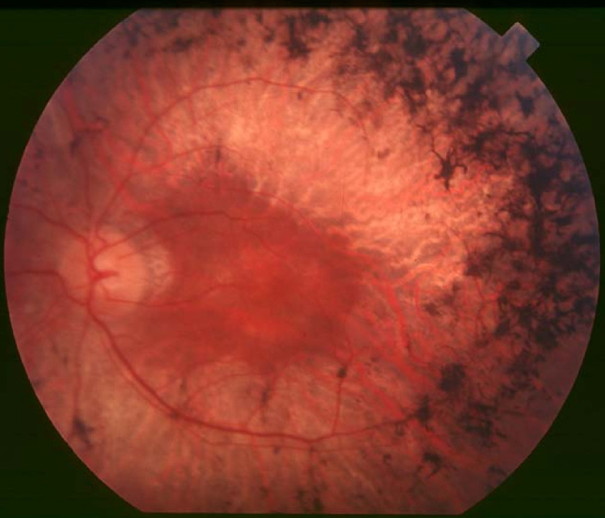
Lam et al. (2014) provided follow-up of the Ashkenazi Jewish family originally reported by Zuchner et al. (2011). Funduscopy showed diffuse pigmentary retinal degeneration with vascular attenuation consistent with RP. Impaired night vision and peripheral field defects developed in the second decade of life; ERG responses were nondetectable in 2 of the sibs and indicated cone-rod dysfunction in the third. By the fourth decade of life, vision had progressively deteriorated to legal blindness with constriction of visual fields to less than 10 degrees.
Zelinger et al. (2011) examined 18 Ashkenazi Jewish patients with RP59 and observed a spectrum of findings, with visual acuities ranging from light perception to 20/20 vision. Funduscopic findings at various disease stages included waxy appearance of the optic nerve head, attenuation of retinal blood vessels, and bone spicule-like pigmentation. Optical coherence tomography (OCT) imaging in early disease showed preserved central retinal photoreceptors but a decline in photoreceptor layer thickness with distance from the fovea, and occasionally the presence of cystoid macular edema. Kinetic visual fields revealed reduced peripheral function in the youngest patients studied and only small central islands of vision remaining later in life. ERG responses were nondetectable in most patients.
Wen et al. (2013) found that patients with RP59 had increased levels of shortened plasma and urinary dolichols compared to controls, and they suggested that this assay could serve as a biomarker. Sabry et al. (2016) noted that patients with RP59 do not present with serum glycoprotein hypoglycosylation, but show abnormal serum and urine dolichols, as demonstrated by Wen et al. (2013).
Congenital Disorder of Glycosylation Type Ibb
Sabry et al. (2016) reported a boy, born of unrelated parents, with a fatal multisytem disorder. The infant had intrauterine growth retardation, axial hypotonia, peripheral hypertonia, enlarged liver, micropenis, and cryptorchidism. He had transient elevation of liver enzymes, renal failure, and seizures. He made no eye contact and had poor feeding with failure to thrive. Ophthalmologic examination at age 2 months showed pale papillae and there was no response on ERG; he also had sensorineural deafness. The infant died at age 8 months during an episode of status epilepticus. Laboratory studies showed hypoglycosylation of plasma proteins; patient fibroblasts showed increased levels of truncated dolichol-linked oligosaccharides, and microsomes derived from the patient showed low levels of dolichol-phosphate. The biochemical findings were consistent with congenital disorder of glycosylation type I.
InheritanceThe transmission pattern of RP59 in the family reported by Zelinger et al. (2011) was consistent with autosomal recessive inheritance.
MappingZelinger et al. (2011) performed homozygosity mapping in Ashkenazi Jewish patients with autosomal recessive RP and identified a 1.7-Mb shared homozygous region on chromosome 1p36.11.
Molecular GeneticsIn an Ashkenazi Jewish family in which 3 of 4 sibs had RP, Zuchner et al. (2011) screened all known RP genes but found no mutations. Whole-exome sequencing identified a single variant in the DHDDS gene (K42E; 608172.0001) that was present in homozygosity in the affected sibs but not present in their unaffected sib and for which the unaffected parents were heterozygous. Zuchner et al. (2011) stated that variant was likely to have arisen from an ancestral founder, as it was detected in heterozygosity in 8 of 717 Ashkenazi Jewish controls but was not found in 6,977 confirmed non-Ashkenazi white controls; the variant was also found once in 5,893 additional white controls for whom genomewide genotype data were not available.
In 15 (12%) of 123 Ashkenazi Jewish (AJ) probands with RP, Zelinger et al. (2011) identified homozygosity for the K42E founder mutation. The K42E mutation was found in heterozygosity in 1 of 322 ethnically matched controls, indicating a carrier frequency of 0.3% in the AJ population; it was not detected in an additional set of 109 AJ patients with RP, in 20 AJ patients with other inherited retinal diseases, or in 70 patients with retinal degeneration of other ethnic origins.
Sabry et al. (2016) demonstrated that the K42E variant was unable to complement the growth defect in yeast lacking the ortholog RER2, consistent with a loss of function. Yeast transfected with the mutation also showed hypoglycosylation of carboxypeptidase Y. These defects could be restored with wildtype DHDDS.
Congenital Disorder of Glycosylation Type Ibb
By sequencing genes required for dolichol biosynthesis in a boy with congenital disorder of glycosylation type Ibb, Sabry et al. (2016) identified compound heterozygosity for nonsense (608172.0004) and splice site (608172.0005) mutations in the DHDDS gene, which segregated with the disorder in the family. Patient cells showed 20 to 25% residual normal DHDDS mRNA, likely from the leaky splice site mutation, and 35% residual DHDDS activity compared to controls. The patient also carried the homozygous F304S polymorphism in the ALG6 gene (604566), which is considered to be a disease modifier that exacerbates the disease in patients with mutations in other genes of the glycosylation pathway. Sabry et al. (2016) noted that the phenotype was much more severe than that reported in the patients with RP59.
Animal ModelBy morpholino-knockdown of Dhdds in zebrafish, Zuchner et al. (2011) observed virtually identical photoreceptor defects as those observed with N-linked glycosylation-interfering mutations in the light-sensing protein rhodopsin (RHO; 180380), which result in RP4 (613731) in humans.